How Kidneys Balance Salt And Water

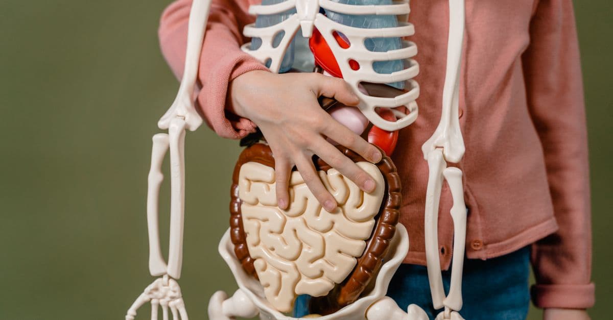
Photo: Leeloo The First / Pexels
Each kidney holds roughly one million microscopic filtering units called nephrons, and together they filter around 180 liters of blood plasma per day in an average adult male (about 150 liters in an average adult female), yet a person typically excretes only 1 to 2 liters of urine daily (Oregon State University Human Anatomy & Physiology, open textbook). The gap between those two numbers, more than 99% of what gets filtered, is quietly reabsorbed back into the bloodstream, is the whole story of how kidneys keep salt and water in balance. This article walks through that filter-then-reclaim system as pure physiology, not medical guidance; anyone with a diagnosed kidney condition should rely on their own clinician for interpretation.
What a nephron actually is, and why it filters almost everything
A nephron is the basic structural and functional unit of the kidney, a microscopic tubule that filters blood plasma and then selectively reclaims most of what it filtered. Each nephron begins at the glomerulus, a tuft of capillaries wrapped inside a cup-shaped structure called Bowman's capsule, and ends several millimeters later at a collecting duct that merges with thousands of others to funnel urine toward the bladder. Understanding the kidney means understanding this single repeating unit, multiplied roughly a million times per kidney.
Blood enters the glomerulus under comparatively high pressure, generated by the kidney's afferent arteriole being wider than its efferent arteriole, a plumbing asymmetry that forces fluid outward through the capillary walls rather than letting it simply pass through. Water, electrolytes (sodium, potassium, chloride, bicarbonate), glucose, amino acids, and small waste products like urea and creatinine all cross into Bowman's capsule. Red blood cells, white blood cells, platelets, and large plasma proteins like albumin are normally held back by the filtration barrier's size and charge selectivity.
This first-stage output is called the glomerular filtrate, and physiologically it looks a great deal like blood plasma with the cells and most proteins removed. The rate at which this filtrate is produced is known as the glomerular filtration rate (GFR), and it is the single most common lab-derived number nephrologists use to estimate how well kidneys are functioning overall (National Kidney Foundation; NCBI StatPearls, "Physiology, Glomerular Filtration Rate"). A healthy adult GFR runs at roughly 90 to 120 milliliters per minute, which is where the 180-liter daily figure comes from when you multiply it out across 24 hours.
The reason the kidney filters so much more than it excretes is not inefficiency. It is a deliberate two-step design: filter broadly and indiscriminately first, at a scale that would be wasteful if none of it were recovered, then reclaim almost everything of value as the filtrate travels onward through the tubule. That second step, tubular reabsorption, is where the real regulation of salt and water happens, and it is the subject of the rest of this article.
The proximal tubule does the heavy lifting, unconditionally
After leaving Bowman's capsule, filtrate enters the proximal convoluted tubule, the first and longest segment of the nephron's tubule. This is where the bulk of reabsorption happens, and it happens whether the body is dehydrated, overhydrated, or perfectly balanced. Roughly two-thirds of filtered sodium, and a proportional amount of water that follows it osmotically, is reclaimed here through a dense array of transport proteins embedded in the tubule wall.
The proximal tubule works largely through active transport. Sodium-potassium ATPase pumps on the basolateral membrane (the side facing the blood) actively push sodium out of the tubule cell and into the surrounding tissue, lowering the sodium concentration inside the cell. That gradient then pulls sodium in from the filtrate side through various co-transporters, which piggyback glucose, amino acids, and other useful solutes along with it. Water follows osmotically through aquaporin-1 water channels, which are constitutively present in this segment rather than hormonally regulated.
A useful way to think about the proximal tubule is as the kidney's bulk-recovery stage, analogous to a recycling plant's first conveyor belt that pulls out the large, obvious, easily sorted material before finer sorting happens downstream. It does not fine-tune anything to the body's momentary needs; it simply reclaims a fixed, large fraction of everything regardless of context. That consistency matters, because it means the kidney's actual regulatory decisions, the ones that respond to dehydration or fluid overload, happen further along the tubule, in segments built specifically for adjustable, hormone-responsive control.
By the time filtrate leaves the proximal tubule, its total volume has shrunk substantially, but its osmolarity (solute concentration) is still close to that of blood plasma, because both water and solutes were reabsorbed roughly in proportion to each other. The real concentrating or diluting work is done later, downstream, in a structure built specifically for that purpose: the loop of Henle.
The loop of Henle: how a hairpin turn concentrates urine
The loop of Henle is a hairpin-shaped segment of the tubule that dips from the kidney's outer region (the cortex) down into its inner region (the medulla) and back up again. Its job is unlike the proximal tubule's: instead of reabsorbing a fixed fraction of everything, it builds a standing salt concentration gradient in the surrounding kidney tissue, one that gets progressively saltier the deeper into the medulla you go. That gradient is what later allows the collecting duct to concentrate urine well beyond the salt concentration of blood plasma, when the body needs to conserve water.
The mechanism behind this is called countercurrent multiplication, and it depends on the loop's two limbs having very different permeability properties (TeachMePhysiology, "Loop of Henle - Counter Current Multiplication"). The descending limb is highly permeable to water but does not actively transport salt, so as filtrate moves down into the increasingly salty medulla, water is pulled out osmotically and the remaining fluid becomes progressively more concentrated. The ascending limb does the opposite: it is essentially impermeable to water, but it actively pumps sodium and chloride out into the surrounding tissue, which is what builds and maintains the medullary salt gradient in the first place.
Why is it called "countercurrent"?
It is called countercurrent because fluid in the descending limb flows in the opposite direction to fluid in the adjacent ascending limb, and this opposing flow arrangement is what lets a relatively modest pumping force at any single point get multiplied into a large overall concentration gradient by the time fluid completes the loop. Without the opposing flow, the same pumps could not build nearly as steep a gradient.
The clinical relevance of this segment shows up directly in pharmacology: loop diuretics, a class of medications that includes furosemide, work by blocking the sodium-potassium-chloride co-transporter in the thick ascending limb, which disrupts the gradient-building step and produces a strong diuretic effect (increased urine output) precisely because this segment normally reabsorbs so much salt. That is also why loop diuretics are more potent than other diuretic classes that act on segments handling smaller absolute quantities of sodium.
By the time filtrate exits the loop of Henle and enters the distal tubule, it has been substantially diluted relative to the surrounding medullary tissue, because so much salt was pumped out without water following it. What happens next, in the distal tubule and collecting duct, determines whether the final urine stays dilute or gets concentrated back down using that medullary gradient the loop just built.
The distal tubule and collecting duct: where hormones make the final call
If the proximal tubule is bulk recovery and the loop of Henle is gradient construction, the distal convoluted tubule and collecting duct are where the kidney makes its real-time decisions. This is the only part of the nephron where reabsorption rates are actively adjusted minute to minute in response to the body's actual hydration and sodium status, largely through two hormones: aldosterone and antidiuretic hormone.
Aldosterone is a steroid hormone produced by the adrenal glands (small glands sitting atop each kidney) that increases sodium reabsorption in the distal tubule and collecting duct by upregulating sodium channels and pumps in those cells. Because water tends to follow sodium osmotically, more sodium reabsorption also means more water retention and, secondarily, more potassium excretion, since the transport machinery aldosterone activates often exchanges reabsorbed sodium for excreted potassium.
Antidiuretic hormone (ADH), also called vasopressin, works differently. It is released by the posterior pituitary gland and acts specifically on the collecting duct's water permeability rather than on sodium transport. ADH binds receptors on collecting duct cells and triggers the insertion of aquaporin-2 water channel proteins into the cell membrane facing the tubule lumen. More aquaporins means more water can be pulled out of the tubule fluid and back into the blood, using the concentrated medullary tissue the loop of Henle built as the osmotic driving force.
How does ADH control how concentrated urine becomes?
ADH increases the number of aquaporin-2 water channels inserted into the collecting duct's cell membranes. More channels mean more water is reabsorbed into the blood rather than excreted, producing concentrated urine during dehydration. When ADH release is suppressed, fewer channels are inserted, water stays in the tubule, and urine comes out dilute.
Without ADH, the collecting duct is largely water-impermeable, and dilute filtrate simply passes through unchanged, producing large volumes of pale, dilute urine. With high ADH, the same segment becomes highly water-permeable, and because it sits inside the salty medullary environment the loop of Henle built, water gets drawn out efficiently, producing small volumes of dark, concentrated urine. This single hormone, acting on a single water channel protein, is the kidney's primary lever for matching water output to hydration status on a minute-to-minute basis.
The renin-angiotensin-aldosterone system: the feedback loop behind it all
None of this hormonal fine-tuning happens on its own initiative. It is triggered by a sensing-and-response cascade called the renin-angiotensin-aldosterone system (RAAS), which begins inside the kidney itself. Specialized cells called the juxtaglomerular apparatus, located where the nephron's distal tubule passes close to its own glomerulus's arterioles, monitor blood pressure and the rate of sodium delivery to that point in the tubule.
When the juxtaglomerular apparatus detects low blood pressure, low blood volume, or reduced sodium delivery, it releases an enzyme called renin into the bloodstream. Renin acts on a liver-produced protein called angiotensinogen, cleaving it into angiotensin I, which is then converted by an enzyme called angiotensin-converting enzyme (ACE, largely in the lungs) into angiotensin II, a potent hormone with two simultaneous effects: it directly constricts blood vessels, raising blood pressure quickly, and it stimulates the adrenal glands to release aldosterone, which raises blood pressure more slowly by increasing sodium and water retention in the kidney.
A useful analogy here is a thermostat system, but one with several sensors feeding a shared response rather than a single sensor reading a single number. A home thermostat does not need to know whether a room is cold because a window is open or because the furnace failed; it just responds to the temperature reading. RAAS works similarly: it does not need to distinguish whether low blood pressure came from dehydration, blood loss, or low dietary sodium, because the appropriate response, conserve sodium and water, raise blood pressure, is largely the same regardless of cause.
RAAS is also the direct pharmacological target of two major drug classes used in cardiovascular medicine: ACE inhibitors, which block the conversion of angiotensin I to angiotensin II, and angiotensin receptor blockers (ARBs), which block angiotensin II from binding its receptor. Both reduce the downstream vasoconstriction and aldosterone release that RAAS would otherwise trigger, which is why they lower blood pressure. This is general physiology and pharmacology background, not a recommendation about any specific medication or condition.
How the nephron's segments compare
The table below summarizes what each major nephron segment reabsorbs, how it is regulated, and what its defining physiological role is.
| Nephron segment | Primary function | Regulated by hormones? | Approximate share of filtered sodium/water handled |
|---|---|---|---|
| Glomerulus | Filters plasma under pressure; blocks cells and large proteins | No | Produces ~180 L/day filtrate (male average) |
| Proximal tubule | Bulk reabsorption of sodium, water, glucose, amino acids | No (constitutive) | ~65-70% of filtered sodium and water |
| Loop of Henle | Builds medullary salt gradient via countercurrent multiplication | No (passive/active transport, not hormone-driven) | Further concentrates fluid; enables later water reabsorption |
| Distal tubule | Fine-tunes sodium and potassium balance | Yes (aldosterone) | Smaller, adjustable fraction |
| Collecting duct | Final water reabsorption; sets urine concentration | Yes (ADH/vasopressin, aldosterone) | Determines final urine volume/concentration |
The nephron moves from bulk, unconditional reabsorption in the proximal tubule to precise, hormone-controlled adjustment in the distal segments, which is why disorders of the distal tubule and collecting duct often show up as electrolyte or fluid-balance problems even when overall filtration looks normal.
What happens when this system is disrupted
Chronic kidney disease is, at its physiological core, a progressive loss of functioning nephrons. As nephron number declines, the kidney's total filtration capacity falls, and the surviving nephrons cannot fully compensate no matter how well their hormonal signaling still works, because RAAS and ADH can only adjust reabsorption within tubules that still exist and still filter blood. This is why blood tests measuring creatinine, a muscle metabolism byproduct that is normally filtered out efficiently and not significantly reabsorbed, are used clinically as an indirect marker of GFR: rising blood creatinine reflects declining filtration capacity, not a direct toxin buildup problem on its own (National Kidney Foundation).
Diuretic medications illustrate the segmented nephron model in a pharmacological context, since different diuretic classes are named for and act on distinct tubule segments. Loop diuretics act on the thick ascending limb of the loop of Henle and are the most potent class, because that segment normally handles a large absolute quantity of sodium reabsorption. Thiazide diuretics act on the distal convoluted tubule, handling a smaller fraction and producing a milder effect. Potassium-sparing diuretics act on the collecting duct, often by blocking aldosterone's effects directly, which is why they carry a distinct risk profile around potassium retention compared with the other two classes. None of this is medical advice about diuretic use; it illustrates how segment-specific transport mechanisms map onto segment-specific drug targets.
Disorders of ADH signaling also demonstrate how tightly the water-balance system depends on a single hormone-receptor-channel pathway. When ADH secretion or its receptor signaling fails, the collecting duct cannot insert enough aquaporin-2 channels regardless of how dehydrated the body becomes, producing excessive dilute urine output, a condition called diabetes insipidus (a name that reflects only the excessive dilute urine output and is otherwise unrelated to diabetes mellitus and blood sugar regulation, despite the similar name).
What people commonly get wrong about kidney function
A common misconception treats the kidney like a simple filter, straining "bad stuff" out of blood the way a coffee filter separates grounds from liquid, letting the good fluid through and holding back the waste. The kidney actually does close to the opposite: it filters almost everything out of blood plasma indiscriminately first, including useful water, sodium, and glucose, and only afterward selectively reclaims more than 99% of that filtrate. The strainer image gets the mechanism backward; recovery, not exclusion, is the more energy-intensive and more important step.
A second misconception holds that drinking more water is always beneficial to kidney function. In a person with normally functioning kidneys, the ADH-driven collecting duct system already matches water reabsorption and excretion tightly to the body's actual physiological needs, so water intake beyond what the body needs is simply excreted as dilute urine rather than providing some additional cleansing effect. This is a description of normal physiological regulation, not guidance about how much water anyone should drink; hydration needs vary by individual and by medical circumstance, and that determination belongs to a clinician, not a general science explainer.
A third misconception treats sodium balance and water balance as separate systems that happen to be discussed together. Physiologically, they are mechanistically inseparable, because water moves osmotically in response to sodium concentration gradients throughout the nephron, and the two major regulatory hormones discussed here, aldosterone and ADH, both ultimately shift the sodium-water relationship rather than manage either one alone. This is why conditions that primarily affect sodium handling, such as certain adrenal or pituitary hormone disorders, reliably show up as fluid balance problems as well, and why lab panels routinely report sodium and fluid status together rather than as unrelated numbers.
The takeaway
Kidneys balance salt and water through a two-stage design: the glomerulus filters blood plasma broadly and indiscriminately, producing about 180 liters of filtrate a day, and the tubule downstream reclaims more than 99% of it through segments with sharply different jobs, the proximal tubule's fixed bulk recovery, the loop of Henle's gradient-building countercurrent multiplication, and the distal tubule and collecting duct's hormone-tuned final adjustment via aldosterone and ADH. The renin-angiotensin-aldosterone system ties this together as a feedback loop that responds to blood pressure and sodium signals without needing to identify their underlying cause, the same way a thermostat responds to temperature without diagnosing why the room got cold. Understanding this filtration-then-reclamation model explains why creatinine is used to estimate kidney function, why different diuretic drugs target distinct nephron segments, and why sodium and water balance are, physiologically, the same problem viewed from two angles.
Read next:
- How the liver detoxifies blood
- How the body stores and releases glucose
- How your body knows it is thirsty
FAQ
Q: How much blood plasma do kidneys filter per day?
Kidneys filter roughly 180 liters of blood plasma per day in an average adult male, and about 150 liters per day in an average adult female, according to standard human physiology references. Despite this volume, more than 99% is reabsorbed, leaving only about 1 to 2 liters excreted as urine daily.
Q: What is the renin-angiotensin-aldosterone system?
RAAS is a hormonal feedback pathway that begins when specialized kidney cells detect low blood pressure or reduced sodium delivery, triggering renin release, which leads to angiotensin II formation and aldosterone release, together raising blood pressure and increasing sodium and water retention.
Q: How does antidiuretic hormone (ADH) control urine concentration?
ADH triggers the insertion of aquaporin-2 water channel proteins into the kidney's collecting duct cells, increasing how much water is reabsorbed back into the blood. More ADH produces concentrated urine during dehydration; less ADH produces dilute urine when the body has excess fluid to shed.
Q: What does the loop of Henle actually do?
The loop of Henle builds a salt concentration gradient in the kidney's medulla through countercurrent multiplication, using a water-permeable descending limb and a salt-pumping, water-impermeable ascending limb. That gradient is what allows the collecting duct to concentrate urine later, when ADH signals it to do so.
Q: Why do different diuretic medications affect the body differently?
Because they target distinct nephron segments, each handling a different share of filtered sodium through different transport mechanisms. Loop diuretics act on the loop of Henle and are the most potent; thiazide diuretics act on the distal tubule; potassium-sparing diuretics act on the collecting duct, often by blocking aldosterone's effect directly.
Follow new articles
Add SciHub101 to your feed reader to see new science and technology stories.
About the Author
SciHub101 is an independent science and technology publication founded and edited by Buğra Sözeri, a software engineer and app developer based in Türkiye. AI tools assist with drafting and research. Source documentation in the existing archive is uneven, and articles are being reviewed and corrected. The Editorial Team byline identifies editorial responsibility; it does not certify specialist or independent human review. Where we compare products, anything built by our own team is labelled as such — see the editorial guidelines and About page for how corrections and disclosures work.
Related Articles

How The Body Keeps Blood pH Stable
Blood pH holds at 7.35 to 7.45 through three layers: chemical buffers in seconds, lungs in minutes, and kidneys over days. See how each works and fails.
Best AI Workout Planners With Calendar Logging (2026)
Best AI workout planners with calendar logging (2026): MyWorkoutCalendar, Fitbod, Hevy, Strong, JEFIT, and Freeletics compared on free access and limits.