The Hershey-Chase Experiment: The Blender That Settled the DNA Debate
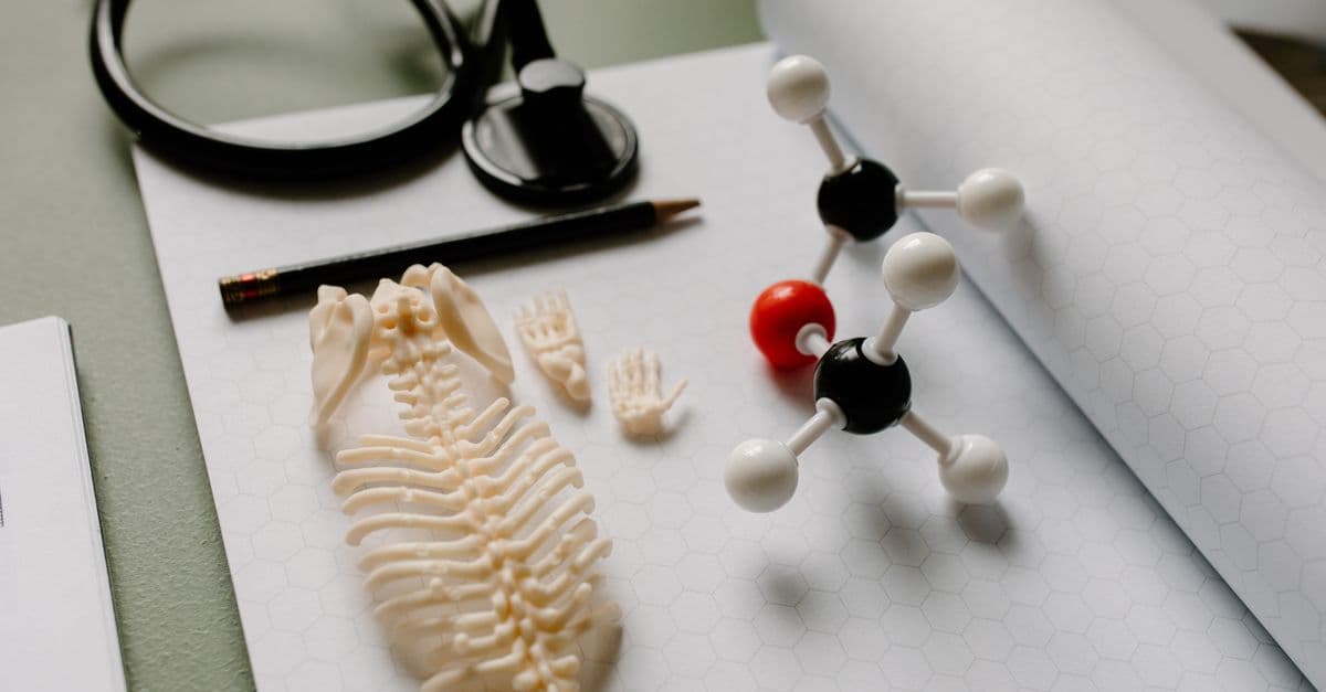
Photo: Tara Winstead / Pexels
The central question of mid-twentieth-century biology was whether DNA or protein carried hereditary information. By 1952, the evidence favored DNA — but enough scientists resisted that the question remained open. Alfred Hershey and Martha Chase resolved it with an experiment built around two radioactive isotopes, a virus that infects bacteria, and a kitchen blender. The logic was clean, the result was decisive, and the tool that separated the answer from the noise was so ordinary that Hershey reportedly called it a "Waring blender" in the lab's supply orders.
The State of the Debate in 1952
The question of whether genes were made of protein or DNA had been contested since the late 1800s. Proteins were the favored candidate through much of the first half of the twentieth century. The reasoning was intuitive: proteins are built from twenty different amino acids, giving them a large combinatorial alphabet capable of encoding complex information. DNA was thought to be a simpler, more repetitive molecule — a structural scaffold rather than an information carrier.
Source: Hershey AD, Chase M. "Independent functions of viral protein and nucleic acid in growth of bacteriophage." Journal of General Physiology 36(1): 39–56 (1952). https://doi.org/10.1085/jgp.36.1.39
Oswald Avery, Colin MacLeod, and Maclyn McCarty had published in 1944 that DNA was the transforming principle in pneumococcal bacteria — the substance that transferred heritable properties between bacterial strains. See The Griffith Transformation Experiment for the origin story of that question. But Avery's result, while compelling, left a technical opening: critics argued that his DNA preparations might contain trace amounts of protein, and that the protein was the true active agent.
What was needed was an experiment with a biological system that physically separated protein from DNA during normal function — where you could watch them go to different places and then ask which one did what.
Source: Scalar/Harvard. "Chapter 5: The Hershey-Chase experiment generates wide acceptance that the genetic material is DNA." https://scalar.fas.harvard.edu/the-river-divides-into-thousands-of-branches/chapter-5-the-hershey-chase-experiment-generates-wide-acceptance-that-the-genetic-material-is-dna
Bacteriophages provided that system.
The Setup: A Virus Built for the Experiment
A bacteriophage (or simply "phage") is a virus that infects bacteria. The T2 phage, which infects Escherichia coli, has a structure that makes it almost purpose-built for the question Hershey and Chase were asking. T2 consists of two components and two components only: a protein coat (called the capsid) surrounding a core of DNA. When T2 infects a bacterium, the phage attaches to the bacterial surface and injects its contents inside. New phages are then assembled inside the bacterium using the bacterial machinery, and the bacterium eventually lyses (bursts open), releasing hundreds of new T2 phages.
Source: Embryo Project Encyclopedia. "The Hershey-Chase Experiments (1952), by Alfred Hershey and Martha Chase." https://embryo.asu.edu/pages/hershey-chase-experiments-1952-alfred-hershey-and-martha-chase
The question was: when T2 infects a bacterium, which part goes in? If DNA enters the bacterium and directs the production of new phages, DNA is the genetic material. If protein enters and does the directing, protein is the genetic material. If both enter together, the experiment is ambiguous.
The key insight was to label each component — protein and DNA — with a different radioactive isotope that would allow tracking independently.
- Sulfur-35 (³⁵S) labels protein, because the amino acids cysteine and methionine contain sulfur. DNA contains no sulfur.
- Phosphorus-32 (³²P) labels DNA, because the phosphate backbone of DNA is rich in phosphorus. Proteins contain essentially no phosphorus.
By growing phages in bacteria fed with ³⁵S, Hershey and Chase produced phages with radioactively labeled protein coats. By growing phages in bacteria fed with ³²P, they produced phages with radioactively labeled DNA. The two types were separate: one batch tracked protein, the other tracked DNA.
Source: Cold Spring Harbor Laboratory. "Blending history and science." https://www.cshl.edu/blending-history-and-science/
The Protocol: Infect, Blend, Centrifuge
The experimental procedure had three steps.
Step 1: Infect bacteria with labeled phages. Hershey and Chase mixed each labeled phage preparation with unlabeled E. coli and allowed infection to begin. They waited long enough for the phages to inject their contents into the bacteria, but not so long that the bacteria had lysed and released new phages.
Step 2: Shear the phage coats off the bacteria with a Waring blender. After a few minutes of infection, they poured the mixture into a Waring blender and ran it for several minutes. The mechanical shear forces of the blender knocked the phage protein coats — which attach to the outside of the bacterial cell — off the bacterial surface. The contents that had been injected into the bacteria during infection remained inside.
Source: Biology LibreTexts. "5.2: The Hershey-Chase Experiments." https://bio.libretexts.org/Bookshelves/Introductory_and_General_Biology/Biology_(Kimball)/05:_DNA/5.02:The_Hershey-_Chase_Experiments
Step 3: Centrifuge to separate bacteria from phage ghosts. The blended mixture was then centrifuged at low speed. Bacteria (large, heavy) pelleted at the bottom. Phage coats that had been knocked off (small, light) remained in the supernatant — the liquid above the pellet.
Hershey and Chase measured radioactivity in both the pellet and the supernatant for both the ³⁵S-labeled and ³²P-labeled preparations.
| Experiment | Supernatant (phage coats) | Pellet (bacteria) |
|---|---|---|
| ³⁵S-labeled phages (protein) | ~80% of ³⁵S | ~20% of ³⁵S |
| ³²P-labeled phages (DNA) | ~20% of ³²P | ~75–80% of ³²P |
The protein, marked by ³⁵S, stayed mostly outside — in the supernatant with the phage coats. The DNA, marked by ³²P, went mostly inside — pelleting with the bacteria. Furthermore, when the infected bacteria were allowed to complete the infection cycle and lyse, the new phages they produced contained ³²P from the labeled DNA. The genetic information required to build new phages had traveled with the DNA.
Source: ScienceInsights. "What Was the Hershey-Chase Experiment? Results Explained." https://scienceinsights.org/what-was-the-hershey-chase-experiment-results-explained/
The Results: DNA Goes In, Protein Stays Out
The result was not 100% clean — approximately 20% of the ³²P stayed in the supernatant, and approximately 20% of the ³⁵S entered the bacteria. Hershey and Chase did not hide this. They acknowledged that the separation was imperfect and that the blender did not remove every phage coat or prevent every protein molecule from entering with the DNA.
Source: Hershey AD, Chase M. "Independent functions of viral protein and nucleic acid in growth of bacteriophage." Journal of General Physiology 36(1): 39–56 (1952). https://doi.org/10.1085/jgp.36.1.39
What made the result decisive was not that it was perfect, but that the asymmetry was unambiguous. DNA went in; protein did not. And only the DNA was needed to produce functional new phages. This showed that phage DNA carried all the information necessary to direct phage reproduction. Protein was structural — a delivery vehicle — not informational.
The new phages assembled inside the infected bacteria inherited their genetics from the parental DNA, not from the protein. The protein coat was a vehicle; the DNA was the message.
This was confirmed by an additional line of evidence: the fraction of ³⁵S that entered the bacteria was not passed on to the progeny phages. New phages made inside the infected bacteria were labeled with ³²P (from the parental DNA) but not significantly with ³⁵S. If protein were the genetic material, the new phage protein would have been templated from the old phage protein — and some ³⁵S would have appeared in the progeny. It did not.
The Controversy: Martha Chase and Scientific Credit
The Hershey-Chase experiment appeared under both names in the 1952 publication in the Journal of General Physiology. Alfred Hershey shared the 1969 Nobel Prize in Physiology or Medicine with Salvador Luria and Max Delbrück "for their discoveries concerning the replication mechanism and the genetic structure of viruses."
Source: Nobel Prize Committee. "Nobel Prize in Physiology or Medicine 1969." https://www.nobelprize.org/prizes/medicine/1969/hershey/facts/
Martha Chase was not included. She received no Nobel Prize, and for decades received limited recognition for her contribution to one of the most important experiments in twentieth-century biology. Historians of science have since documented that her role in the experiment was substantial — she performed many of the key procedures — and that her exclusion from the Nobel was part of a broader pattern of women's contributions being overlooked in mid-century science.
Chase left active research in the 1960s and spent her later years outside academia. She died in 2003. In recent decades, biologists have worked to restore her name to its proper place in the history of the discovery.
Source: UCR Digital Library Hub. "Hershey Chase Experiment." https://digilib.ucr.edu/hershey-chase-experiment
The name "Hershey-Chase experiment" includes both scientists. The Nobel did not.
The skeptics who remained
Even after Hershey and Chase published in 1952, a small number of scientists continued to argue for protein as the genetic material. The objections were increasingly technical — focused on the approximately 20% of protein that entered the bacteria — but they persisted until the Watson-Crick structure of DNA was published in April 1953. The double-helix structure, with its complementary base pairs providing an obvious copying mechanism, reframed the entire debate. Once you could see how DNA could encode and copy information, the protein hypothesis lost its last intellectual footing.
Source: Wikipedia. "Hershey–Chase experiment." https://en.wikipedia.org/wiki/Hershey%E2%80%93Chase_experiment
Replication and Reanalysis: What Later Work Added
The Hershey-Chase result was replicated by multiple laboratories in the 1950s using T2 and related phages (T4, T7). The result held consistently: phage DNA, not phage protein, entered the bacterial host. This was confirmed to be mechanically true — electron microscopy, developed through the 1950s, eventually showed the hollow protein tail structure of T2 phages that acts as a syringe, injecting DNA while the protein coat remains outside.
Source: Omic.ly. "Deoxyribonucleic acid (DNA) is the genetic material." https://www.omic.ly/hershey-chase-experiment/
Modern structural biology has resolved the injection mechanism in atomic detail. T4 phages (a close relative of T2) use a contractile sheath that drives a tube through the bacterial cell wall, forcing DNA through the tube and into the host cytoplasm. The protein coat — called the "ghost" after injection — remains on the outside, exactly as Hershey and Chase's centrifugation data showed.
The principle established by the experiment — that nucleic acids carry genetic information and protein does not — is now foundational. It applies not just to bacteriophages but to all viruses. The genetic material of influenza, SARS-CoV-2, HIV, and every other characterized virus is nucleic acid (DNA or RNA), never protein.
Source: National Institute of Allergy and Infectious Diseases. "Understanding Viruses." https://www.niaid.nih.gov/research/emerging-infectious-diseases-viruses
Bacteriophage research today
The T2 and T4 phages studied by Hershey and Chase are members of a broader class that has remained central to molecular biology. The study of phage genetics at Cold Spring Harbor Laboratory — where Hershey and Chase worked — produced many of the foundational discoveries in molecular biology, including restriction enzymes, genetic recombination, and gene regulation.
Phage biology has also returned to clinical relevance through phage therapy: the use of bacteriophages to treat antibiotic-resistant bacterial infections. As antibiotic resistance grows, phages are being reconsidered as targeted antibacterial agents.
Source: Principi N et al. "Bacteriophage therapy: advantages and limitations." Frontiers in Pharmacology 10: 513 (2019). https://www.frontiersin.org/articles/10.3389/fphar.2019.00513/full
Where It Stands Today
The Hershey-Chase experiment occupies a fixed position in introductory genetics and molecular biology curricula worldwide because it exemplifies a methodological virtue that is easy to describe but hard to achieve: using the biology of the system to provide its own separation. The blender did not perform magic — it exploited the natural geometry of phage infection, where protein attaches outside and DNA is injected inside. Hershey and Chase recognized that geometry and used it.
The experiment also provides a clean illustration of the use of radioactive tracers in biological research. The two isotopes — ³⁵S for protein, ³²P for DNA — were already available from the Manhattan Project-era expansion of nuclear physics. Hershey and Chase were among the first biologists to use them this way. Radioactive tracers went on to become a standard tool in biochemistry throughout the 1950s and 1960s.
Source: The Bumbling Biochemist. "The Hershey-Chase Experiment." https://thebumblingbiochemist.com/life-in-the-lab/the-hershey-chase-experiment/
For how the DNA structure that made these results fully interpretable came to be understood, see The Meselson-Stahl Experiment, which showed four years later how that DNA copies itself. For the foundational observation that first raised the question of what the genetic material was, see The Griffith Transformation Experiment, from which the chain of inquiry leading to Hershey and Chase began.
The key contribution of Hershey and Chase was not just the result, but the irreversibility of its logic. Protein could not enter the bacteria and build new phages; DNA did. The physical separation, achieved by a kitchen blender and a centrifuge, made the argument impossible to ignore.
Why the Blender Mattered: Methodology That Changed Biology
The result of the Hershey-Chase experiment — DNA goes in, protein stays out — is what gets remembered. The methodology that produced it is what made the result convincing, and understanding that methodology explains why the experiment became a template for how biological questions should be asked.
The design rested on three interlocking choices, each of which had to be correct for the experiment to work.
The first choice was the isotopes. Phosphorus-32 (³²P) and sulfur-35 (³⁵S) are not arbitrary radioactive labels. They were selected because of a precise biochemical exclusivity: DNA is built on a phosphate backbone, so ³²P incorporates cleanly into DNA and not into protein; the amino acids cysteine and methionine carry sulfur atoms, so ³⁵S incorporates into protein and not into DNA. These two elements do not compete. There is no ambiguity about which molecule each label tracks. If Hershey and Chase had used a label that appeared in both protein and DNA, the experiment would have been uninterpretable. The isotope selection was not a technical detail — it was the logical foundation of the whole design.
Source: Cold Spring Harbor Laboratory. "Blending history and science." https://www.cshl.edu/blending-history-and-science/
The second choice was the blender. Think of the blender step as a deliberate exploitation of the infection's geometry. When T2 phage injects its contents into a bacterium, the protein coat remains anchored to the outside of the bacterial cell wall — like a syringe that stays on the skin's surface after injecting fluid beneath it. The blender's agitation sheared those coats free, separating "what was outside" from "what went inside" with mechanical precision. A gentler mixing step would not have detached the coats. A harsher one would have broken the bacteria open, mixing the contents and destroying the separation. The blender speed and duration were empirically calibrated to thread that gap.
The third choice was the centrifuge. After blending, the mixture contained a mixture of intact bacteria (which had received the injected material) and free phage protein coats (now detached). Centrifugation at low speed exploited the size and density difference between them: bacteria are roughly a thousand times larger than a phage coat and pellet to the bottom of the tube, while the lighter phage ghosts remain suspended in the supernatant. The researchers could then measure radioactivity in each fraction separately, asking precisely: where is the ³²P, and where is the ³⁵S?
Tracer methodology — the use of radioactively labeled molecules to follow specific substances through a biological process without disrupting that process — was relatively new in biology in 1952. The isotopes ³²P and ³⁵S had become widely available in the years following the Manhattan Project, when the Oak Ridge National Laboratory began producing radioisotopes for scientific distribution. Hershey and Chase were among the first biologists to use them in a genetic context.
Source: Creager, A.N.H. Life Atomic: A History of Radioisotopes in Science and Medicine. University of Chicago Press, 2013. Discussed in context of isotope availability post-WWII.
The design's elegance is also what makes it a standard teaching model in introductory molecular biology courses. Unlike many foundational experiments, the logic of Hershey-Chase can be reconstructed from scratch by a student who understands two facts: that ³²P goes into DNA and not protein, and that T2 phage injects material into bacteria while leaving something outside. Given those two facts, the experimental structure follows almost inevitably. The blender and centrifuge are the tools that make the biology's own geometry visible. That is the standard that later experiment designers would aspire to: not forcing a biological system to answer a question, but designing a readout that lets the system answer it on its own terms.
Source: Hershey AD, Chase M. "Independent functions of viral protein and nucleic acid in growth of bacteriophage." Journal of General Physiology 36(1): 39–56 (1952). https://doi.org/10.1085/jgp.36.1.39
FAQs
Q: Why was a Waring blender the right tool for this experiment? A: The blender provided mechanical shear force sufficient to strip phage protein coats from the outside of bacterial cells without breaking the bacteria open or damaging the DNA inside. It was not a precise instrument — it was a practical one. The key was that the agitation was strong enough to detach external protein but gentle enough to leave the bacteria intact so they could be pelleted by centrifugation.
Q: Why did Hershey and Chase use T2 phage specifically? A: T2 was a well-characterized phage already being studied at Cold Spring Harbor Laboratory. Its infection cycle was understood — the sequence and timing of attachment, injection, replication, and lysis were known — which allowed Hershey and Chase to time their blending step precisely. T2 also has a simple two-component structure (DNA and protein), with no lipid envelope or RNA, which made the experimental logic cleaner.
Q: What did the experiment not prove? A: It did not prove the molecular mechanism by which DNA encodes protein sequences. It showed that DNA carries the genetic information required to produce new phages, but not how that information is read out or translated. That question — the mechanism of gene expression — was answered over the following decade through work on messenger RNA, codons, and the ribosome.
Q: Why wasn't Martha Chase included in the Nobel Prize? A: The Nobel Prize in Physiology or Medicine 1969 was awarded for a body of work in phage genetics that extended well beyond the 1952 blender experiment. The committee recognized Hershey, Luria, and Delbrück for decades of contributions to phage biology. Chase's contribution was specific to the 1952 experiment. The committee's decision to exclude her has been criticized by historians of science, particularly given that her experimental work was central to the result.
Q: How did the experiment contribute to the discovery of the double helix? A: Directly and indirectly. Hershey and Chase demonstrated in April 1952 that DNA was the genetic material of phages — confirming and strengthening Avery's 1944 result. Watson and Crick published their double-helix structure in April 1953. The Hershey-Chase result was part of the evidence base that motivated the race to determine DNA's structure; knowing that DNA carried genetic information made its three-dimensional structure a problem of the highest priority.
Q: Are there equivalent experiments showing RNA is the genetic material in RNA viruses? A: Yes. Heinz Fraenkel-Conrat and Bea Singer demonstrated in 1957 that the RNA of tobacco mosaic virus (TMV), not its protein, was sufficient to infect tobacco plants. When they separated TMV RNA from TMV protein and applied RNA alone to plants, the plants developed infection symptoms and produced complete new virus particles. This extended the Hershey-Chase principle to RNA viruses.
The Takeaway
The Hershey-Chase experiment worked because it asked biology to provide its own answer. T2 phage naturally separates its DNA from its protein during infection — the DNA goes inside the bacterium, the protein stays outside. Hershey and Chase did not force this separation; they used a blender to exaggerate and exploit it, then measured which component went where. The result settled a question that had been debated for half a century: the genetic material is nucleic acid, not protein. That answer has not changed.
Read next:
Weekly digest
Stay ahead of the curve
Get the best science, AI, and tech stories delivered to your inbox every week. No spam — unsubscribe any time.
About the Author
SciHub101 is an independent science and technology publication founded and edited by Buğra Sözeri, a software engineer and app developer based in Türkiye. Articles are drafted against peer-reviewed literature and primary sources, fact-checked, and edited by a human before publication. Where we compare products, anything built by our own team is labelled as such — see the editorial guidelines and About page for how corrections and disclosures work.
Related Articles
The Hodgkin-Huxley Squid Axon Experiment: How We Cracked the Action Potential
Hodgkin and Huxley used squid giant axons and voltage-clamp to decode the action potential's ionic machinery. Their 1952 model still powers neuroscience today.
The Hafele-Keating Experiment: Atomic Clocks, Airplanes, and Time Dilation
In October 1971, physicists flew atomic clocks around the world and proved time dilation is real — clocks gained and lost nanoseconds exactly as Einstein predicted.
The Avery-MacLeod-McCarty Experiment: DNA Is the Transforming Principle
In 1944, Avery, MacLeod, and McCarty proved DNA carries hereditary information by identifying it as Griffith's transforming principle. The experiment that made the DNA era possible.
Watson and Crick: The Double Helix, Photo 51, and the Most Consequential Discovery in Biology — SciHub101
Watson and Crick solved DNA's double helix structure in 1953 using Franklin's X-ray data without her knowledge. Crick shouted 'the secret of life' in a Cambridge pub. The full story is complicated.
Rosalind Franklin: Photo 51 and the Discovery That Was Stolen in Plain Sight — SciHub101
Rosalind Franklin's life, discoveries, and verified quirks: Photo 51, refusal to marry, death at 37, and how her X-ray crystallography work was used without her knowledge.
DNA Replication and Genetics Explained
How DNA replicates, the central dogma, mutations, epigenetics, and how consumer genetics tests like 23andMe actually work.