The Loewi Frog Heart Experiment: A Dream That Proved Chemical Neurotransmission
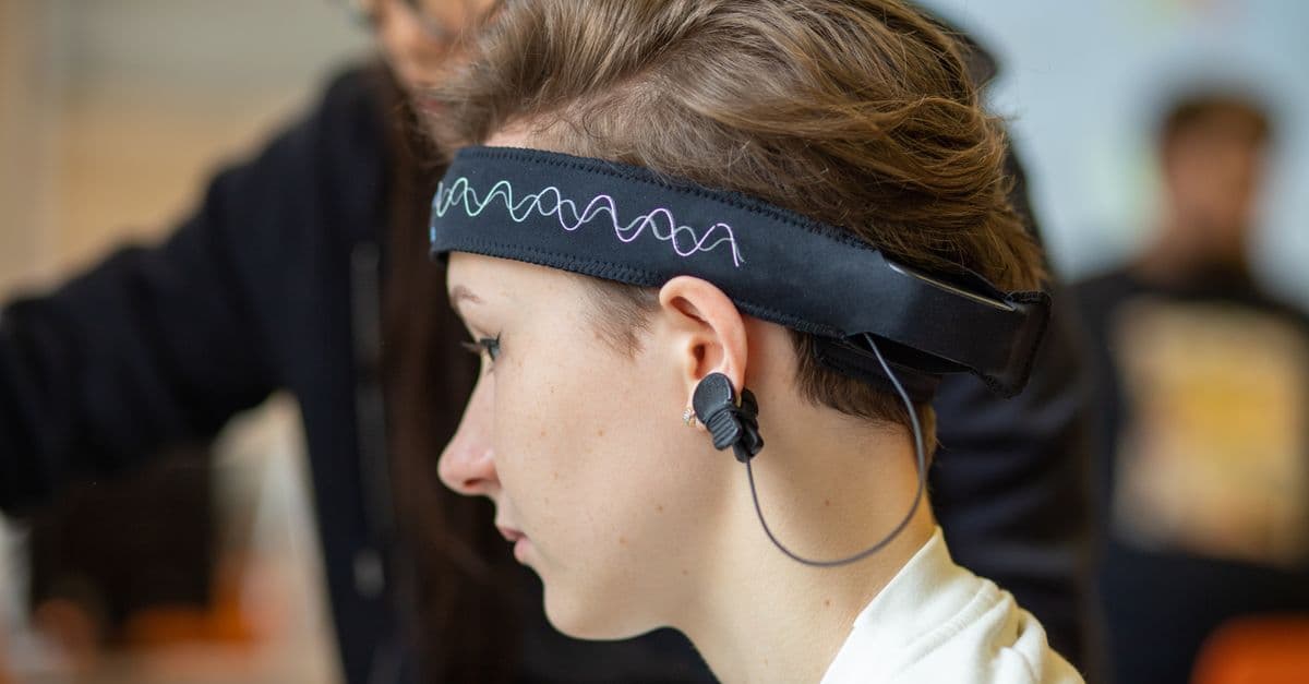
Photo: Михаил Крамор / Pexels
The debate that occupied physiologists for decades in the early twentieth century sounds almost philosophical now: when one nerve cell signals to another, does it do so electrically — a spark jumping a gap — or chemically, by releasing a substance that drifts across? In the early hours of Easter Sunday, 1921, Otto Loewi woke from a dream in which the experimental answer had come to him whole. He scrawled notes on a scrap of paper, fell back asleep, and woke to find his notes illegible. He lay awake the following night until the same idea returned, then drove straight to his laboratory before dawn.
The Problem Loewi Set Out to Solve
By 1920, physiologists had accumulated strong indirect evidence that the nervous system controlled the heart through specific chemical pathways. It was known that the alkaloid muscarine mimicked the slowing effect of vagal stimulation on the heart, and that adrenaline mimicked the accelerating effect of sympathetic nerve stimulation. Henry Dale had isolated acetylcholine from ergot extracts in 1914 and noted that its effects were similar to vagal stimulation.
Vagus nerve — the tenth cranial nerve, running from the brainstem through the chest and abdomen. Its cardiac branch slows the heart rate and reduces the force of contraction when stimulated.
But mimicry is not proof. The question was whether the nerve itself released a substance during normal activity. The dominant alternative — favored by many prominent physiologists including those in the electrical transmission camp — held that the nerve impulse crossed the junction directly as an electrical signal. There was no experimental test that could distinguish the two possibilities definitively, largely because no one had devised a way to catch any putative chemical in the act.
Source: Loewi, O. (1936), Nobel Lecture, "The Chemical Transmission of Nerve Action," NobelPrize.org — Loewi's own account of the intellectual background and the experimental approach.
The key obstacle was the synapse. Even in 1920, anatomists knew there was a physical gap between neurons and between nerve endings and the tissues they controlled. The question was whether signals jumped this gap as electrical current or as molecules. Loewi's stroke of insight was that the frog heart gave him a way to run a chemical messenger through a test — an entirely separate, unmistakable test.
The Dream and the Design
Loewi described the genesis of the experiment in his Nobel lecture with disarming specificity. On the night before Easter Sunday 1921, he woke and wrote down an idea that had come to him in a dream. He fell back asleep without reading what he had written. In the morning, the notes were illegible — "a few symbols and scratch marks," he later said. For the entire day he was unable to reconstruct the idea. That night, he lay awake until 3 a.m. when the idea returned, complete. He got up, dressed, and went immediately to his laboratory.
The idea was straightforward. If a nerve releases a chemical when stimulated, that chemical should be present in the fluid bathing the organ being stimulated. If the fluid is then transferred to a second organ, the chemical should produce the same effect there — without any nerve stimulation at all.
Source: Loewi, O. (1921), "Über humorale Übertragbarkeit der Herznervenwirkung," Pflügers Archiv, 189, 239–242 — the original publication describing the experiment. "Über humorale Übertragbarkeit" translates roughly as "On the Humoral Transferability of Cardiac Nerve Action."
The frog heart was the ideal preparation. It beats spontaneously without any neural input — a useful property of cardiac muscle. The vagus nerve slows it down, the accelerator nerve speeds it up. Both nerves can be stimulated electrically with precision. And the whole organ can survive for hours in a simple salt solution (Ringer's solution), making transfer experiments feasible.
The Experimental Setup
Loewi removed two hearts from frogs. The first heart was kept with its vagus nerve intact and was placed in a small chamber bathed in Ringer's solution. The second heart had no attached nerves. Both were beating spontaneously.
Ringer's solution — a physiological saline solution with ion concentrations approximating those in frog blood, developed by Sidney Ringer in the 1880s. It keeps isolated tissues alive and functional for hours.
He then electrically stimulated the vagus nerve of the first heart and watched its beating slow, as expected. He collected the Ringer's solution that had been bathing the first heart and applied it to the second heart. The second heart also slowed — at the same rate, with the same magnitude — even though no nerve had been stimulated anywhere near it.
The implication was clear. Vagal stimulation had released something into the bathing solution. That something was a chemical messenger — a substance that could travel in fluid and act on cardiac muscle directly, without any nerve. Loewi called it Vagusstoff (German: vagus substance). He followed the same logic for the accelerator nerve, collecting fluid after sympathetic stimulation and applying it to the second heart, which then sped up. He called this substance Acceleransstoff.
Source: Loewi, O. (1921), Pflügers Archiv, 189, 239–242 — first demonstration of Vagusstoff; followed by Loewi, O. (1922), Pflügers Archiv, 193, 201–213 — extension of the experiments to the accelerator nerve and additional pharmacological characterization.
The Pharmacological Confirmation
The transfer experiment was compelling, but Loewi was a rigorous pharmacologist. He needed to rule out the possibility that the effect was caused by potassium ions or other small molecules that might leach out of the heart tissue during vagal stimulation, rather than a specific nerve-released substance.
Over the following years, he conducted a series of pharmacological tests. Atropine, a drug known to block the cardiac effects of vagal stimulation, also blocked the effects of the transferred Vagusstoff. Physostigmine, a drug that amplified the effects of vagal stimulation by preventing breakdown of the active substance, also amplified the effect of Vagusstoff on the second heart. Eserine (another name for physostigmine) treatment allowed smaller amounts of vagal fluid to produce larger effects.
These pharmacological fingerprints matched exactly the profile that Henry Dale had described for acetylcholine.
Source: Dale, H.H. & Dudley, H.W. (1929), "The presence of histamine and acetylcholine in the spleen of the ox and the horse," Journal of Physiology, 68, 97–123 — Dale's group isolating acetylcholine from animal tissues, providing the chemical identification of what Loewi had found as Vagusstoff.
The final identification came when Otto Loewi and his colleagues showed that Vagusstoff was destroyed by an extract of horse serum at a rate consistent with enzymatic hydrolysis — the same reaction by which acetylcholinesterase (the enzyme that breaks down acetylcholine) was later shown to operate. By 1933, the chemical identity was established: Vagusstoff was acetylcholine.
Acetylcholine — a neurotransmitter released at neuromuscular junctions and at many synapses in the brain and autonomic nervous system. It acts on muscarinic receptors (which mediate heart slowing and glandular secretion) and nicotinic receptors (which mediate muscle contraction).
Why the Experiment Was Initially Resisted
The chemical transmission hypothesis was not immediately accepted. The strongest objection came from the speed argument: chemical diffusion across a synaptic gap should be too slow to account for the millisecond precision of neural signaling. John Eccles — who later shared the Nobel Prize with Hodgkin and Huxley — was among those who argued vigorously for electrical transmission well into the 1940s.
The speed objection was not unreasonable. Simple diffusion of molecules across a large gap is slow. If the synaptic cleft were even a few micrometers wide, the time for transmitter to diffuse from the presynaptic membrane to the postsynaptic receptor would be on the order of 10 milliseconds — far too long to account for the rapid responses seen in motor reflexes. The "electrical spark" model, in which current flows directly across a junction as it does in an electrical circuit, had no such speed problem.
The resolution came from anatomy. Electron microscopy in the 1950s revealed that the synaptic gap (the synaptic cleft) is extremely narrow — about 20 nanometers wide. At such a scale, diffusion of acetylcholine from the release point to the postsynaptic receptor takes less than a microsecond, fast enough to account for the observed synaptic delays of about 0.5 milliseconds. The delay is actually dominated by the time required to release the transmitter from the presynaptic terminal, not by diffusion time.
Source: Palade, G.E. & Palay, S.L. (1954), "Electron microscope observations of interneuronal and neuromuscular synapses," Anatomical Record, 118, 335–336 — among the earliest electron micrographs revealing the synaptic cleft dimensions and the presence of synaptic vesicles.
Electron microscopy also revealed something Loewi could not have imagined: the presynaptic terminal is packed with tiny membrane-bound spheres, each about 40–50 nanometers in diameter, which are now known as synaptic vesicles. These vesicles contain high concentrations of neurotransmitter and fuse with the presynaptic membrane when an action potential arrives, releasing their contents into the cleft in a rapid burst. The mechanism of vesicle fusion — which involves specialized proteins called SNAREs — was worked out in the 1980s and 1990s and earned the 2013 Nobel Prize in Physiology or Medicine for James Rothman, Randy Schekman, and Thomas Südhof.
Eccles himself eventually performed experiments (for which he shared the 1963 Nobel Prize with Hodgkin and Huxley) demonstrating that even his preferred CNS synapses worked by chemical transmission. He acknowledged Loewi's priority publicly and unreservedly.
Source: Eccles, J.C. (1963), Nobel Lecture, "The ionic mechanism of excitatory and inhibitory synaptic action," NobelPrize.org — Eccles's own account of his conversion from the electrical transmission hypothesis to the chemical transmission hypothesis, with explicit acknowledgment of Loewi's foundational work.
The Timeline
| Year | Event |
|---|---|
| 1914 | Henry Dale isolates acetylcholine from ergot; notes vagal-like cardiac effects |
| 1921 | Loewi performs the dream-inspired frog heart experiment; publishes in Pflügers Archiv |
| 1922 | Extension to accelerator nerve; Acceleransstoff (later identified as noradrenaline) demonstrated |
| 1926 | Loewi shows Vagusstoff is destroyed by serum esterase, suggesting it is an ester |
| 1929 | Dale & Dudley isolate acetylcholine from spleen tissue |
| 1933 | Chemical identity of Vagusstoff as acetylcholine definitively established |
| 1936 | Loewi and Dale share Nobel Prize in Physiology or Medicine |
| March 1938 | Anschluss: Loewi arrested by Gestapo; forced to transfer Nobel Prize funds to Nazi account |
| September 1938 | Loewi released; flees to England; joins Dale in Oxford |
| 1940 | Loewi emigrates to the United States; eventually settles in New York |
| 1954 | Electron microscopy reveals synaptic cleft dimensions confirming chemical transmission feasibility |
| 1963 | John Eccles shares Nobel Prize with Hodgkin and Huxley; acknowledges chemical transmission even in CNS |
| 1961 | Otto Loewi dies in New York, age 88 |
The Political Rupture: Arrest, Exile, and the Nobel Prize Money
The story of Loewi's scientific triumph is intertwined with a historical catastrophe. In March 1938, Nazi Germany annexed Austria. On the night of March 11–12, Loewi was awakened by Gestapo officers and arrested. He was held in Vienna for three months in what contemporaries described as brutal conditions.
The specific charge was technically that he had violated a law forbidding citizens from accepting the Nobel Prize — a prohibition that Nazi Germany had imposed in retaliation for the 1935 peace prize being awarded to Carl von Ossietzky, a critic of the regime. In practice, the arrest was part of a broader program targeting Jewish academics and intellectuals. Loewi was Jewish.
As a condition of his release, the Gestapo required Loewi to transfer his Nobel Prize money — approximately 78,000 Swedish kronor — to a Nazi-controlled bank account. He complied. He was released in September 1938 and left Austria immediately, traveling to England where Henry Dale — his longtime friend and collaborator, co-recipient of the 1936 Nobel — arranged a position for him.
Source: Otto Loewi — Biographical, NobelPrize.org — official biographical note describing his arrest, imprisonment, and forced emigration.
Loewi spent two years in England before moving to the United States in 1940. He obtained a position at New York University College of Medicine, where he continued research and teaching until his death in 1961. He never returned to Austria. The episode illustrates a pattern — the flight of Jewish scientists from Nazi Europe — that reshaped the landscape of twentieth-century science, transferring much of Europe's research capacity to American institutions.
What Vagusstoff Revealed About the Nervous System
Before Loewi, the nervous system was often conceived as a purely electrical system — a network of wires carrying currents. The demonstration that neurons communicate by releasing molecules completely changed this picture. The synapse is not a junction but a chemical factory: the presynaptic terminal synthesizes, stores in small membrane-bound vesicles, and releases specific molecules; the postsynaptic membrane has receptor proteins that detect those molecules and translate their presence into a new electrical or chemical signal.
This picture has implications that Loewi could not have anticipated in 1921. It means the strength of synaptic transmission can be regulated by controlling the amount of transmitter released, the density of postsynaptic receptors, the rate at which the transmitter is broken down, or the availability of molecules that mimic or block the transmitter. These are the mechanisms by which drugs act on the nervous system. Almost every psychiatric and neurological medication works by altering synaptic chemistry: antidepressants block serotonin reuptake; antipsychotics block dopamine receptors; anesthetics potentiate GABA receptors; caffeine blocks adenosine receptors. The entire pharmacology of the central nervous system rests on the foundation that Loewi's frog heart transfer experiment established.
Source: Cooper, J.R., Bloom, F.E. & Roth, R.H. (2003), The Biochemical Basis of Neuropharmacology, 8th ed., Oxford University Press — the standard reference on neurotransmitter systems and drug mechanisms, rooted in the chemical transmission framework.
The number of neurotransmitters identified since Loewi's discovery has grown to over 100 distinct chemical messengers, each with its own receptor subtypes, synthesis pathways, reuptake mechanisms, and pharmacological profiles. Acetylcholine itself acts through five muscarinic receptor subtypes (G protein-coupled receptors mediating slow, modulatory effects) and multiple nicotinic receptor subtypes (ligand-gated ion channels mediating fast, direct excitation). The cardiac slowing that Loewi observed comes from acetylcholine binding to M2 muscarinic receptors on the sinoatrial node, opening potassium channels and slowing the pacemaker rhythm.
Understanding this receptor diversity has transformed medicine. Drugs that selectively block muscarinic receptors (atropine, scopolamine) treat motion sickness and slow heart rate during anesthesia. Drugs that block nicotinic receptors at the neuromuscular junction (curare and its derivatives) are used as muscle relaxants in surgery. Drugs that inhibit acetylcholinesterase — the enzyme that breaks down acetylcholine after it acts — include the main class of medications for Alzheimer's disease, where acetylcholine-producing neurons degenerate early in the disease course.
Source: Rang, H.P. et al. (2019), Rang and Dale's Pharmacology, 9th ed., Elsevier — standard pharmacology reference detailing the receptor pharmacology of acetylcholine and its clinical applications.
How the Experiment Has Been Reproduced and Extended
Loewi's basic fluid-transfer experiment has been reproduced in undergraduate physiology laboratories for over a century. The preparation — isolated frog hearts in Ringer's solution, vagal stimulation, fluid transfer — is technically simple enough to teach and robust enough to work reliably, making it one of the few Nobel Prize-winning experiments that can be replicated in an afternoon with introductory-level equipment.
More sophisticated replications have gone far beyond the original design. In the 1960s and 1970s, radiolabeled acetylcholine (synthesized with carbon-14 or tritium-labeled choline) allowed researchers to directly track the release and degradation of the transmitter during nerve stimulation, confirming the relationship Loewi had inferred from functional observations. Mass spectrometry has since allowed quantification of acetylcholine release at the attomole level — a sensitivity that would have seemed impossible to Loewi working with bulk fluid transfers.
The concept of Loewi's experiment — that a physiologically active substance released by one tissue can affect a second tissue through diffusion — has also expanded conceptually into the field of paracrine signaling, where local chemical messengers (prostaglandins, nitric oxide, histamine) act on nearby cells without entering the bloodstream. Nitric oxide, identified as a vascular signaling molecule in the 1980s, is in some ways a modern Vagusstoff: a gaseous chemical released by endothelial cells that acts on adjacent smooth muscle cells to cause vasodilation. Robert Furchgott, Louis Ignarro, and Ferid Murad received the 1998 Nobel Prize in Physiology or Medicine for this discovery.
Source: Furchgott, R.F. & Zawadzki, J.V. (1980), "The obligatory role of endothelial cells in the relaxation of arterial smooth muscle by acetylcholine," Nature, 288, 373–376 — the paper that established the endothelium-derived relaxing factor (later identified as nitric oxide), a modern parallel to Loewi's humoral transmission concept.
The Autonomic Nervous System: Loewi's Larger Context
The organ Loewi chose — the heart — is controlled by the autonomic nervous system, the division of the nervous system that regulates internal organs without conscious involvement. The autonomic system has two main branches: the parasympathetic (which promotes "rest and digest" states, including heart slowing, increased digestion, and pupil constriction) and the sympathetic (which drives "fight or flight" states, including heart speeding, reduced digestion, and pupil dilation). Loewi's Vagusstoff came from the parasympathetic vagus nerve; his Acceleransstoff came from sympathetic fibers.
Autonomic nervous system — the division of the peripheral nervous system that controls involuntary functions: heart rate, blood pressure, digestion, respiration, pupil diameter, and secretion of glands. It operates continuously without conscious awareness.
Loewi's demonstration that both branches of the autonomic system use chemical transmission immediately suggested that different chemicals might mediate the two branches — which turned out to be correct. The parasympathetic system uses acetylcholine at both its ganglionic synapses (where preganglionic and postganglionic neurons communicate) and at its target organs. The sympathetic system uses acetylcholine at ganglionic synapses but noradrenaline (norepinephrine) at most target organs — Loewi's Acceleransstoff. The chemical divergence between the two branches explains why drugs can selectively activate or block one branch without affecting the other.
This chemical specificity has enormous pharmacological importance. Beta-blockers (drugs like propranolol and metoprolol) reduce heart rate and blood pressure by blocking the noradrenaline receptors (beta-adrenergic receptors) that mediate sympathetic acceleration of the heart — acting on the same transmitter pathway Loewi identified as Acceleransstoff. Anticholinergic drugs (atropine, ipratropium) block acetylcholine's muscarinic receptors, blocking the parasympathetic pathway Loewi identified as Vagusstoff. Together, these two drug classes form the foundation of cardiovascular pharmacology.
Source: Katzung, B.G., Masters, S.B. & Trevor, A.J. (2012), Basic and Clinical Pharmacology, 12th ed., McGraw-Hill — standard clinical pharmacology reference, covering beta-blockers and anticholinergics as direct applications of the Loewi-Dale chemical transmission framework.
The autonomic ganglia themselves — where the pre- and postganglionic neurons communicate — use nicotinic acetylcholine receptors, the same type that mediate neuromuscular transmission. This means that drugs blocking ganglionic nicotinic receptors affect both sympathetic and parasympathetic pathways simultaneously. Ganglionic blockers were the first effective antihypertensive drugs, used in the 1950s, but their lack of selectivity (blocking both branches) caused severe side effects. The subsequent development of selective beta-blockers was a direct consequence of understanding the chemical specificity of each autonomic pathway, with Loewi's experiment as the conceptual foundation.
What People Get Wrong About the Loewi Experiment
"Loewi discovered acetylcholine." He discovered chemical neurotransmission and named the cardiac transmitter Vagusstoff. Henry Dale had isolated acetylcholine as a compound in 1914, and the chemical identification of Vagusstoff as acetylcholine came through collaborative work in the late 1920s and early 1930s. The Nobel Prize recognized both men for their complementary contributions.
"The dream literally gave him the complete method." Loewi's own account suggests the dream provided the conceptual insight — that fluid transfer could serve as a test. The experimental design, the choice of frog hearts, the pharmacological controls: all of that followed from his deep expertise as a pharmacologist. Dreams can surface ideas that waking analytical thought has not connected; they do not substitute for the technical knowledge needed to execute the test.
"The experiment proved chemical transmission everywhere in the nervous system." Loewi's experiment demonstrated chemical transmission at the cardiac vagal ending — one specific type of synapse. Generalizing to all synapses, especially in the central nervous system, required decades of additional work. John Eccles initially maintained that central synapses used electrical transmission as late as the mid-1940s. The universal chemical transmission principle was established only through the combined evidence of the 1950s.
"Acetylcholine is just a heart-slowing drug." Acetylcholine is one of the most broadly acting neurotransmitters in the nervous system. It controls the neuromuscular junction (where all skeletal muscle contractions are initiated), mediates arousal in the cortex, influences memory formation in the hippocampus, and acts throughout the autonomic nervous system. Its cardiac slowing effect — the one Loewi studied — is only one function among dozens.
FAQs
Q: What exactly did Loewi transfer from the first heart to the second?
A: The Ringer's solution bathing the first heart after vagal stimulation. This saline contained Vagusstoff — what we now know as acetylcholine — released by the nerve endings during stimulation. Applying this fluid to the denervated second heart slowed it, demonstrating that the substance was chemically active independently of any nerve.
Q: Why did Loewi use frog hearts rather than mammalian hearts?
A: Frog hearts can beat spontaneously for many hours in simple Ringer's solution at room temperature, making them ideal for isolated organ experiments. Mammalian hearts require oxygenated, temperature-controlled perfusion and are far more technically demanding to keep alive outside the body.
Q: How did Loewi know the slowing wasn't caused by potassium or other leaked substances?
A: Through pharmacological fingerprinting. The slowing caused by vagal fluid was blocked by atropine and potentiated by physostigmine — exactly as vagal nerve stimulation itself was. Potassium or other non-specific substances do not have this pharmacological profile. The fingerprint matched acetylcholine precisely.
Q: What happened to Loewi's Nobel Prize money?
A: The Gestapo forced him to transfer it to a Nazi-controlled bank account as a condition of his release from prison in 1938 after the Austrian Anschluss. He received none of it.
Q: What is the connection between Loewi's work and modern antidepressants?
A: Loewi established that synaptic transmission is chemical. That insight opened the door to the entire field of neuropharmacology. If nerves communicated electrically, drugs could not selectively block or mimic specific transmitter actions. Because they communicate chemically, drugs targeting specific transmitter systems — serotonin, dopamine, noradrenaline — can selectively alter brain function. Modern antidepressants, antipsychotics, and anxiolytics all depend on this framework.
Q: Was the "dream" story verified or embellished?
A: Loewi told the dream story consistently in his Nobel lecture (1936), his autobiography, and numerous other accounts over decades. No contemporary correspondence from 1921 corroborates or contradicts it. The story is plausible — the design problem was one Loewi had been thinking about for years, and incubation during sleep is a documented aspect of creative problem solving. Whether it happened exactly as described cannot be independently confirmed.
Q: What were the longer-term consequences of the Loewi-Dale framework for understanding the brain?
A: The framework established that the brain is a chemical machine as much as an electrical one. It made possible the discovery of dopamine (1957, by Arvid Carlsson, who received the 2000 Nobel Prize), serotonin, noradrenaline, GABA, glutamate, and dozens of neuropeptides, each mediating specific aspects of brain function. It enabled the rational design of psychoactive drugs and the mechanistic understanding of how natural compounds from plants (caffeine, morphine, cocaine, nicotine) act on the brain — they work by mimicking or blocking specific neurotransmitter systems that Loewi's experiment proved must exist.
Q: How does Loewi's experiment relate to the study of electrical synapses?
A: Chemical synapses are not the only type. Gap junctions — direct protein connections between adjacent cells that allow ions to pass from one cell's cytoplasm directly to another's — mediate electrical synaptic transmission and do exist in some nervous systems. These were discovered after chemical transmission was established and are now known to play roles in neural synchronization, cardiac conduction, and lens transparency. Loewi's experiment established the primacy of chemical transmission for most neuronal communication but did not eliminate the existence of direct electrical coupling, which does occur in specific biological contexts.
The Takeaway
Loewi's experiment is one of the rare cases in science where the entire conceptual content of a discovery fits inside a single, elegant observation: fluid from a stimulated heart slows an unstimulated one. Everything else — the identification of acetylcholine, the mapping of synaptic vesicles, the pharmacology of the central nervous system, the mechanism of every psychiatric drug ever developed — is downstream of that fluid transfer on an Easter morning in 1921.
Read next:
Weekly digest
Stay ahead of the curve
Get the best science, AI, and tech stories delivered to your inbox every week. No spam — unsubscribe any time.
About the Author
SciHub101 is an independent science and technology publication founded and edited by Buğra Sözeri, a software engineer and app developer based in Türkiye. Articles are drafted against peer-reviewed literature and primary sources, fact-checked, and edited by a human before publication. Where we compare products, anything built by our own team is labelled as such — see the editorial guidelines and About page for how corrections and disclosures work.
Related Articles

The Loftus Misinformation Effect: How Memory Gets Rewritten
Elizabeth Loftus's 1974 car-crash study showed a single word can alter memory. Here's how she proved it, what it means for eyewitness testimony, and why courts still struggle with the findings.
The Lind Scurvy Trial: The First Controlled Clinical Trial
James Lind's 1747 experiment on HMS Salisbury tested six treatments for scurvy on 12 sailors. Only citrus worked. Discover how it became the blueprint for clinical trials.

The Hodgkin-Huxley Squid Axon Experiment: How We Cracked the Action Potential
Hodgkin and Huxley used squid giant axons and voltage-clamp to decode the action potential's ionic machinery. Their 1952 model still powers neuroscience today.
Pavlov's Classical Conditioning: The Methodology and Science Behind Conditioned Reflexes
Ivan Pavlov's conditioned reflex research was built on surgical precision, quantitative measurement, and decades of systematic trials. Here is how the methodology actually worked and why it still shapes behavioral science.

Why Do We Cry? The Science of Tears, Emotional Expression, and Stress Relief
Tears are among the most visible and emotionally laden expressions in human behavior. They fall from the eyes during emotional moments — joy, sadness, grief, anger, or profound relief — yet tears also serve practical functions: lubricating the eyes during blinking, protecting the cornea from irritants, and fighting infection. Humans produce three types of tears, each with a distinct chemical composition and function. Basal tears are produced continuously throughout the day to maintain corneal health. Reflex tears flow in response to irritants like onions, cold wind, or dust. Emotional tears, uniquely abundant in humans compared to other primates, contain elevated levels of stress hormones including cortisol and prolactin, suggesting that crying is a physiological response to emotional stress. Research indicates that emotional crying may indeed relieve stress, though the evidence is more nuanced than pop-psychology claims. The tear-related hypothesis — that the physical act of crying somehow removes stress from the body — has become increasingly credible as neuroscience reveals the neurochemical basis of emotional tears and their relationship to the sympathetic nervous system, emotion regulation, and social signaling.
Why Do We Get Hiccups? The Neuroscience of Involuntary Spasms and Evolutionary Mysteries
A hiccup is one of the most peculiar and involuntary motor acts in the human behavioral repertoire. The diaphragm, the muscle responsible for drawing air into the lungs, suddenly contracts involuntarily, causing an abrupt inhalation. The vocal cords snap shut milliseconds after the inhalation begins, blocking airflow and producing the characteristic 'hic' sound. The entire sequence lasts roughly 35 milliseconds and repeats in regular intervals (typically 5–60 times per minute), often continuing for seconds or minutes despite your conscious desire for them to stop. Hiccups are triggered by a variety of stimuli: sudden temperature changes in the stomach (hot or cold food/drink), gastric distension, swallowing irritating substances, sudden emotional changes, or no obvious trigger at all. The neural circuit underlying hiccuping involves the phrenic nerve (controlling the diaphragm), the vagus nerve (carrying sensory information from the stomach), and brainstem centers regulating respiration, suggesting that hiccups represent a malfunction or partial dysfunction of normal respiratory control. The evolutionary function of hiccups is obscure; some researchers propose that hiccups are an evolutionary vestige of amphibian breathing mechanisms, retained in mammals but serving no current function. Other researchers propose that hiccups may serve to clear the esophagus or coordinate swallowing, though evidence is limited. What is clear is that hiccups are involuntary, difficult to consciously control, and generally self-limited, resolving within minutes to hours.