How Bones Work: Living Tissue, Remodeling, Marrow, and Why Bone Beats Steel by Weight
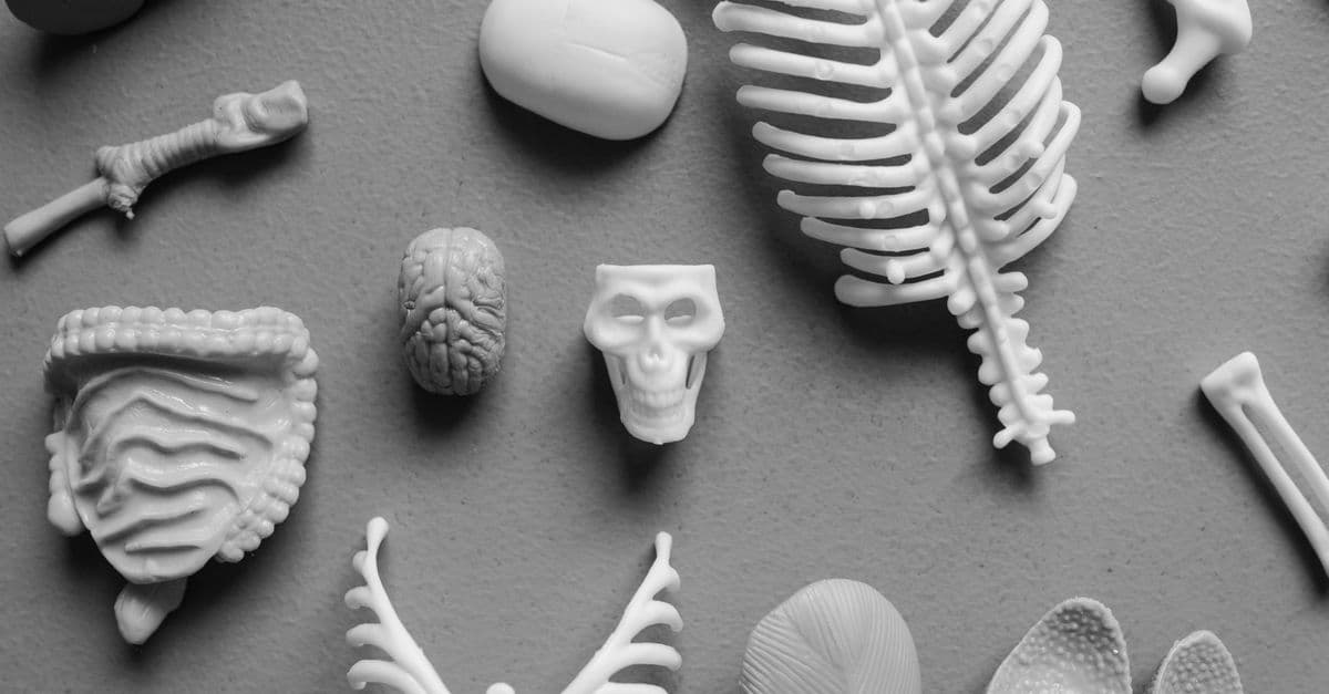
Photo: Tara Winstead / Pexels
Most people think of bone as the body's permanent scaffolding — inert, hard, fixed. The opposite is true. Bone is a metabolically active tissue with its own blood vessels, nerves, and immune cells, continuously dismantling and rebuilding itself in a coordinated cycle driven by two cell types that work in opposition. Every atom of calcium in your skeleton is replaced over roughly 10 years. Bone responds to mechanical load by growing stronger, and to unloading (bed rest, spaceflight) by rapidly weakening. It simultaneously serves as structural support, calcium reservoir, and blood cell factory. Understanding bone requires discarding the idea that it is anything like the dried, whitened specimens in biology classrooms.
The Composition of Bone: A Composite Material
Bone's remarkable combination of strength, toughness, and relative lightness comes from a two-component composite structure — an engineering principle independently discovered by humans much later in the form of reinforced concrete and fiberglass.
Collagen — specifically type I collagen, the most abundant protein in the body. Collagen fibers give bone its tensile strength and flexibility. Collagen alone is tough but bendable, like a thick rope. If you soak a bone in acid (which dissolves the mineral component), what remains is a rubbery, flexible collagen scaffold that can be bent without breaking.
Hydroxyapatite — a calcium phosphate mineral with the formula Ca₁₀(PO₄)₆(OH)₂. Hydroxyapatite crystals deposit within and around the collagen fibers, providing hardness and compressive strength. Hydroxyapatite alone is hard but brittle. If you heat a bone to ash (which destroys the collagen), what remains is white and rigid but shatters easily.
Together, collagen and hydroxyapatite produce a material stronger than either component alone — the mineral resists compression, the collagen resists tension and distributes stress to prevent crack propagation. Compact bone has a compressive strength of approximately 130–170 MPa and a tensile strength of approximately 50–130 MPa. High-strength structural steel has a tensile strength of about 400 MPa, but bone's density is roughly one-sixth that of steel, making it competitive in strength-to-weight terms.
Source: Rho, J.Y., Kuhn-Spearing, L. and Zioupos, P., Medical Engineering and Physics (1998) — Mechanical properties and the hierarchical structure of bone, from nanoscale collagen-mineral composite to macroscale architecture.
A third component — water — makes up roughly 25% of wet bone weight and is essential to its mechanical properties. Dehydration significantly reduces bone toughness, which is why wet bone fractures more cleanly than dried bone, and why the bone specimens in anatomy labs behave differently from living bone.
The Architecture of Bone: Compact and Spongy
Look at the cross section of a long bone like the femur (thigh bone) and you see two distinct architectural zones.
Compact bone (cortical bone) — the dense outer shell, making up about 80% of total bone mass. Under a microscope, compact bone is organized into cylindrical units called osteons (Haversian systems). Each osteon is a series of concentric rings of bone tissue (lamellae) surrounding a central canal (the Haversian canal) that contains blood vessels and nerves. Between osteons run connecting Volkmann's canals, creating a branching internal vascular network that keeps every part of the bone within diffusion distance of a blood supply.
Spongy bone (trabecular bone, cancellous bone) — the interior honeycomb of thin bony struts (trabeculae) that fills the ends of long bones, the vertebral bodies, the pelvis, and other bones. Spongy bone looks fragile but is architecturally sophisticated: its trabecular struts align along the lines of principal mechanical stress — the directions in which force is most commonly applied. This orientation makes spongy bone extraordinarily efficient, providing support with minimal material. The spaces between trabeculae are filled with bone marrow.
| Property | Compact bone | Spongy bone |
|---|---|---|
| Location | Outer shaft of long bones | Ends of long bones, vertebrae, pelvis |
| Porosity | 5–10% | 50–90% |
| Mass fraction | ~80% of skeleton | ~20% of skeleton |
| Primary function | Structural strength, load-bearing | Energy absorption, houses marrow |
| Organization | Osteons (Haversian systems) | Trabecular network |
The periosteum — a dense connective tissue membrane — covers the outer surface of compact bone everywhere except at joint surfaces. It contains osteoprogenitor cells (bone stem cells), blood vessels, and sensory nerves. The periosteum is why bone fractures are painful even before the bone has begun to heal: nerves in the periosteum transmit pain signals rapidly.
Source: Clarke, B., Clinical Journal of the American Society of Nephrology (2008) — Normal bone anatomy and physiology: comprehensive review of bone structure, cell types, and remodeling.
The Cells That Run the Skeleton
Four cell types maintain bone tissue:
Osteoblasts — bone-building cells derived from mesenchymal stem cells. Osteoblasts synthesize and secrete the collagen matrix (called osteoid) and then direct mineral deposition into it. As they become surrounded by their own secreted matrix, they transition into osteocytes.
Osteocytes — the most numerous bone cells, embedded within small cavities (lacunae) in the bone matrix, connected to each other and to surface cells by tiny channels (canaliculi) through which they extend cytoplasmic processes. Osteocytes are the mechanosensory cells of bone — they detect strain in the matrix caused by mechanical loading and send signals that either stimulate or inhibit remodeling. They are essentially strain gauges embedded in the structure they regulate.
Osteoclasts — large multinucleated cells derived from the same monocyte/macrophage lineage as immune cells. Osteoclasts dissolve bone by secreting hydrochloric acid (to dissolve hydroxyapatite) and proteases (to digest collagen) onto the bone surface in a sealed resorption pit called a Howship's lacuna. They are the demolition crew.
Bone lining cells — flattened, quiescent osteoblasts that cover resting bone surfaces and regulate the movement of minerals in and out of bone fluid.
Source: Harada, S. and Rodan, G.A., Nature (2003) — Osteoblast and osteoclast differentiation, signaling, and the control of bone mass.
Bone Remodeling: The Perpetual Renovation Cycle
Bone remodeling — the coordinated cycle of bone resorption followed by new bone formation — serves two functions simultaneously: it repairs microscopic damage before cracks can grow large enough to cause fracture, and it adapts bone geometry and density to current mechanical demands. It happens throughout life in small discrete units called basic multicellular units (BMUs).
A remodeling cycle in compact bone proceeds over roughly 3–4 months:
- Activation — osteocytes detect damage or mechanical signals and recruit osteoclast precursors.
- Resorption — osteoclasts dissolve a tunnel through compact bone (or a trench across trabecular bone surface), releasing calcium and phosphate into the bloodstream. This takes 2–3 weeks.
- Reversal — osteoclasts leave. A brief pause. Signals are sent to recruit osteoblasts.
- Formation — osteoblasts fill the resorption cavity with new osteoid, then mineralize it over several months.
- Mineralization and quiescence — new bone is complete; the BMU terminates.
At any moment, approximately 1–2 million BMUs are active throughout an adult skeleton. The entire mineral content of the adult human skeleton turns over every 7–10 years. In children, remodeling is faster because growth demands it; in elderly individuals, the balance tips toward resorption, explaining why bone density declines with age.
The communication between osteoclasts and osteoblasts is mediated by a critical signaling pathway: RANK/RANKL/OPG. Osteoblasts express RANKL (receptor activator of NF-κB ligand) on their surface; osteoclast precursors express RANK. When RANKL binds RANK, osteoclasts differentiate and become active. Osteoblasts also produce osteoprotegerin (OPG), a decoy receptor that binds RANKL and blocks osteoclast activation — a natural check on bone resorption. The ratio of RANKL to OPG is a master switch for bone balance. Understanding this pathway produced the drug denosumab (a monoclonal antibody against RANKL), now used clinically to treat osteoporosis and bone metastases from cancer.
Source: Boyle, W.J., Simonet, W.S. and Lacey, D.L., Nature (2003) — Osteoclast differentiation and activation: the RANK/RANKL/OPG signaling system.
Bone Marrow: The Blood Cell Factory
The hollow interior of most long bones and the spaces within spongy bone contain bone marrow — the tissue responsible for producing every type of blood cell in the adult body. This process is called hematopoiesis.
Red marrow — hematopoietically active marrow, present in virtually all bones at birth and in the flat bones, vertebrae, sternum, ribs, and the ends of long bones in adults. Red marrow contains hematopoietic stem cells (HSCs) — multipotent cells that give rise to all blood cell lineages. Each HSC can self-renew (produce more HSCs) or differentiate along pathways producing red blood cells, the five types of white blood cells described in how blood works explained, and platelets. The bone marrow produces roughly 200 billion red cells, 100 billion white cells, and 150 billion platelets every day.
Yellow marrow — fatty, metabolically inactive marrow that gradually replaces red marrow in the shafts of long bones as children grow. By adulthood, only the bones listed above retain red marrow. Yellow marrow can be converted back to red marrow when demand increases dramatically — severe blood loss, high altitude adaptation, or certain blood diseases.
The bone marrow niche — the specific microenvironment in which HSCs reside and from which their behavior is regulated. This includes osteoblasts (which help maintain HSCs in a quiescent state), blood vessels (which provide oxygen and signals for differentiation), and various stromal support cells. The discovery that osteoblasts directly regulate HSC behavior revealed an unexpected connection between the skeletal and hematopoietic systems.
Source: Morrison, S.J. and Scadden, D.T., Nature (2014) — The bone marrow niche for haematopoietic stem cells: structure, components, and regulatory signals.
Bone marrow transplantation (more precisely, hematopoietic stem cell transplantation) works by replacing a patient's diseased or destroyed marrow with donor HSCs. The donor cells home to the marrow niche via bloodstream circulation, guided by signaling molecules, and establish a new hematopoietic system. The skeletal architecture of the host bone is not transplanted — only the stem cells are.
Bone as the Body's Calcium Bank
Ninety-nine percent of the body's calcium is stored in bone — approximately 1,000 grams in an adult. This calcium is not static. The bones continuously exchange calcium with the bloodstream in both directions, with bone remodeling serving as a slow exchange mechanism and a faster ion-exchange mechanism operating at bone surfaces.
Blood calcium concentration must be kept within a narrow range (approximately 2.1–2.6 mmol/L) because calcium is essential for muscle contraction, nerve signaling, and cardiac function. When blood calcium drops, three mechanisms raise it:
Parathyroid hormone (PTH) — secreted by four small parathyroid glands behind the thyroid. PTH stimulates osteoclast activity (releasing calcium from bone), stimulates the kidneys to reabsorb calcium from urine rather than excreting it, and stimulates the kidneys to convert vitamin D to its active form (calcitriol).
Calcitriol (active vitamin D) — increases calcium absorption from food in the small intestine.
Calcitonin — released by thyroid C cells when blood calcium is high. It inhibits osteoclasts, reducing bone resorption and lowering blood calcium. Calcitonin's importance in humans is debated — people who have had their thyroid removed do not develop calcium dysregulation, suggesting other mechanisms compensate.
Vitamin D deficiency disrupts this system. Without calcitriol, calcium absorption from the gut falls, blood calcium threatens to drop, and PTH rises to compensate — pulling more calcium from bone. Sustained vitamin D deficiency causes rickets in children (soft, deformed bones) or osteomalacia in adults (soft bones with aching pain). Both conditions are really failures of bone mineralization, not just calcium shortage.
Source: Holick, M.F., New England Journal of Medicine (2007) — Vitamin D deficiency: mechanisms, consequences, and global prevalence.
How Mechanical Loading Builds Bone
The most direct stimulus for bone formation is mechanical stress. This is Wolff's Law (formulated by German anatomist Julius Wolff in 1892): bone adapts its structure and density to the loads it habitually bears. Osteocytes embedded in the matrix detect deformation (strain) when bone is loaded and respond by signaling to osteoblasts to deposit new bone in the overloaded region.
The practical implication: weight-bearing exercise (walking, running, resistance training) stimulates bone formation; prolonged bed rest or spaceflight causes rapid bone loss. Astronauts on the International Space Station lose 1–2% of bone mineral density per month in weightlessness — approximately what a postmenopausal woman might lose per year on Earth. This loss is concentrated in the weight-bearing bones (lumbar spine, hip, legs) rather than the arms.
High-impact exercise is more anabolic than low-impact exercise for bone. Swimming is excellent cardiovascular exercise but provides minimal bone stimulus because the body is buoyant. Tennis players develop measurably greater bone density in their dominant arm compared to the non-dominant arm — a within-person comparison that cleanly isolates the mechanical loading effect.
Source: Rubin, C.T. and Lanyon, L.E., Journal of Bone and Joint Surgery (1984) — Regulation of bone formation by applied dynamic loads: frequency and magnitude thresholds for bone adaptation.
Osteoporosis and the Tipping Balance
Osteoporosis — a condition in which bone mineral density has decreased sufficiently that fracture risk is significantly elevated, defined by a bone mineral density (measured by DEXA scan) more than 2.5 standard deviations below the young adult mean (T-score below -2.5). It affects an estimated 200 million people worldwide and causes approximately 8.9 million fractures annually.
The primary driver is an imbalance in the remodeling cycle: resorption outpaces formation. This occurs for several reasons:
Estrogen deficiency — estrogen normally suppresses RANKL expression by osteoblasts, restraining osteoclast activation. Menopause removes this restraint, causing accelerated bone resorption. Women can lose 3–5% of bone density per year in the first several years after menopause.
Age-related osteoblast dysfunction — osteoblast progenitor cells gradually shift toward fat cell differentiation rather than bone cell differentiation with aging, reducing the formation side of the equation.
Calcium and vitamin D insufficiency — particularly common in housebound elderly people with limited sun exposure and reduced dietary intake.
Treatment options target the RANK/RANKL/OPG pathway (denosumab), inhibit osteoclast activity via a different mechanism (bisphosphonates like alendronate), or stimulate osteoblast activity (teriparatide, a PTH analogue). Each approach works, and each has specific indications. The endocrine system's hormonal axes — particularly PTH, estrogen, and cortisol — are all deeply connected to bone health, explaining why endocrine disorders frequently manifest as skeletal problems.
Source: Consensus Development Conference, American Journal of Medicine (1993) — Diagnosis, prophylaxis, and treatment of osteoporosis: foundational definition and classification criteria.
FAQs About Bones
Q: How many bones does the adult human body have?
A: 206, but this varies slightly between individuals due to extra ribs, extra carpal bones, or variation in how certain small bones fuse. Infants are born with approximately 270–300 bones; many fuse during childhood and adolescence. The final adult count of 206 is typically reached around age 25 when the last growth plates close.
Q: Is it true that bones are stronger than steel?
A: On a weight-for-weight basis, cortical bone rivals steel in compressive and tensile strength, while being roughly six times less dense. Steel has a higher absolute tensile strength, but a bone structure as heavy as a steel structure of equivalent strength would be far heavier — which is the relevant comparison for a load-bearing skeleton in a moving organism.
Q: Do bones really heal themselves?
A: Yes. Fracture healing proceeds through distinct phases: a hematoma forms at the break, then cartilage tissue (a soft callus) bridges the gap, then osteoblasts convert the cartilage to bone (hard callus), and finally remodeling reshapes the repair to restore original architecture. Smaller fractures can heal within 6–8 weeks; large ones take longer. The process requires adequate nutrition (protein, calcium, vitamin D) and mechanical stability.
Q: Why do older people break bones more easily?
A: Both bone density and bone quality decline with age. Density loss (fewer mineral crystals per volume) is the better-known factor, but changes in collagen cross-linking and microarchitecture reduce the energy absorbed before fracture even at similar densities. Fall risk also increases with age due to muscle weakness, balance changes, and medication effects — a bone at lower density that never faces a fall may never fracture.
Q: What does bone marrow have to do with immunity?
A: All immune cells originate in bone marrow. B lymphocytes (which produce antibodies) both originate and mature in bone marrow. T lymphocytes originate in bone marrow but mature in the thymus. Natural killer cells, macrophages, neutrophils, and other immune cells all derive from bone marrow hematopoietic stem cells. Bone is literally the source of the immune system.
Q: Can you strengthen bones with diet alone without exercise?
A: Diet provides the raw materials (calcium, phosphate, vitamin D, protein, vitamin K2) necessary for bone building, but without mechanical stimulus, osteoblasts receive no signal to deposit new bone. Diet is necessary but not sufficient. The combination of adequate nutrition plus weight-bearing exercise is the most effective approach to maintaining and building bone density.
Q: Why do growth plates matter for children's bone injuries?
A: Growth plates (physes) are regions of cartilage near the ends of long bones where longitudinal growth occurs. They are mechanically weaker than surrounding bone, so fractures in children often occur through the growth plate rather than the shaft. Damage to the growth plate can disrupt growth and cause limb length discrepancy or angular deformity — which is why growth plate injuries in children receive more careful evaluation than similar-appearing injuries in adults.
Source: Diméglio, A. and Canavese, F., Journal of Pediatric Orthopedics (2012) — Growth plate physiology, Salter-Harris fracture classification, and long-term outcomes of growth plate injuries.
The Takeaway
Bone is not the body's inert infrastructure — it is a metabolically active tissue whose daily operations include blood cell production, calcium regulation, structural adaptation to load, and autonomous microfracture repair. Its mechanical superiority over purely ceramic or purely fibrous materials comes from the collagen-hydroxyapatite composite, a solution biology arrived at hundreds of millions of years before materials engineers. The remodeling cycle driven by osteoblasts and osteoclasts ensures that the skeleton you carry at 40 is genuinely different, atom by atom, from the one you carried at 30 — and whether it is stronger or weaker depends substantially on the mechanical loads you apply to it and the nutritional environment you provide.
Read next:
Follow new articles
Add SciHub101 to your feed reader to see new science and technology stories.
About the Author
SciHub101 is an independent science and technology publication founded and edited by Buğra Sözeri, a software engineer and app developer based in Türkiye. AI tools assist with drafting and research. Source documentation in the existing archive is uneven, and articles are being reviewed and corrected. The Editorial Team byline identifies editorial responsibility; it does not certify specialist or independent human review. Where we compare products, anything built by our own team is labelled as such — see the editorial guidelines and About page for how corrections and disclosures work.
Related Articles
How We Taste and Smell: The Science of Flavor, 400 Odor Receptors, and the Nose-Brain Connection
Pinch your nose while eating a strawberry and the flavor nearly disappears — what remains is just sweetness with a hint of acid. Flavor, it turns out, is about 80% smell. The tongue detects only five basic tastes; the nose handles everything else through 400 distinct olfactory receptors, making smell the most molecularly diverse sensory system in the human body. Here is how these two ancient senses combine to produce the richness of what we call taste.
How the Body Regulates Temperature: The Hypothalamic Thermostat, Sweating, Shivering, and Fever
The human body maintains core temperature within a range of less than 1°C (1.8°F) despite external temperatures swinging 60°C or more across seasons, and despite internal heat generation that — in hard exercise — rises to 20 times the resting rate. This precision requires a thermostat, an active cooling system, an emergency heating system, and dedicated fat that burns itself to generate warmth. None of it is conscious. Here is how thermoregulation actually works.

How Bones and Joints Work: The Living Skeleton
Bone is not dead material — it's living tissue constantly being broken down and rebuilt, completely renewing itself roughly every 10 years.

Why Do We Have Eyebrows? The Science and Evolution
Eyebrows serve a dual purpose that touches on both biology and behavior. They channel sweat, rain, and debris away from the eyes, protecting our vision in wet or dusty environments. But more importantly, eyebrows communicate emotion and intent across distances, making them crucial for social bonding and face recognition. Humans evolved progressively smaller brow ridges and more mobile eyebrows than our archaic ancestors, supporting increasingly complex facial expression and nonverbal conversation.

Why Do Our Ears Pop? The Science of Pressure Equalization
Every airplane climb or diving descent reminds us: the human ear is exquisitely sensitive to pressure change. The familiar pop occurs because the middle ear—a small air-filled chamber behind the eardrum—must equalize its pressure to match the ambient environment. When you yawn or swallow, the Eustachian tube opens to let air flow in or out, restoring balance. Altitude, diving, colds, and even weather shifts can create pressure mismatches that strain the tympanic membrane; when equalization finally succeeds, you feel the sudden relief of the pop.
Why Do Our Limbs Fall Asleep? The Science of Paresthesia
Sustained pressure compresses the nerves running through your limbs, blocking the electrical signals that carry sensation to your brain and creating numbness. When you shift position and release the pressure, those compressed nerves 'wake up' with a flood of erratic firing that produces the familiar 'pins and needles' tingling sensation called paresthesia. This temporary phenomenon is harmless and resolves in seconds, but understanding what happens reveals how your peripheral nervous system works and when similar sensations might signal something more serious.