How Does the Human Eye Work? The Physics and Neuroscience of Vision
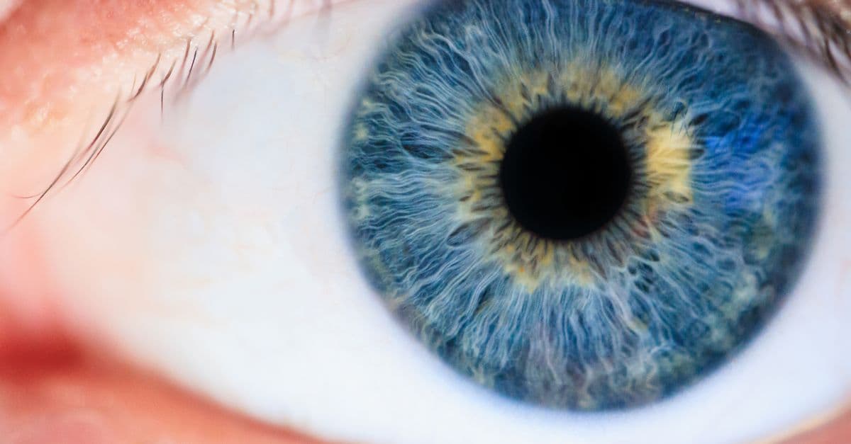
Photo: Michael Morse / Pexels
When light from the sun reflects off a red apple and enters your eye, it undergoes one of the most elegant transformations in physics and biology. The cornea — a transparent dome on the eye's surface — bends (refracts) the light rays. The lens, suspended behind the cornea, fine-tunes the focus. The light converges on the retina, a tissue thinner than a human hair, positioned at the back of the eye. Within the retina, molecules called opsins, arrayed in 126 million photoreceptor cells, absorb the photons. A single photon can excite an opsin, triggering a cascade of biochemical reactions that hyperpolarize the cell, silencing its baseline neurotransmitter release. This silence propagates through interneurons to a ganglion cell, which fires an action potential. Within milliseconds, electrical signals travel along the optic nerve to the visual cortex at the back of the skull. There, the signal activates neurons in one of 30 distinct visual processing areas. The brain reconstructs shape, color, motion, and depth from this neural cascade — a reconstruction so vivid and immediate that you are unaware of the processing and believe you are simply "seeing" the apple. This illusion of direct visual perception is one of the brain's most convincing tricks.
The Optical Pathway: From Cornea to Retina
Refraction — the bending of light as it passes from one transparent medium to another, due to a change in the speed of light in different materials; the amount of bending depends on the refractive index (a measure of how much the material slows light compared to vacuum).
The eye works on the same principle as a photographic camera, but with performance criteria that silicon-based optical systems struggle to match. Light travels in straight lines until it encounters a boundary between two materials with different densities. At that boundary, the light bends. The cornea, the clear outer surface of the eye, has a refractive index of 1.38 (compared to air's 1.0). When light from the apple enters the eye, it bends sharply at the air-cornea boundary, converging inward by roughly 48 diopters of optical power (a diopter is a measure of refractive strength: a +1 diopter lens focuses parallel light 1 meter away).
The light then passes through the aqueous humor, a clear fluid filling the space between the cornea and the lens. The aqueous humor has a refractive index nearly identical to the cornea (1.33) and supplies the cornea and lens with oxygen and nutrients. Behind the aqueous humor sits the crystalline lens, a biconvex structure made of layers of protein (crystallins) arranged with near-crystalline precision. The lens is suspended by ligaments (the zonula) attached to a ring of muscle (the ciliary muscle) surrounding the lens. This muscle is controlled by the parasympathetic nervous system, allowing precise, involuntary control of lens shape and focal distance.
The lens alone provides roughly 15–20 diopters of optical power, but its power is variable: when the ciliary muscle relaxes, the lens flattens and its power decreases, focusing light from distant objects. When the ciliary muscle contracts, the lens bulges and its power increases, focusing light from nearby objects. This process is called accommodation — the adjustment of lens shape to focus on objects at varying distances. The amplitude of accommodation (the maximum change in lens power) is greatest in infants and declines steadily with age. By age 50, accommodation is nearly absent, which is why presbyopia (age-related difficulty focusing on nearby objects) is universal — the lens has stiffened, losing elasticity, and the ciliary muscle can no longer change its shape significantly.
The cornea and lens together bend light rays such that they converge onto the retina, a distance of roughly 24 millimeters from the corneal surface (the axial length of the eye). In an eye with correct refractive geometry, the focal point (the point where light rays converge most sharply) falls precisely on the retina. In myopic eyes (nearsighted), the cornea is too curved or the eye is too long, causing the focal point to fall in front of the retina; distant objects focus before reaching the retina, appearing blurred. In hyperopic eyes (farsighted), the opposite occurs: the focal point falls behind the retina, causing difficulty focusing on near objects.
The light that reaches the retina has been compressed enormously. Light entering the pupil (the variable aperture of the eye, controlled by the iris) is focused into a much smaller image. The magnification from the pupil to the focal point on the retina is roughly 1/4 — four times smaller. Yet the neural encoding that follows preserves spatial information through topographic maps: adjacent photoreceptors activate adjacent neurons in the optic nerve and in visual cortex, maintaining the spatial relationships of the retinal image throughout the visual system.
Source: Artal, P. (2014), "Optics of the eye and its impact in vision: a review," Progress in Retinal and Eye Research, Vol. 36 — a comprehensive overview of eye optics, refractive error, and visual performance.
The Retina: Converting Light into Neural Signals
The retina is not a simple camera sensor but a complex neural tissue, a specialized part of the brain that happens to be located in the eye. It contains roughly 126 million photoreceptors and another 50 million neurons (bipolar cells, horizontal cells, amacrine cells, and ganglion cells), all arranged in layers and connected by synapses. Light must pass through the ganglion cells and bipolar cells to reach the photoreceptors — a seemingly backward arrangement that neuroscientists call the "inverted retina." Despite its inefficiency, this arrangement provides evolutionary advantages in terms of metabolic support and neural integration.
Photoreceptors — specialized neurons containing opsins that absorb photons and convert light energy into electrical signals via a biochemical cascade.
There are two types of photoreceptors. Rod cells, roughly 120 million of them, are exquisitely sensitive to light — a single photon can produce a measurable electrical signal. Rods lack color selectivity; they contain a single opsin (rhodopsin) tuned to wavelengths around 507 nanometers (blue-green light). At night, under dim starlight, vision is rod-mediated, and color perception vanishes — this is why your nighttime vision is grayscale. Rod cells are slower to respond than cone cells and do not recover quickly from saturation (when the light is so bright that the rod reaches maximum neural response and cannot respond further).
Cone cells, roughly 6 million of them, are less sensitive than rods but more rapid in response and capable of color discrimination. There are three cone subtypes, each containing a different opsin: S-cones (short wavelength, sensitive to blue light around 420 nm), M-cones (medium wavelength, sensitive to green light around 530 nm), and L-cones (long wavelength, sensitive to red light around 560 nm). In bright light, color vision is cone-mediated. Cones are concentrated in the central retina, especially in the fovea — a specialized pit at the center of the retina where vision is sharpest because photoreceptors are most densely packed and other retinal layers are thinned.
The conversion of light into electrical signals begins with the absorption of a photon by an opsin molecule. Opsins are G-protein coupled receptors (GPCRs), a large family of proteins that span the cell membrane. A photon causes the opsin to undergo an isomerization — a change in the three-dimensional shape of the retinoid (vitamin-A-derived) chromophore bound within the opsin. This shape change sets off a cascade of molecular events:
- The activated opsin triggers a G-protein called transducin.
- Transducin activates a phosphodiesterase enzyme.
- Phosphodiesterase degrades cyclic GMP (cGMP), a second messenger.
- cGMP normally holds ion channels in the photoreceptor membrane open, allowing cations to flow inward.
- The reduction in cGMP closes these channels, hyperpolarizing the cell (making its membrane potential more negative).
- Hyperpolarization reduces the release of glutamate, the photoreceptor's neurotransmitter.
This cascade is called the phototransduction cascade. It amplifies the signal from a single photon into a measurable change in cell potential and neurotransmitter release. A single photon can close roughly 200 cation channels, even though only one opsin is activated — an amplification factor that enables rod vision to detect starlight.
The phototransduction cascade is extraordinarily efficient. In bright light, all cGMP channels close, all transducin molecules deactivate, and the cascade shuts down. The photoreceptor returns to baseline. This on-off cycling occurs millions of times per second, allowing the visual system to track the finest details of motion and the subtlest changes in illumination.
Source: Yau, K. W., & Hardie, R. C. (2009), "Phototransduction motifs and variations," Cell, Vol. 139, No. 2 — a definitive review of phototransduction mechanisms in vertebrate photoreceptors.
From Photoreceptors to the Brain: The Retinal Neural Circuitry
The hyperpolarization and reduced neurotransmitter release from a photoreceptor propagates through the retina's inner layers. The photoreceptor synapses with bipolar cells, which transmit the signal to amacrine cells (interneurons for lateral integration) and horizontal cells (feedback interneurons that modulate photoreceptor release). Finally, bipolar cells synapse with ganglion cells, the only retinal neurons that fire action potentials and transmit signals out of the eye via the optic nerve.
This arrangement — photoreceptors → bipolar cells → ganglion cells — is a simple feedforward chain, but the retina adds sophisticated lateral processing through horizontal cells and amacrine cells. Horizontal cells connect photoreceptors laterally, allowing photoreceptors to pool information from their neighbors. This spatial pooling enhances sensitivity in dim light (rods can share signals across large areas) but reduces spatial resolution. Amacrine cells similarly integrate information across photoreceptors and bipolar cells, enhancing motion detection and directional selectivity. The result is that the retina does not simply transmit a pixel-by-pixel representation of the visual scene; it computes edge contrast, motion direction, and local luminance changes before sending signals to the brain.
Ganglion cells are the output neurons of the retina. There are roughly 1.5 million ganglion cells in each eye (compared to 126 million photoreceptors) — a massive compression of visual information before it even leaves the eye. This compression is not random; it reflects the prioritization of visually important features. Center-surround receptive fields are the hallmark of retinal ganglion cells: each ganglion cell has a small central region (the center of its receptive field) where light increases or decreases the cell's firing rate, and a larger surrounding region (the surround) where light has the opposite effect. This organization enhances contrast and edge detection.
Receptive field — the region of space (or, in the retina, the region of the visual scene) that, when stimulated, affects the activity of a particular neuron; the organization of the receptive field (center-surround, direction preference, color opponency) determines the feature that neuron encodes.
Ganglion cells also encode color. Some ganglion cells, called P-cells (parvocellular), integrate signals from L and M cones to encode color in a narrow central region and receive spatial-opponent processing from the surround. Others, called M-cells (magnocellular), integrate rod and broad-spectrum cone signals, encoding brightness and motion but not fine color. A third population, the intrinsically photosensitive retinal ganglion cells (ipRGCs), contain their own opsins and respond directly to light, independent of photoreceptor input — these cells are thought to mediate circadian rhythm regulation and pupil responses rather than conscious vision.
The 1.5 million ganglion cells in each eye converge into the optic nerve, a bundle of axons roughly 1.5 millimeters in diameter. The optic nerve exits the eye at a point called the optic disk, where no photoreceptors are present — this is the anatomical basis of the blind spot. Surprisingly, the blind spot is not experienced as a gap in your visual field. The brain fills in the blind spot using information from the surrounding retina and from the contralateral eye (the eye on the opposite side). This "filling-in" is so effective that you must deliberately search for your blind spot to perceive it; under normal circumstances, your vision appears complete.
Source: Masland, R. H. (2012), "The neuronal organization of the retina," Neuron, Vol. 76, No. 2 — a comprehensive review of retinal circuitry and the properties of retinal neurons.
Central Vision: The Optic Nerve and Visual Pathways
The optic nerve carries signals from each eye to the brain, but the routing is not straightforward. At the optic chiasm (a structure at the base of the brain where the optic nerves meet), axons from the nasal (inner) half of the retina of one eye cross over to the opposite side of the brain, while axons from the temporal (outer) half remain on the same side. This crossing ensures that the visual world is split vertically: the right visual field (the right half of what you see from both eyes) is processed by the left hemisphere, and the left visual field is processed by the right hemisphere. This crossing is nearly complete in humans — roughly 53 percent of ganglion cell axons cross, a proportion that varies slightly among individuals and is thought to relate to binocular depth perception and handedness.
After crossing the optic chiasm, ganglion cell axons travel to the lateral geniculate nucleus (LGN) of the thalamus, a relay station that preserves the retinotopic map (the spatial relationship of the retina is maintained in the LGN). The LGN receives not only feedforward input from the retina but also feedback from the visual cortex, allowing the cortex to modulate the flow of sensory information to itself. This feedback circuit is thought to enhance signals from attended locations and suppress signals from unattended regions — a mechanism of selective attention operating at the level of the thalamus.
The LGN sends signals to the primary visual cortex (V1), located at the very back of the brain in the occipital lobe. V1 contains roughly 200 million neurons arranged in six layers, each specialized for different aspects of visual processing. Some neurons are tuned to edge orientation; others to color, spatial frequency, or eye-of-origin (binocular depth). V1 performs the remarkable feat of integrating monocular signals from two retinas and extracting binocular disparity — the slight difference in the location of an object's image on each retina, which is the primary cue for stereoscopic depth perception.
From V1, visual information diverges into two major pathways. The ventral stream (the "what" pathway) proceeds ventrally to the inferior temporal cortex, specializing in object recognition and color. The dorsal stream (the "where" pathway) proceeds dorsally to the posterior parietal cortex, specializing in spatial processing and motion. Information flowing through these streams integrates in higher visual areas (V2, V3, V4, V5/MT, etc.), ultimately reaching association cortex in the prefrontal and temporal regions, where conscious visual perception arises.
Retinotopic map — a topographic representation of the visual field in which the spatial relationships of the visual scene are preserved; adjacent locations in the visual field activate adjacent neurons in the visual cortex.
This hierarchical architecture solves a profound problem: vision is not achieved by a single image in the brain but by parallel processing of different visual attributes across 30 distinct cortical areas. Color, form, motion, and depth are computed separately and then integrated. The brain constructs a unified visual percept from these parallel streams, a solution that seems necessary because no single neural circuit can simultaneously optimize all aspects of visual processing.
Source: Wandell, B. A., Dumoulin, S. O., & Brewer, A. A. (2007), "Visual field maps in human cortex," Neuron, Vol. 56, No. 2 — describing the retinotopic organization of visual cortex and the parallel visual streams.
Limits and Variations in Human Vision
Myopia (nearsightedness) results from excessive corneal curvature or excessive eye length, causing the focal point of light to fall in front of the retina. The prevalence of myopia has increased dramatically in recent decades, particularly in East Asian populations. Studies suggest that environmental factors, especially prolonged near-focus activities (reading, computer work) and reduced time outdoors, contribute to myopia development in genetically susceptible individuals. The mechanism may involve the release of dopamine in the retina during outdoor light exposure; dopamine is thought to inhibit the elongation of the eye during development. Outdoor time appears protective against myopia progression.
Hyperopia (farsightedness) results from insufficient corneal curvature or a short eye, causing the focal point to fall behind the retina. People with hyperopia can focus on distant objects but struggle with near vision. Unlike myopia, hyperopia may partially correct itself with accommodation (lens thickening), but the accommodative load becomes unsustainable in adulthood or with presbyopia.
Astigmatism occurs when the cornea or lens is not perfectly spherical — it refracts light more strongly in one meridian (direction) than another. The result is that lines oriented in one direction appear sharp while lines in perpendicular directions appear blurred. Astigmatism is common and can coexist with myopia or hyperopia.
Color blindness, or color vision deficiency, results from loss or malfunction of one or more cone types. The most common form is red-green color blindness (protanopia or deuteranopia), affecting roughly 8 percent of males and 0.5 percent of females. This occurs due to mutations in the genes encoding L-cones or M-cones on the X chromosome. People with red-green color blindness perceive reds and greens as shades of yellow or brown, though research suggests their subjective experience is quite different from the colorblind simulation images (saturated colors shifted) that sighted people are shown.
Presbyopia — the age-related decline in accommodation due to hardening of the crystalline lens and weakening of the ciliary muscle; affects all individuals typically beginning around age 40–50.
The blind spot, present in all sighted humans, is sometimes perceived as a limitation, but it reflects a design trade-off: the optic nerve must exit somewhere, and the exit point is necessarily a region lacking photoreceptors. The blind spot typically spans about 5 degrees of visual angle. Its location is offset from the center of vision and rarely noticed because the brain fills in the missing information and because the other eye typically has an unobstructed view of that region.
Source: Curcio, C. A., & Allen, K. A. (1990), "Topography of ganglion cells in human retina," Journal of Comparative Neurology, Vol. 300, No. 1 — describing the distribution and density of photoreceptors and blind-spot anatomy.
Source: Neitz, J., Geist, T., & Jacobs, G. H. (1989), "Color vision in the dog," Visual Neuroscience, Vol. 3, No. 2 — examining color perception mechanisms and variation across individuals and species.
Source: Sharpe, L. T., Stockman, A., Jägle, H., & Nathans, J. (1999), "Opsin genes, cone photopigments, color vision, and blindness," Progress in Retinal and Eye Research, Vol. 18, No. 6 — a comprehensive review of the molecular basis of color vision and color blindness.
Source: Holden, B. A., Fricke, T. R., Wilson, D. A., Jong, M., Naidoo, K. S., Sankaridurg, P., ... & Resnikoff, S. (2016), "Global prevalence of myopia and high myopia and temporal trends from 2000 through 2050," Ophthalmology, Vol. 123, No. 5 — documenting the increase in myopia prevalence globally.
Frequently Asked Questions
Q: Why is the retina inverted? A: This is one of the most frequently questioned aspects of eye anatomy. The inverted arrangement (light must pass through ganglion cells and bipolar cells before reaching photoreceptors) seems inefficient, yet it persists. The likely explanation is evolutionary: the retina evolved from an invagination (folding inward) of the brain tissue itself, retaining the original interior-outside orientation. The apparent disadvantage is offset by metabolic benefits: the dense network of blood vessels supporting the retina sits on the outer surface (in the retinal pigment epithelium), providing efficient oxygen and nutrient supply to the photoreceptors, which are the most metabolically demanding cells in the body. Additionally, horizontal cells and amacrine cells located in this arrangement enable sophisticated lateral processing that enhances contrast and motion detection. The inverted retina is not a suboptimal quirk but a design that balances multiple functional requirements.
A: When light enters the eye and is focused onto the retina, the image is inverted — it appears upside-down and mirror-reversed on the retina, just as an image appears inverted in a camera. However, the brain interprets this inverted image as upright. The inversion occurs at the level of the retina, and the brain's neural processing corrects for it automatically through learned experience in early development. A classic experiment in which subjects wore inverting prism glasses showed that after adaptation (typically 1–2 weeks of continuous wear), the visual world appeared upright again, even though the eyes were receiving inverted images — demonstrating that the perceived orientation is not intrinsic to the image but a construction of the brain.
Q: How does the brain know which eye is looking at what? A: The optic nerves from both eyes project to the lateral geniculate nucleus (LGN) and visual cortex, where the inputs are segregated into eye-of-origin columns — regions of cortex responsive to one eye or the other. This organization allows the brain to compare the slightly different images from the two eyes and extract binocular disparity, the primary cue for stereoscopic depth. Additionally, cortical neurons in visual areas beyond V1 often integrate inputs from both eyes, allowing the brain to fuse the two images into a unified percept. When the eyes are misaligned (strabismus), the fusion process fails, and the brain may suppress input from the weaker eye, leading to amblyopia (lazy eye) if not corrected in childhood.
Q: Can the human eye see ultraviolet or infrared light? A: The human eye is sensitive to visible light (roughly 380–740 nanometers wavelength) because opsins (the light-absorbing proteins in photoreceptors) have peak absorption in that range. Ultraviolet light (below 380 nm) is absorbed and scattered by the lens and cornea before reaching the photoreceptors; humans cannot perceive UV light naturally. However, some individuals who have had the lens surgically removed (due to cataracts or injury) report seeing into the near-ultraviolet range, suggesting that the lens is the primary UV filter. Infrared light (above 740 nm) has too little energy to activate opsins; the eye perceives infrared only if the photons have sufficient intensity to generate heat, which then stimulates warm-sensing neurons in the skin. Many insects and birds can see into the ultraviolet range because their lens transmits UV light and their opsins absorb it, a difference that provides advantages in flower recognition and prey detection.
Q: What causes the blind spot, and why don't you notice it? A: The blind spot occurs where the optic nerve exits the eye, at the optic disk. This region contains no photoreceptors, so no visual information can be extracted from it. You don't notice the blind spot in everyday vision because the brain fills in the region using information from the surrounding retina and the other eye. This filling-in is not a conscious process but a perceptual phenomenon arising from the way the visual cortex reconstructs the visual field. The blind spot can be perceived with specific stimuli: if you cover one eye and focus on a fixation point, a carefully placed stimulus (such as a moving dot) disappears when it enters the blind spot region and reappears when it exits.
Q: How do contact lenses and eyeglasses correct vision? A: Contact lenses and eyeglasses work by adding optical power to the eye's refractive system. Glasses sit roughly 12 millimeters in front of the cornea; contacts sit on the corneal surface. Both refract light to shift the focal point of the eye closer to (for myopia) or farther from (for hyperopia) the retina. A minus (concave) lens diverges light rays outward, reducing the eye's total refractive power, which moves the focal point backward in myopic eyes. A plus (convex) lens converges light rays inward, increasing refractive power, which moves the focal point forward in hyperopic eyes. Bifocal and progressive lenses have regions of different power to correct both distance and near vision (presbyopia). Astigmatism correction requires cylindrical lenses that have different curvatures in different meridians, matching the eye's irregular curvature.
Q: Is color blindness the same as seeing in black and white? A: No, color blindness is not the same as complete absence of color perception. Most people with red-green color blindness can still distinguish colors but perceive reds and greens as shades of yellow, brown, or gray. They may struggle to differentiate ripe versus unripe fruit, or distinguish traffic-light colors. Complete color blindness (seeing only in grayscale) is extremely rare, occurring only with damage to all three cone types or severe cortical processing deficits. The subjective experience of color-blind individuals remains poorly understood; the common colorblind simulation images shown to sighted people may not accurately represent what people with color blindness actually perceive, partly because their visual cortex may adapt over development to extract maximum information from available color signals.
Read Next
Follow new articles
Add SciHub101 to your feed reader to see new science and technology stories.
About the Author
SciHub101 is an independent science and technology publication founded and edited by Buğra Sözeri, a software engineer and app developer based in Türkiye. AI tools assist with drafting and research. Source documentation in the existing archive is uneven, and articles are being reviewed and corrected. The Editorial Team byline identifies editorial responsibility; it does not certify specialist or independent human review. Where we compare products, anything built by our own team is labelled as such — see the editorial guidelines and About page for how corrections and disclosures work.
Related Articles

How Does the Sense of Balance Work? The Vestibular System Explained
Balance is not a single sense but a symphony of three systems working in concert: the vestibular system (inner ear acceleration sensors), proprioception (body position awareness), and vision. The vestibular system contains the semicircular canals (which detect rotation) and the otolith organs (which detect linear acceleration and head tilt). Damage to any component creates the sensation of spinning, nausea, and falls. Understanding how the brain fuses these signals explains vertigo, motion sickness, and why astronauts become disoriented in microgravity where the vestibular system receives no gravitational input.

How Do Muscles Grow? The Science of Hypertrophy, Protein Synthesis, and Muscle Adaptation
When you lift a weight, you are not building muscle — you are damaging it. Within hours, the muscle responds by activating protein synthesis, repairing the damage with new protein, and strengthening the contractile machinery. Over weeks of repeated stimulus, the cross-sectional area of muscle fibers expands, a process called hypertrophy. The physiology of muscle growth involves three fundamental mechanisms: mechanical tension (the force produced by contracting muscle against resistance), muscle damage (the microtears that occur when muscle is stressed), and metabolic stress (the accumulation of metabolites like lactate during intense activity). Satellite cells, dormant muscle stem cells surrounding each fiber, activate in response to growth signals and fuse to existing fibers, donating nuclei that increase the capacity for protein synthesis. Progressive overload — gradually increasing the weight, volume, or intensity of exercise — is essential because muscles adapt to stress; once adaptation occurs, growth plateaus until a new, greater stimulus is applied. Research reveals that muscle growth is constrained by genetics, nutritional status (particularly protein intake), recovery (sleep and time between sessions), and age, yet the fundamental mechanisms are conserved across these variables.

How the Eye Works: Light, the Retina, and How the Brain Builds Sight
Your eye contains 126 million photoreceptors but only one million output fibers to the brain — meaning the retina compresses visual data by a factor of 126 before it leaves the eye. What you see is a model your brain built, not a photograph it took.
How Does the Human Eye Actually Work?
Your eye captures light, converts it to electrical signals, and sends them to the brain — which then constructs everything you see. Here's the elegant biology behind vision.
How the Human Eye Works — From Light to Vision
How light becomes sight: eye anatomy, rods and cones, phototransduction, the visual cortex, color vision, and why eyes evolved 8+ times.

How the Eye Sees Color: The Science of Vision
Your eyes can distinguish around 10 million colors, yet the entire mechanism runs on just three types of cone cell. Here's how that's possible.