How the Eye Works: Light, the Retina, and How the Brain Builds Sight
Photo: lil artsy / Pexels
Right now, photons are striking the back of your eye at the speed of light, being absorbed by 126 million specialized cells that convert them into electrical signals, compressed through a single cable containing only one million fibers, and reconstructed by roughly a third of your brain's cortex into the scene you perceive as seamless reality. The eye is not a camera. It is the front end of an interpretation system that decides — actively, continuously, and often incorrectly — what the world looks like. Understanding how it works means confronting the fact that vision is construction, not capture.
The Eye's Optical System: Getting Light to the Right Place
Before any cell responds to light, the eye has to focus it precisely onto the retina at the back of the eyeball — a task accomplished by two optical elements working in series.
Cornea — the transparent, curved dome at the front of the eye that provides approximately 70% of the eye's total optical power through refraction.
The cornea performs most of the eye's focusing, but it cannot change shape — it is fixed. Fine-tuning focus across different distances falls to the lens, a transparent, biconvex disc made of crystalline proteins that can change curvature through a process called accommodation. The ciliary muscle surrounding the lens contracts to allow the lens to round up (for close objects) and relaxes to let it flatten (for distant ones). A 20-year-old lens changes shape easily; by age 45, the lens stiffens and accommodation range shrinks — which is why people in their mid-40s begin holding books at arm's length before eventually needing reading glasses.
Source: Atchison, D.A. & Smith, G., Optics of the Human Eye (2000) — Authoritative account of corneal and lens contributions to total ocular power and the mechanics of accommodation.
Light passes from the lens through the vitreous humor — a gel-like substance filling the back four-fifths of the eyeball — before striking the retina. The vitreous is 99% water but maintains its gel structure via a collagen-hyaluronic acid matrix. As people age, this matrix breaks down and collagen fibers clump, producing floaters — the drifting specks and threads many people see against bright backgrounds. They are real objects casting shadows on the retina, not visual hallucinations.
The iris regulates light entry via the pupil, contracting in bright light (to a minimum diameter of about 2 mm) and dilating in dim conditions (to a maximum of roughly 8 mm). This gives the eye a dynamic range adjustment of about 16-fold in area, which sounds significant but covers only a fraction of the 10-billion-fold range of light intensities the full visual system can handle. The rest comes from the photoreceptors themselves.
The Retina: 126 Million Sensors in a Sheet Thinner Than a Credit Card
The retina is a sheet of neural tissue approximately 0.2 to 0.5 mm thick lining the back two-thirds of the eye. It contains five major classes of neurons — photoreceptors, bipolar cells, ganglion cells, horizontal cells, and amacrine cells — arranged in distinct layers, and it performs substantial image processing before a single signal leaves the eye.
Retina — a light-sensitive layer of neural tissue at the back of the eye that converts photons into electrical signals and performs initial visual processing before transmitting signals to the brain via the optic nerve.
The photoreceptors come in two types: rods and cones.
| Feature | Rods | Cones |
|---|---|---|
| Number in human eye | ~120 million | ~6 million |
| Location | Peripheral retina | Concentrated at fovea |
| Light sensitivity | Extremely high (single photon) | Lower — require brighter light |
| Color discrimination | None (one pigment: rhodopsin) | Yes — three pigment types |
| Function | Night and peripheral vision | Color, fine detail, daytime vision |
| Response speed | Slower | Faster |
Source: Cleveland Clinic (2022) — "Photoreceptors: Rods and Cones" — Confirms photoreceptor counts, distribution, and functional distinctions.
Rods contain a single photosensitive pigment, rhodopsin, made of a protein (opsin) bound to a light-absorbing molecule called retinal. When a photon strikes rhodopsin, the retinal molecule changes shape (from 11-cis to all-trans), triggering a cascade of molecular events that closes ion channels in the rod's membrane, hyperpolarizing the cell and ultimately altering the neurotransmitter signal it sends to bipolar cells. A fully dark-adapted rod can respond to a single photon — a sensitivity that makes the eye's low-light performance rival the theoretical physical limit.
Source: Rieke, F. & Baylor, D.A., Reviews of Modern Physics (1998) — "Single-photon detection by rod photoreceptors" — confirms single-photon sensitivity of dark-adapted rods.
Cones fall into three subtypes based on their peak light sensitivity: S-cones (short wavelength, ~420 nm, perceived as violet-blue), M-cones (medium wavelength, ~530 nm, green), and L-cones (long wavelength, ~560 nm, red). Color perception emerges from the brain comparing the relative activation rates of these three populations — a system called trichromacy. People with red-green color blindness typically have S-cones and M-cones but lack functional L-cones (or vice versa), so the brain loses the ability to distinguish those spectral regions.
The Fovea: The Eye's High-Resolution Window
The retina is not uniformly sensitive. Visual acuity — the ability to resolve fine detail — peaks sharply at a small pit in the center of the retina called the fovea centralis, which covers roughly 2 degrees of visual angle but contains about 50% of all cone photoreceptors.
Fovea — a 1.5 mm pit at the center of the retina containing only cone photoreceptors at extremely high density (~200,000 per mm²) and providing maximum visual acuity.
The fovea has no rods. It is optimized entirely for fine detail and color in bright conditions. Each foveal cone connects through a private wire — its own dedicated bipolar and ganglion cell — to the optic nerve, preserving maximum spatial resolution. Away from the fovea, multiple rods converge onto a single ganglion cell, sacrificing resolution for light sensitivity. This is why peripheral vision is terrible at detail and excellent at detecting motion in dim light.
The practical consequence: your eye is constantly moving in small, rapid jumps called saccades — typically 3 to 5 per second during normal viewing — to drag the fovea across a scene and build a high-resolution mental map from a series of snapshots. The smooth, stable view you experience is entirely a brain construction. During each saccade, visual processing is suppressed (a phenomenon called saccadic suppression) so you are not aware of the blur. Your visual experience of the world is a continuous-seeming montage, not a live feed.
Source: Purves, D. et al., Neuroscience, 6th ed. (2018) — Chapters on retinal organization, foveal cone density, saccadic eye movements, and saccadic suppression.
The Blind Spot: The Hole Your Brain Papers Over
Every human eye has a region with zero photoreceptors — the optic disc, where the roughly one million axons of the retinal ganglion cells bundle together and exit the eye as the optic nerve. At this point, there are no rods, no cones, and no other light-sensing cells.
The blind spot sits roughly 15 degrees to the nasal side of the fovea. You can demonstrate it easily: close your left eye, hold your right index finger about 20 cm from your face, and move a small object on the right side of your visual field while staring straight ahead — the object disappears at a specific location. The brain does not show you a black hole or a grey smudge. It fills the gap seamlessly with pattern-matched content from the surrounding visual field.
This filling-in is one of the clearest demonstrations that vision is active inference. The brain does not passively display what the retina sends. It generates a model of the visual scene and uses retinal input to update that model, patching missing data wherever it exists — the blind spot being only the most dramatic case.
Source: Ramachandran, V.S. & Gregory, R.L., Nature (1991) — "Perceptual filling in of artificially induced scotomas in human vision" — demonstrates active filling-in of the blind spot with texture and color.
From Retina to Brain: The Visual Processing Pipeline
The optic nerve from each eye carries signals to a relay station in the thalamus called the lateral geniculate nucleus (LGN), which routes them to the primary visual cortex (V1) at the back of the brain. V1 processes basic features — edges, orientations, colors, motion direction. From V1, information flows along two major pathways.
The ventral stream ("what" pathway) runs from V1 toward the temporal lobe and identifies objects, faces, and words. Damage here produces conditions like visual agnosia — the inability to recognize objects by sight despite having intact retinas and V1. Patients can see edges and shapes but cannot interpret what they are looking at.
The dorsal stream ("where" pathway) runs from V1 toward the parietal lobe and handles spatial location, movement, and guiding actions in space. Someone with dorsal stream damage can identify objects normally but cannot locate them reliably in space or coordinate reaching movements.
Source: Goodale, M.A. & Milner, A.D., Trends in Neurosciences (1992) — "Separate visual pathways for perception and action" — foundational paper establishing the ventral/dorsal stream distinction.
The visual cortex is not a passive display. Roughly half the neurons in V1 respond not just to input from the eyes but also to top-down expectations from higher brain areas. When you expect to see a face, face-tuned neurons become active before the relevant input arrives. The brain is constantly predicting what the eyes will show, and perception is the resolution of those predictions against incoming data. This is why the same image can look radically different in different contexts — what you see depends partly on what you expect to see.
Color Vision: Three Filters and an Interpretation
Color is not a property of light. Light has wavelength. Color is what the brain constructs from comparing three different outputs — those three cone types — when light of particular wavelength mixes hits the retina. Two physically different spectral distributions of light can produce identical cone activation patterns and appear the same color. This is called metamerism, and it is why color-matching in printing and photography works at all.
The brain's interpretation of color is not even fixed by the retinal signals alone. The visual system applies a continuous adjustment called color constancy to compensate for changes in illumination. A white piece of paper looks white under sunlight (which is blue-shifted) and under incandescent light (orange-shifted) because the brain estimates the illuminant and corrects for it. The 2015 viral "dress" photograph — famously perceived by different people as blue-and-black or white-and-gold — went viral precisely because the visual system applied different illuminant estimates to the same image, generating genuinely different conscious experiences from identical retinal input.
Source: Foster, D.H., Vision Research (2011) — "Color constancy" — comprehensive review of the neural and perceptual mechanisms of illuminant-independent color perception.
Why the Brain Sees What the Eye Cannot — Filling In the Gaps
Your eye has a hole in it. The optic disc — where the roughly one million ganglion cell axons bundle together and exit as the optic nerve — contains zero photoreceptors. At the precise location where those fibers leave, the retina cannot detect light at all. The blind spot is not a quirk or a rounding error; it is a region approximately 5 degrees wide and 7 degrees tall in each visual field, large enough to conceal roughly 18 full moons stacked side by side. You are reading this sentence through a visual field that contains two sizeable holes, and you are not aware of either one.
Blind spot (optic disc) — the region of the retina where the optic nerve exits, containing no photoreceptors; corresponds to a fixed gap in the visual field of each eye, located roughly 15 degrees nasal to the fovea.
The brain does not render the blind spot as darkness, as a grey smudge, or as any noticeable absence. It fills the gap seamlessly — reconstructing plausible visual content based on the surrounding image. If the background behind the blind spot is a uniform blue, the blind spot appears blue. If there is a grid pattern, the grid continues across it without interruption. The filling-in uses the same predictive machinery the brain applies to every other region of the visual field: it generates its best model of what should be there, given what is visible nearby.
Source: Ramachandran, V.S. & Gregory, R.L., Nature (1991) — "Perceptual filling in of artificially induced scotomas in human vision" — demonstrates that the visual system fills the blind spot with surrounding texture and color, not just a neutral default, suggesting active construction rather than passive suppression.
The blind spot is the most anatomically obvious example of a far broader phenomenon: the visual system operates almost entirely on gaps. Your eyes perform three to five saccades per second — rapid ballistic jumps that redirect the fovea across a scene. During each saccade, the retinal image smears across the photoreceptors in a blur. Instead of perceiving constant blurring, you experience a stable, uninterrupted scene. This is because the brain suppresses visual processing during saccades — a mechanism called saccadic masking — and bridges each jump by holding the previous stable frame in memory while the eyes reposition. What you experience as continuous vision is actually a series of still images joined by an editing process you never consciously observe.
Change blindness reveals how shallow that constructed continuity really is. In classic experiments, participants fail to notice substantial changes — a person's sweater switching color, a central object disappearing, even the face of a person they are talking to being swapped mid-conversation — if those changes occur during a saccade, a film cut, or a brief visual disruption. The brain does not maintain a detailed pixel-by-pixel memory of the visual scene between fixations. It maintains a sparse, interest-weighted model, and it assumes that anything it did not explicitly encode has remained the same. Most of the time that assumption is correct. When it is wrong, the error can be spectacular.
Color constancy is perhaps the subtlest version of gap-filling. The wavelength distribution of light reflecting off any surface changes dramatically with the illuminant — a white page under bluish daylight and under orange incandescent light sends very different spectral signals to the retina. Yet the page looks white under both. The visual system continuously estimates the illuminant from contextual cues (the overall color distribution across the scene, shadows, highlights) and applies a correction — effectively subtracting the illuminant's color signature from every surface. This makes surface colors appear stable across wildly different lighting conditions, which is enormously useful for identifying objects, but it is not passive physics. It is neural computation.
The 2015 viral image of the dress — perceived by some viewers as blue and black, by others as white and gold from identical retinal input — became famous precisely because it exposed this mechanism failing to reach consensus. The image was photographed in a way that provided ambiguous illuminant cues: some viewers' visual systems interpreted it as a shadowed dress in bright light (white and gold); others interpreted it as a dress under blue illumination (blue and black). Both perceptions were internally consistent. Neither was more "correct." The dress illustrated, with unusual clarity, that color perception is not a measurement but a verdict — the brain's best guess about surface color given an uncertain illuminant, a guess that reasonable visual systems can make differently.
Source: Brainard, D.H. & Hurlbert, A.C., Current Biology (2015) — "#TheDress: Colour constancy, illumination and individual differences" — analysis of the dress phenomenon as a case study in illuminant estimation under ambiguous conditions, demonstrating that individual differences in prior assumptions about daylight explain the perceptual split.
Q: Why do eyes glow red in flash photographs?
A: The camera flash illuminates the retina through the dilated pupil in dim conditions, and the highly vascular retina reflects red light back through the pupil toward the camera. The eye's natural red-eye reduction feature — bright pre-flashes that constrict the pupil before the main exposure — works by limiting how much of the retina is visible through the smaller aperture.
Q: What is the actual resolution of the human eye?
A: Measured in pixels equivalent, the human fovea captures about 5–7 megapixels of detail, but the full visual field (including the low-acuity periphery) cannot be compared to a single camera frame because the eye builds its high-resolution view through constant saccades. The effective "useful" field is much smaller than it feels.
Q: Why does it take time for eyes to adjust in the dark?
A: Dark adaptation involves two phases. Cones adapt within the first 5–10 minutes, reaching their sensitivity limit. Rods take 20–30 minutes to reach full dark-adapted sensitivity because rhodopsin regeneration — the reconversion of bleached retinal back to its light-absorbing form — is a slow chemical process. Full dark adaptation requires staying in darkness for the entire period.
Q: Can you actually go blind from looking at the sun?
A: Yes. Solar retinopathy occurs when intense UV and visible radiation from the sun (or from viewing a solar eclipse without protection) destroys photoreceptors in the fovea. There are no pain receptors in the retina, so the damage occurs without warning. It can result in permanent central vision loss. The damage is photochemical, not thermal — even brief unprotected viewing during an eclipse carries real risk.
Q: What causes floaters?
A: Floaters are clumps of collagen fibers within the vitreous humor that cast shadows on the retina. As the vitreous gel degrades with age, the collagen network partially collapses. Most floaters are benign, but a sudden large increase in floaters, especially with flashes of light, can indicate a retinal tear — a medical emergency.
Q: Why does reading small text get harder with age?
A: The lens stiffens with age, reducing its ability to change curvature (accommodation). This makes it progressively harder to focus on close objects — a condition called presbyopia, which typically begins in the early-to-mid 40s and affects nearly everyone to some degree by age 50.
The Takeaway
Vision is an act of construction, not reception. The eye captures compressed, gap-ridden, constantly interrupted samples of photon flux and sends them to a brain that spends enormous computational resources turning those samples into a stable, continuous, richly colored world. The blind spot is patched. Colors are corrected for lighting. High-resolution detail exists only where the fovea has recently pointed. What you see is your brain's best current model of what is out there — updated 3 to 5 times per second, filled in at the edges, and shaped at every step by expectation as much as by light.
Read next:
Follow new articles
Add SciHub101 to your feed reader to see new science and technology stories.
About the Author
SciHub101 is an independent science and technology publication founded and edited by Buğra Sözeri, a software engineer and app developer based in Türkiye. AI tools assist with drafting and research. Source documentation in the existing archive is uneven, and articles are being reviewed and corrected. The Editorial Team byline identifies editorial responsibility; it does not certify specialist or independent human review. Where we compare products, anything built by our own team is labelled as such — see the editorial guidelines and About page for how corrections and disclosures work.
Related Articles

How the Kidneys Work: Filtration, the Nephron, and Blood Pressure Control
Your kidneys filter your entire blood supply roughly 40 times every day — about 180 liters of fluid — and return 99% of it to the bloodstream while excreting only 1 to 2 liters as urine. The precision of that selective reclamation process is what keeps you alive.

How Muscles Work: Sliding Filaments, Fiber Types, and Why Muscles Grow
Every movement you make — from blinking to deadlifting — comes down to two proteins sliding past each other using energy from ATP. The mechanism is staggeringly elegant, and the reason muscles grow stronger is not what most people think.
How Does the Human Eye Work? The Physics and Neuroscience of Vision
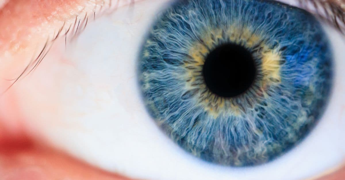
The human eye is a biological optical device, a precision camera that rivals instruments costing thousands of dollars, yet operates flawlessly for decades with zero conscious effort. Light enters through the cornea, passes through the aqueous humor, and is focused by the crystalline lens onto the retina — a thin sheet of neural tissue at the back of the eye containing two types of photoreceptors: 120 million rod cells (monochromatic, exquisitely sensitive in dim light) and 6 million cone cells (color-sensitive, active in daylight). Each photoreceptor contains millions of opsins, protein molecules that undergo a chemical transformation when struck by a photon, initiating a neural cascade that ultimately fires signals through the optic nerve to the visual cortex. The brain constructs vision from 30 discrete visual areas, each specialized for a different attribute: color, motion, depth, form. Yet we perceive a single, seamless, three-dimensional world, a construction so compelling that we believe we are seeing reality directly, unaware that the image in our visual cortex is inverted, compressed, and extensively processed.

How the Eye Sees Color: The Science of Vision
Your eyes can distinguish around 10 million colors, yet the entire mechanism runs on just three types of cone cell. Here's how that's possible.
How Does the Human Eye Actually Work?
Your eye captures light, converts it to electrical signals, and sends them to the brain — which then constructs everything you see. Here's the elegant biology behind vision.
How Color Perception Works — Biology of Color
How human eyes and brain construct color from light. Cones, trichromacy, color blindness, animal vision, and why color isn't a property of objects.