How the Brain Works: Neurons, Regions, and the Mind
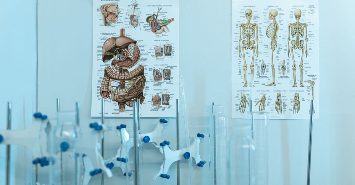
Photo: RDNE Stock project / Pexels
The human brain weighs about 1.4 kg (3 lbs) — roughly the density of firm tofu — yet it runs the most complex information-processing system on Earth. It contains approximately 86 billion neurons, each one connected to thousands of others, forming a network with an estimated 100 trillion connection points. That network consumes 20% of your body's total energy, never fully shuts off, and physically rewires itself every time you learn something new. Understanding how it does all of that starts at the smallest scale: a single cell.
What a Neuron Actually Does
Neuron — a specialized cell that transmits information as electrical and chemical signals. Neurons are the basic computational units of the brain and nervous system.
A neuron looks nothing like a typical cell. It has a central body (the soma), a long output cable called the axon, and a dense tree of receiving branches called dendrites. The dendrites collect incoming signals from other neurons; the soma integrates those signals; and if they collectively cross a voltage threshold, the axon fires an electrochemical pulse called an action potential — an all-or-nothing spike of electrical activity that travels down the axon at speeds of up to 120 meters per second (roughly 430 km/h).
The signal does not jump directly from one neuron to the next. It crosses a tiny gap called a synapse (typically 20–40 nanometers wide) by triggering the release of chemical messengers called neurotransmitters. Glutamate excites the receiving neuron; GABA inhibits it; dopamine, serotonin, acetylcholine, and dozens of others modulate how neurons respond to subsequent signals. The brain is not primarily electrical — it is chemical at every junction that matters.
Source: Kandel et al., Principles of Neural Science, 6th edition (2021) — Comprehensive account of neuron structure, action potential generation, and synaptic transmission mechanisms.
The 86 billion figure comes from a meticulous 2009 study by Brazilian neuroscientist Suzana Herculano-Houzel, who dissolved human brains into a cell soup and counted nuclei rather than trying to image neurons in situ — a method that corrected decades of estimates that had placed the count at 100 billion. But neurons are not alone. The brain contains roughly an equal number of glial cells (astrocytes, oligodendrocytes, microglia), which support, insulate, and maintain neurons, clear waste, and regulate blood flow. For a long time dismissed as mere scaffolding, glia are now understood to actively shape neural signaling.
Source: Herculano-Houzel, S., The Journal of Comparative Neurology (2009) — Isotropic fractionation method establishes ~86 billion neurons and ~85 billion non-neuronal cells in the adult human brain.
The Energy Budget: Why Your Brain Never Switches Off
The brain is metabolically voracious. It accounts for roughly 2% of adult body weight but consumes about 20% of the body's resting energy — the equivalent of running a 12-watt light bulb continuously. Almost all of that energy comes from glucose, which neurons cannot store. Any interruption in blood glucose for more than a few minutes causes loss of consciousness; a few minutes more and neurons begin to die.
Where does the energy go? In the cerebral cortex, approximately 75% of the brain's energy budget goes to signaling — firing action potentials, pumping ions back across membranes to reset after each spike, and recycling neurotransmitters at synapses. The remaining ~25% maintains the basic survival of cells. The ion pumps (specifically the sodium-potassium ATPase) are the biggest single consumer: every action potential slightly scrambles the ion balance across the cell membrane, and the pump has to spend ATP to fix it.
Source: Attwell, D. & Laughlin, S.B., Journal of Cerebral Blood Flow & Metabolism (2001) — Quantitative analysis of the brain's energy budget in rodents, extrapolated to humans; establishes signaling as the dominant energy consumer.
Source: BrainFacts.org / Society for Neuroscience (2019) — "How Much Energy Does the Brain Use?" — confirms 20% energy consumption figure for human brain at rest.
Crucially, the brain uses nearly as much energy during sleep as it does while awake. The idea of "resting" the brain at night is a popular misconception. During sleep, particularly slow-wave and REM stages, the brain is actively consolidating memories, pruning weak synapses, and — through the glymphatic system — flushing out metabolic waste products including amyloid-beta, the protein associated with Alzheimer's disease. Sleep is not downtime; it is maintenance. For a deeper look at the sleep-brain relationship, see Why We Sleep: The Science.
The Major Regions and What They Handle
The brain is not a homogeneous mass. Evolution layered structure upon structure over hundreds of millions of years, and the result is a set of specialized regions that communicate constantly but handle distinct operations.
| Region | Location | Primary Functions |
|---|---|---|
| Cerebral cortex | Outermost layer | Thought, language, perception, voluntary movement |
| Prefrontal cortex | Front of cortex | Planning, decision-making, impulse control |
| Hippocampus | Medial temporal lobe | Forming new long-term memories, spatial navigation |
| Amygdala | Medial temporal lobe | Emotional processing, threat detection, fear |
| Cerebellum | Rear base of brain | Fine motor coordination, balance, timing |
| Brainstem | Base, connects to spinal cord | Breathing, heart rate, sleep-wake transitions |
| Thalamus | Central relay hub | Routes sensory signals to appropriate cortical areas |
| Basal ganglia | Deep beneath cortex | Motor control, habit formation, reward processing |
The cerebral cortex — the deeply wrinkled outer layer — is divided into four lobes. The frontal lobe handles executive function and voluntary movement; the parietal lobe processes touch and spatial awareness; the occipital lobe handles vision; the temporal lobe handles hearing and language comprehension. The wrinkling (called gyrification) is an evolutionary solution to a packing problem: a smooth cortex the size of a human one would not fit inside a human skull. The folds increase surface area to roughly 2,500 cm² (about the size of a page of a broadsheet newspaper) while fitting inside a skull with a volume of roughly 1,400 mL.
The hippocampus deserves special attention. Shaped like a seahorse (the name comes from Greek), it acts as a temporary holding area and indexing system for new explicit memories before they are gradually distributed to the cortex for long-term storage. Patients with severe hippocampal damage, most famously the patient H.M. (Henry Molaison), can retain memories from before the damage but cannot form new long-term memories. H.M. lived the same day, every day, from the perspective of new memory formation, for the final 55 years of his life. The hippocampus is also covered in more depth in How Memory Works in the Brain.
Source: Scoville, W.B. & Milner, B., Journal of Neurology, Neurosurgery and Psychiatry (1957) — Landmark case study of patient H.M. following bilateral hippocampectomy, establishing the hippocampus as essential for new memory formation.
How Neurons Communicate: The Synapse in Detail
The synapse is where computation actually happens. When an action potential arrives at the axon terminal, it triggers voltage-gated calcium channels to open. Calcium ions flood in, causing vesicles (tiny membrane bubbles) packed with neurotransmitter molecules to fuse with the cell membrane and release their contents into the synaptic cleft. The neurotransmitters drift across the 20–40 nm gap and bind to receptor proteins on the receiving dendrite, which may open ion channels or trigger internal signaling cascades.
Think of the synapse like a chemical key-and-lock system run at millisecond speed: the neurotransmitter is the key, the receptor is the lock, and opening the lock either helps or hinders the next neuron from firing. Each neuron integrates hundreds or thousands of these signals simultaneously, summing excitatory and inhibitory inputs across its dendrite tree and firing only if the net depolarization at the soma reaches threshold (roughly -55 millivolts from a resting potential of about -70 mV).
The strength of a synapse is not fixed. It can be potentiated (strengthened) or depressed (weakened) based on how frequently and how recently it has been used. This property — synaptic plasticity — is the physical substrate of learning and memory, and is explored at length in the context of memory formation in How Memory Works in the Brain.
Source: Bhatt, D.L. et al., Neuron (2009) — Reviews synaptic plasticity mechanisms including long-term potentiation and long-term depression as the cellular basis of learning.
Neuroplasticity: The Brain That Rewires Itself
For most of the 20th century, the scientific consensus held that the adult human brain was essentially fixed — neurons you were born with, no replacements, no new connections after a critical developmental window. That consensus was wrong.
Neuroplasticity — the brain's capacity to reorganize itself by forming new neural connections and pruning others in response to experience, learning, or injury.
The evidence for adult neuroplasticity is now overwhelming. Taxi drivers in London — who must memorize an enormous street network called "The Knowledge" — show measurably enlarged posterior hippocampi compared to non-drivers, with the enlargement correlating with years of driving experience. Musicians who practice string instruments develop expanded cortical representations for the fingers of their left hand. Stroke patients can recover lost motor or language function as neighboring cortical areas take over the role of damaged regions.
Source: Maguire, E.A. et al., PNAS (2000) — Structural MRI study showing significantly larger posterior hippocampal volume in London taxi drivers relative to controls, correlating with navigation experience.
At the cellular level, neuroplasticity operates through several mechanisms. New synaptic spines (the small protrusions on dendrites that receive synaptic input) can grow within hours of learning. Existing synapses change in strength via long-term potentiation (LTP) — the mechanism by which repeated activation of a synapse makes it stronger and more responsive. The NMDA receptor is the molecular trigger: it acts like a coincidence detector, opening only when the receiving neuron is already partially depolarized AND the presynaptic neuron fires — capturing the "neurons that fire together, wire together" principle formalized by Donald Hebb in 1949.
There is also neurogenesis — the birth of new neurons — in the adult brain, though it appears limited to a few regions. The most studied site is the dentate gyrus of the hippocampus, where new neurons are generated throughout adulthood and appear to play a role in pattern separation (distinguishing similar memories from each other).
Source: Gage, F.H., Science (2002) — Review of adult neurogenesis evidence in mammals, confirming ongoing neuron production in the adult hippocampus.
The Left Brain / Right Brain Myth (and the Real Story of Lateralization)
Popular culture insists on a clean division: logical left brain, creative right brain. The reality is considerably more nuanced.
The brain does exhibit lateralization — certain functions are handled predominantly by one hemisphere. Language production (Broca's area) and language comprehension (Wernicke's area) are located in the left hemisphere in roughly 95% of right-handed people and about 70% of left-handed people. The right hemisphere tends to handle global spatial processing and certain aspects of emotional prosody (reading tone in speech).
But this does not mean hemispheres operate independently. The two hemispheres are connected by a massive bundle of around 200–250 million axons called the corpus callosum, which passes information between them constantly. Split-brain patients — whose corpus callosum was surgically severed to treat intractable epilepsy — show striking dissociations only under highly artificial experimental conditions. In normal daily life, the two hemispheres work as a single system.
Source: Gazzaniga, M.S., Nature Reviews Neuroscience (2005) — Review of split-brain research and lateralization; argues that hemispheric specialization is real but the popular "two separate brains" narrative is a distortion.
The "right-brained = creative, left-brained = analytical" narrative traces to popularizations of this split-brain research in the 1970s, not to the actual findings. A 2013 fMRI study by Nielsen et al. analyzed resting-state connectivity in over 1,000 participants and found no evidence for individuals being strongly "left-brained" or "right-brained" — network connectivity was roughly symmetrical across the population.
How the Brain Regulates Itself: Feedback, Rhythm, and Attention
The brain does not simply receive information and output commands. It is constantly predicting, modulating, and filtering its own activity.
The default mode network (DMN) is a set of regions — including the medial prefrontal cortex, posterior cingulate cortex, and angular gyrus — that activates when the brain is not engaged in focused external tasks: daydreaming, mind-wandering, self-referential thought, imagining the future. For decades it was treated as noise in neuroimaging data. It is now understood as a critical system for internal cognition and narrative self-construction.
Attention is mediated partly by neuromodulatory systems — diffuse projections of acetylcholine (from the basal forebrain), norepinephrine (from the locus coeruleus), and dopamine (from the ventral tegmental area and substantia nigra) that collectively tune the signal-to-noise ratio across the cortex. When you are highly focused, norepinephrine and acetylcholine narrow the bandwidth of cortical processing, amplifying relevant signals and suppressing irrelevant ones. This is why stimulant drugs that boost norepinephrine and dopamine (such as amphetamines) can sharpen focus — they are hijacking a natural gain-control system.
The brain also generates characteristic electrical oscillations at different frequencies — delta (0.5–4 Hz) during deep sleep, theta (4–8 Hz) during memory tasks and REM sleep, alpha (8–12 Hz) during relaxed wakefulness, beta (12–30 Hz) during active thinking, and gamma (30–100 Hz) during high-level cognitive binding. These rhythms are not merely epiphenomenal; they appear to coordinate communication between distant brain regions by establishing shared timing windows.
Source: Buzsáki, G. & Draguhn, A., Science (2004) — Review of neuronal oscillations and their role in coordinating large-scale brain network activity.
What This Means
The brain's defining feature is not raw processing power — it is adaptability. The same tissue that encodes language also learns to navigate a city, recognize faces, compose music, and model the mental states of other people, all without a hardware upgrade, purely through rearranging the strengths of connections among a fixed population of cells. The 20% energy cost is the price of running a system that never stops updating its own wiring diagram. Every conversation you have, every skill you practice, every night of sleep that consolidates the day's experience — all of it is literally changing the physical structure of the 1.4 kg of tissue sitting behind your eyes.
Read next:
What Brain Imaging Revealed That Dissection Never Could
For most of neuroscience's history, understanding the brain meant examining dead tissue. Dissection, staining, and microscopy built the anatomical map — lobes, nuclei, tracts — but they could not answer the question that mattered most: what is each region actually doing in a living, thinking person? That question opened up only when imaging technology caught up with biology, starting in the 1970s with positron emission tomography (PET) and accelerating through the 1990s with functional magnetic resonance imaging (fMRI).
Functional MRI (fMRI) — a neuroimaging technique that measures brain activity indirectly by detecting changes in blood oxygenation. Active neurons consume more oxygen, drawing oxygenated blood to the area. Because oxygenated and deoxygenated hemoglobin have different magnetic properties, MRI can detect this contrast — the BOLD signal (Blood Oxygen Level-Dependent). Think of it as watching where the brain calls for a fuel delivery, rather than watching the neurons themselves fire.
The distinction matters. fMRI does not measure neural activity directly. It measures a vascular response to that activity, with a lag of 4–6 seconds after the neural event and a spatial resolution of roughly 1–3 mm. That is extraordinarily good by historical standards, but it means that the iconic colored brain maps that fill neuroscience press releases are showing where blood flow changed, averaged across many subjects and many seconds — not which neurons fired. A region "lighting up" means increased metabolic demand in that area, not that the area exclusively performs the highlighted function.
PET imaging uses radioactively labeled glucose or oxygen tracers to map metabolic activity, and was the earlier technology. Its advantage is the ability to track specific neurotransmitter systems by labeling receptor-binding molecules — something fMRI cannot do. EEG (electroencephalography) sacrifices spatial resolution entirely but captures electrical activity with millisecond precision, making it invaluable for studying the timing of neural events such as sleep oscillations, seizures, and event-related potentials. The three technologies are complementary: fMRI tells you where, EEG tells you when, PET tells you what neurochemistry.
Source: Logothetis, N.K., Nature (2008) — "What we can do and what we cannot do with fMRI." Critical analysis of the BOLD signal's relationship to underlying neural activity; essential reading on the interpretive limits of fMRI.
One of the most consequential imaging discoveries was entirely accidental. In the early 1990s, neuroimager Marcus Raichle noticed that a set of brain regions — including the medial prefrontal cortex, posterior cingulate cortex, and angular gyrus — consistently deactivated when subjects performed demanding external tasks. The regions were most active when subjects were doing nothing in particular. Raichle named this the default mode network (DMN), and published the formal characterization in 2001. For decades prior, this activity had been dismissed as experimental noise. It turned out to be one of the brain's most metabolically expensive networks, now understood to support self-referential thinking, mental time travel (imagining past and future), and social cognition — the brain's "internal life" running beneath every focused task.
The ambition to map every connection in the human brain — not just its regions — produced connectomics: the project of reconstructing the complete wiring diagram of neural tissue. The Human Connectome Project, launched in 2010 with NIH funding, used high-resolution diffusion MRI to map the major white-matter fiber tracts connecting cortical and subcortical regions across 1,200 healthy adults. Diffusion MRI tracks the preferred direction of water movement through tissue; in myelinated axon bundles, water moves preferentially along the fiber direction, revealing the tract's path. The result is not a synapse-level circuit diagram — that level of resolution requires electron microscopy and is currently feasible only for cubic millimeters of tissue — but a macro-scale connectivity atlas that revealed how tightly hubs like the prefrontal cortex and posterior cingulate are interconnected with nearly everything else.
Source: Van Essen, D.C. et al., NeuroImage (2013) — Overview of the Human Connectome Project; methodology, data types, and scientific goals for mapping large-scale human brain connectivity.
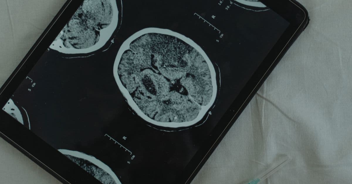
The interpretive limits of these methods are real and worth knowing. The "reverse inference" problem — seeing activity in the amygdala and concluding "the subject felt fear" — is only valid if the amygdala activates exclusively for fear, which it does not. It also responds to novelty, reward, and positive social stimuli. Pop-science coverage routinely commits this error. What imaging has genuinely established is a set of probabilistic associations between regions and cognitive processes, refined across thousands of studies and subject to the same statistical replication challenges as any other field. The map is far more detailed than anything dissection provided — and far less certain than a casual glance at a brain scan image suggests.
Q: How many neurons does the human brain actually have?
A: Approximately 86 billion, based on the isotropic fractionation method developed by Suzana Herculano-Houzel in 2009. Earlier estimates of 100 billion were based on extrapolation rather than direct counting.
Q: Why does the brain use so much energy?
A: About 75% of the brain's energy goes to signaling — firing action potentials and resetting ion gradients after each spike. Ion pumps (especially sodium-potassium ATPase) are the biggest consumers because every action potential partially disrupts the membrane voltage balance that must be continuously restored.
Q: Can the adult brain grow new neurons?
A: Yes, in limited regions. The dentate gyrus of the hippocampus generates new neurons throughout adulthood in a process called neurogenesis. Whether this occurs at meaningful rates in other human brain regions remains an active research question.
Q: What is neuroplasticity in simple terms?
A: Neuroplasticity is the brain's ability to physically reorganize itself — strengthening used connections, pruning unused ones, and in some cases growing new ones — in response to learning, experience, or injury.
Q: Is the left brain / right brain split real?
A: Partly. Certain functions (especially language) are lateralized, meaning they are handled predominantly by one hemisphere. But the two hemispheres communicate constantly via the corpus callosum and work as one integrated system. The idea that people are either "left-brained" or "right-brained" in personality has no support in neuroimaging data.
Q: How fast do signals travel in the brain?
A: It depends on the neuron type. Unmyelinated axons conduct at roughly 0.5–2 meters per second. Heavily myelinated axons (insulated with a fatty sheath that speeds transmission) can reach 70–120 m/s. Myelination is why white matter — the brain's wiring bundles — appears white.
Follow new articles
Add SciHub101 to your feed reader to see new science and technology stories.
About the Author
SciHub101 is an independent science and technology publication founded and edited by Buğra Sözeri, a software engineer and app developer based in Türkiye. AI tools assist with drafting and research. Source documentation in the existing archive is uneven, and articles are being reviewed and corrected. The Editorial Team byline identifies editorial responsibility; it does not certify specialist or independent human review. Where we compare products, anything built by our own team is labelled as such — see the editorial guidelines and About page for how corrections and disclosures work.
Related Articles

How the Human Heart Works: The Cardiac Cycle Explained
The heart beats more than 100,000 times per day without a single conscious command, regulated by an electrical system so precise that it can be measured from electrodes on the skin. Here is how that system works, from the first spark in the SA node to the pressure wave that reaches your fingertips.

How Memory Works in the Brain: Encoding, Storage, and Forgetting
Memory is not a recording. It is a reconstruction — assembled fresh each time you recall it, from fragments stored across the cortex. Understanding how encoding, long-term potentiation, and retrieval actually work explains why memories are reliable enough to be useful and unreliable enough to be distorted.
How the Brain Processes Information
86 billion neurons, 100 trillion synapses, 20 watts of power. How the human brain actually processes information — and why it beats supercomputers.

How the Brain Works — Neuroscience Overview
Neurons, action potentials, brain regions, plasticity, and the science behind consciousness — a clear neuroscience primer.
How the Brain Learns: The Science of Neuroplasticity
Every time you learn something new, your brain physically rewires itself. Here's exactly how synaptic plasticity, long-term potentiation, and pruning turn experience into lasting change.

How the Nervous System Works: Neurons, Signals, and the Brain
Your nervous system transmits signals at up to 120 meters per second — and a single neuron can connect to up to 200,000 other neurons.