How the Brain Processes Information
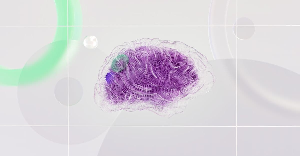
Photo: Google DeepMind / Pexels
The human brain weighs about 1.4 kilograms, accounts for roughly 2% of body mass, and consumes something close to a fifth of the body's resting energy budget. It performs feats of pattern recognition that very large computers still struggle to match, and it does so with components that are slow, noisy and individually unreliable.
This is a tour of what we do know: the cells, the signals, the networks, the energy budget, and the principles that turn three pounds of soft tissue into a thinking organ. It is also, deliberately, a tour of what we do not know — because the gap between "we can describe the parts" and "we can explain the behaviour" is still the central fact about neuroscience.
The numbers, and why the famous one was wrong
For decades, textbooks repeated that the human brain contains roughly 100 billion neurons and ten times as many glial cells. Neither figure had a solid empirical basis; they were estimates that got copied.
In 2009, Frederico Azevedo, Suzana Herculano-Houzel and colleagues published a direct count in the Journal of Comparative Neurology (volume 513, pages 532–541) using a technique called the isotropic fractionator, which dissolves brain tissue into a uniform suspension of cell nuclei that can then be stained and counted. Averaged across the adult male brains they examined, they found:
- 86.1 ± 8.1 billion neurons (NeuN-positive cells)
- 84.6 ± 9.8 billion non-neuronal cells
Two things followed. The 100-billion figure was an overestimate, and the "ten glia per neuron" claim collapsed: the ratio is close to one to one. The authors also noted that the human brain's cellular composition scales much as other primate brains do, which undercut the idea that human cognition rests on an exceptional cell-type ratio.
A caveat worth keeping: these are averages from a small sample, with large standard deviations, and "non-neuronal" is a broader category than "glia." Precision here is lower than round numbers suggest.
Synapse counts are shakier still. Estimates in the range of 100 trillion for the whole brain are widely quoted, but they are extrapolations from electron-microscopy density measurements in small cortical samples, not counts. What direct measurement exists is local and recent: the MICrONS project's reconstruction of roughly one cubic millimetre of mouse visual cortex detected more than 523 million synapses among over 200,000 cells in that single speck of tissue.
| Quantity | Best current figure | Source and caveat |
|---|---|---|
| Neurons, human brain | ~86 billion | Azevedo et al. 2009, isotropic fractionator; small sample |
| Non-neuronal cells | ~85 billion | Same study; broader than "glia" |
| Synapses, human brain | ~100 trillion (order of magnitude) | Extrapolated from local density, not counted |
| Synapses, 1 mm³ mouse visual cortex | >523 million | MICrONS dataset, 2025; directly detected |
| Share of resting energy used | ~20% | Attwell & Laughlin 2001 and later reviews |
| Resting membrane potential | about −70 mV | University of Queensland QBI |
| Firing threshold | about −50 mV | University of Queensland QBI |
| Action potential duration | about 1 ms | University of Queensland QBI |
| Synaptic cleft width | 20–40 nm | University of Queensland QBI |
The neuron as a computing unit
The idea that the brain is built from discrete cells rather than a continuous web was itself a hard-won result, settled largely by Santiago Ramón y Cajal's drawings at the turn of the twentieth century — a story told in our profile of Cajal and the discovery of the neuron.
Each neuron has three functional parts:
- Dendrites — branching inputs that receive signals from other neurons.
- Soma (cell body) — integrates incoming signals and determines whether the cell fires.
- Axon — an output cable, sometimes over a metre long, that carries the signal to other neurons.
A resting neuron sits at a membrane potential of about −70 millivolts, meaning its interior is negative relative to the outside. Excitatory inputs push that potential upward and inhibitory inputs push it down. When the sum crosses a threshold of roughly −50 mV, the neuron fires an action potential: a brief pulse lasting about a millisecond, generated in the axon and propagated along it.
The action potential is all-or-nothing. It either fires fully or not at all, and its amplitude carries no information. Intensity is encoded in the rate of firing and in the precise timing of spikes relative to other neurons — which means a single neuron's output is closer to a stream of identical clicks than to a graded voltage.
The biophysics was worked out by Alan Hodgkin and Andrew Huxley in 1952, using the giant axon of the squid, which is large enough to insert an electrode into. Their model described the pulse in terms of voltage-dependent sodium and potassium conductances: sodium ions rush in and depolarise the membrane, potassium ions flow out and repolarise it, and the local disturbance triggers the same cycle in the adjacent patch of membrane. The work earned a share of the 1963 Nobel Prize in Physiology or Medicine and remains the foundation of computational neuroscience; variants of the Hodgkin–Huxley equations are still simulated today.
Conduction speed depends on insulation. Axons wrapped in myelin — a fatty sheath produced by glial cells, interrupted at regular gaps called nodes of Ranvier — conduct by jumping from node to node, reaching speeds on the order of 100 metres per second. Unmyelinated axons, including most pain fibres, conduct at one or two metres per second. The practical consequence is that the nervous system runs at two very different timescales at once, and that demyelinating conditions such as multiple sclerosis degrade function by slowing conduction rather than by killing cells outright.
The synapse: where the computation actually happens
At the axon terminal the electrical signal becomes chemical. The arriving action potential opens calcium channels, vesicles fuse with the membrane, and neurotransmitter molecules cross a gap of 20–40 nanometres to bind receptors on the receiving cell. Those receptors open ion channels, and the resulting ion flow nudges the next neuron toward or away from its own threshold. The University of Queensland's Brain Institute describes this cycle as an electrical signal converted to a chemical one and back again.
Different transmitters do different jobs:
| Neurotransmitter | Typical effect | Rough role |
|---|---|---|
| Glutamate | Excitatory | The main driver of cortical signalling |
| GABA | Inhibitory | Sculpts and constrains activity; targets of many sedatives |
| Dopamine | Modulatory | Reward prediction, motivation, motor control |
| Serotonin | Modulatory | Mood, appetite, sleep regulation, among others |
| Acetylcholine | Mixed | Neuromuscular junction; attention and arousal in cortex |
| Noradrenaline | Modulatory | Alertness and vigilance |
The excitatory–inhibitory distinction matters more than popular accounts suggest. Roughly 80% of cortical neurons are excitatory and 20% inhibitory, and the inhibitory minority does most of the work of keeping the network stable. Without it, excitation feeds back on itself and the result is a seizure.
The modulatory transmitters are not messages so much as settings. Dopamine does not encode "pleasure"; the dominant interpretation, built on Wolfram Schultz's recordings in monkeys, is that midbrain dopamine neurons signal reward prediction error — the gap between what was expected and what arrived. Our piece on the dopamine system unpacks why that distinction changes how motivation is understood.
Parallel processing, not serial
A modern processor executes a small number of operations at a time, extremely fast. The brain does the opposite: billions of slow, simple operations at once.
Consider the arithmetic. A neuron's membrane takes milliseconds to integrate its inputs, a million times slower than a logic gate. Yet humans recognise a familiar face in a fraction of a second. Classic work by Simon Thorpe and colleagues, published in Nature in 1996, flashed photographs for 20 milliseconds and asked participants to indicate whether an animal was present; the EEG signal distinguishing targets from non-targets emerged about 150 ms after the image appeared. With millisecond-scale components, 150 ms allows only a short chain of processing stages — which implies that most of the work must happen in parallel across many neurons rather than sequentially through many steps.
This is parallel distributed processing, and it explains the brain's characteristic profile of strengths:
- Recognising a face in a crowd under unfamiliar lighting.
- Understanding ambiguous, context-dependent language.
- Generalising from very few examples.
- Degrading gracefully — losing neurons blurs performance instead of crashing it.
And its equally characteristic weaknesses:
- Arithmetic, especially multi-step.
- Holding more than a handful of precise items in working memory.
- Producing identical output twice.
- Reporting its own processing accurately; introspection is a reconstruction, not a log file.
Hierarchy and specialisation
The brain is not homogeneous. Different regions do different things, and the clearest illustration is the visual system.
Signals from the retina reach primary visual cortex (V1), where, as David Hubel and Torsten Wiesel discovered in work that earned a share of the 1981 Nobel Prize, individual cells respond selectively to edges at particular orientations. Downstream areas combine those responses into textures, colours, shapes and motion, and higher areas in the temporal lobe respond to whole objects and faces largely independent of size, position and lighting. Each stage pools from the one before, which is how invariance is built. The process begins with the optics covered in our explainer on how the eye works.
| Region | Primary function | Consequence of damage |
|---|---|---|
| Visual cortex (V1) | Edge and orientation detection | Cortical blindness in the affected field |
| Hippocampus | Encoding new episodic memories | Inability to form new lasting memories |
| Broca's area | Speech production | Effortful, non-fluent aphasia |
| Wernicke's area | Language comprehension | Fluent but semantically empty speech |
| Amygdala | Threat detection, emotional learning | Blunted fear responses |
| Cerebellum | Motor coordination and timing | Ataxia, loss of balance |
| Prefrontal cortex | Planning, inhibition, working memory | Impulsivity, poor planning |
| Default mode network | Rest, self-referential thought | Altered introspection and mind-wandering |
| Brainstem | Breathing, heart rate, arousal | Death |
Nineteenth-century neurology built its first map from exactly this logic: specific damage, specific deficit. The most famous single case, Phineas Gage's survival of a tamping iron through the frontal lobe in 1848, is examined in our article on what the Gage case does and does not show — a useful corrective, because the story has been embellished for 170 years.
Two cautions about localisation. First, a region being necessary for a function is not the same as that region performing the function alone; almost everything interesting is distributed across networks. Second, resting-state imaging revealed that the brain is never idle. The default mode network, described by Marcus Raichle and colleagues in PNAS in 2001, is more active during quiet rest than during many demanding tasks — the subject of our piece on what the default mode network actually does.
Learning: the network rewrites itself
The brain does not just process information. It physically changes in response to it.
The most studied mechanism is long-term potentiation (LTP), reported by Tim Bliss and Terje Lømo in the Journal of Physiology in 1973. Repeated, closely timed activation of a pathway strengthens its synapses for hours or longer; the mirror process, long-term depression, weakens connections that are not used together. At the molecular level LTP typically involves NMDA receptors acting as coincidence detectors — they pass current only when the receiving cell is already depolarised and glutamate is present — followed by insertion of additional AMPA receptors into the postsynaptic membrane and, for lasting changes, new protein synthesis and structural growth of dendritic spines.
This is the cellular grounding for the principle Donald Hebb articulated in 1949, usually paraphrased as "neurons that fire together wire together." The paraphrase is loose: timing matters, and in spike-timing-dependent plasticity the synapse strengthens only when the presynaptic spike reliably precedes the postsynaptic one, and weakens when the order reverses. Correlation is not enough; the network is learning something closer to causal order.
Plasticity operates at several scales simultaneously — synaptic strength, dendritic spine structure, myelination of active pathways, and the slow large-scale remapping seen after injury or sustained training. Our article on neuroplasticity covers what this does and does not mean for adult learning, since the term is routinely oversold.
Memory consolidation depends heavily on sleep. During slow-wave sleep the hippocampus appears to replay sequences of activity recorded during the day, in compressed form, in coordination with cortical slow oscillations — a process thought to transfer memories into longer-term cortical storage. REM sleep is associated with emotional memory processing and with the recombination that may underlie creative insight. Deprive someone of sleep and encoding, consolidation and emotional regulation all degrade within a day. See how sleep affects the brain and, for the storage side, how memory works.
The energy budget
Information processing is expensive, and the brain's energy constraint shapes its design.
The brain is about 2% of body mass but accounts for roughly 20% of resting energy expenditure, a figure analysed in detail by David Attwell and Simon Laughlin in 2001. At a typical adult resting metabolic rate, that share corresponds to something on the order of 20 watts — a convenient comparison figure, though it is arithmetic from the percentage rather than a directly measured quantity, and individual values vary.
Where does it go? Attwell and Laughlin's budget attributed the bulk of cortical energy use to restoring ion gradients after synaptic and action-potential activity — chiefly the sodium-potassium pump working to undo what signalling does. Signalling, in other words, is the cost; maintaining the cells is comparatively cheap.
Three design consequences follow:
- Sparse coding. If spikes are expensive, an efficient code uses few of them. Cortical neurons fire at low average rates, and a given stimulus activates a small fraction of the available cells.
- Analogue where possible. Short-range communication often uses graded potentials rather than spikes; spikes are for distance, where signal regeneration is needed.
- Physical compactness. Wiring consumes volume and energy, which is one widely argued reason why functionally related areas sit near one another and why the cortex is folded.
Thinking harder does not meaningfully change the total. Task-related increases in local blood flow — the signal fMRI measures — are a few percent above baseline, which is why the brain's overall consumption stays roughly flat whether you are solving equations or staring out of a window.
Predictive processing: a candidate unifying theory
A prominent modern framework, associated with Karl Friston and developed by many others, holds that the brain is fundamentally a prediction machine. Rather than passively receiving sensory input and building a representation from it, the brain continuously generates predictions about incoming signals and transmits only the error — the difference between prediction and reality — upward for correction.
The appeal is parsimony. One mechanism accommodates several observations:
- Perception is constructive. What you experience is the brain's current best model, corrected by sensory evidence. Visual illusions are what happens when the model's assumptions are violated deliberately.
- Attention is precision weighting. Attending to something amounts to treating its prediction errors as more reliable, and therefore letting them drive updates more strongly. That framing fits findings like inattentional blindness, covered in our piece on how attention hides obvious things.
- Action closes the loop. The brain can reduce prediction error either by updating the model or by moving the body so the world matches the prediction. Both count as inference.
- Some symptoms become intelligible. Hallucinations have been modelled as priors that overwhelm sensory evidence, and certain features of autism as atypical weighting of prediction errors.
Two honest caveats. First, the strongest version — the free energy principle as a single formal account of all brain function — is difficult to falsify, which is a recurring criticism. Second, "the brain predicts" is compatible with many specific implementations, and which one the cortex actually uses is unsettled. The framework is a productive research programme, not a solved theory.
Mapping the wiring: connectomics
For most of its history neuroscience inferred circuits indirectly. That is changing, because it is now possible to reconstruct every cell and connection in a volume of tissue from electron-microscope images, with machine learning doing the segmentation that humans cannot do at scale.
The milestones to date:
- C. elegans, a nematode with 302 neurons, was mapped by hand over more than a decade and published in 1986 — the first complete connectome of any animal.
- The Janelia Research Campus "hemibrain" reconstruction of part of the Drosophila brain, completed in December 2019 and released in January 2020, contains around 25,000 neurons.
- The FlyWire consortium extended this to the entire adult fruit fly brain — 139,255 neurons — reconstructed from 21 million electron-microscope images and published across a set of papers in 2024.
- The MICrONS project released a cubic-millimetre reconstruction of mouse visual cortex in 2025, with more than 200,000 cells, about 120,000 neurons and over 523 million detected synapses, uniquely combined with functional recordings from roughly 75,000 of those neurons before the tissue was sectioned.
A human connectome at this resolution remains far off: the data volume scales with tissue volume, and a human brain is roughly a million times larger than a cubic millimetre. The nearer-term payoff is different. Having both the wiring and the activity of the same neurons lets researchers test whether circuit structure predicts function — the question that justified building the maps. Our piece on brain mapping and neural circuits follows that work in more detail.
The brain versus the computer
The computer analogy is useful for a paragraph and misleading after that.
| Feature | Brain | Digital computer |
|---|---|---|
| Power draw | ~20 W | Tens to thousands of watts |
| Architecture | Massively parallel, ~86 billion units | Few to thousands of fast cores |
| Memory and processing | Co-located in synapses | Physically separated |
| Reliability of components | Noisy, probabilistic | Essentially deterministic |
| Self-modification | Continuous, at every level | Only via software changes |
| Basic operation time | ~1 millisecond | ~1 nanosecond |
| Clock | None; asynchronous | Centrally clocked |
| Failure mode | Graceful degradation | Abrupt, often total |
The deepest mismatch is the memory–processing split. In a computer, data travels between storage and processor, and that traffic dominates the energy bill. In the brain, the synapse is both the memory and the computing element, which eliminates the transfer entirely. This is precisely what neuromorphic hardware tries to imitate, and why the imitation is difficult.
Artificial neural networks borrowed vocabulary from neuroscience and little else. An artificial "neuron" is a weighted sum followed by a simple nonlinearity; a biological neuron has active dendritic processing, dozens of receptor types, local protein synthesis and neuromodulatory state. Backpropagation, the algorithm that trains artificial networks, requires a precise backward pass of error signals that has no clearly identified biological counterpart. The resemblance is an inspiration, not a model.
Common misconceptions
"We only use 10% of our brain." No. Imaging shows activity throughout the brain across a normal day, and damage to almost any region produces a deficit. An organ this metabolically costly could not carry 90% dead weight through evolution.
"The left brain is logical, the right creative." Some functions are lateralised — language production leans left in most people — but the popular personality version has no support. Both hemispheres participate in nearly every complex task, continuously exchanging signals across the corpus callosum.
"Adults cannot grow new neurons." The honest answer is that this is contested. Sorrells and colleagues reported in Nature in 2018 that signs of hippocampal neurogenesis fall sharply in childhood and are essentially undetectable in adults; Moreno-Jiménez and colleagues reported in Nature Medicine in 2019 that immature neurons remain abundant in healthy adult hippocampus. Much of the discrepancy appears to come down to tissue handling, particularly how long samples sat in fixative. Anyone stating this one confidently in either direction is ahead of the evidence.
"Neurons are the brain's only important cells." Glia were long treated as packing material. Astrocytes regulate blood flow and transmitter clearance, microglia prune synapses, and oligodendrocytes' myelination tunes conduction timing. They are participants, not scaffolding.
"Brain scans show thoughts." fMRI measures blood oxygenation changes with a resolution of seconds and millimetres — an indirect, slow, spatially coarse proxy for the activity of millions of neurons. It is a powerful tool and nothing like a readout.
What we still cannot explain
It is worth ending on the size of the gap.
- The neural code. We know spikes carry information. We do not have a general account of how populations of neurons encode and transform it.
- Binding. Colour, motion, shape and sound are processed in separate areas on different timescales, yet experience is unified. How is not settled.
- Consciousness. Explaining why physical processing is accompanied by subjective experience — the hard problem — remains open, and competing theories differ on what would even count as evidence.
- Scaling from circuits to behaviour. The complete 302-neuron C. elegans connectome has been available since 1986 and still does not let anyone predict the worm's behaviour from first principles. Wiring alone is evidently not enough.
None of this diminishes what is known. It does suggest treating confident one-sentence explanations of the brain with suspicion, including some of the ones above.
FAQs
Q: How many neurons are in the human brain? A: About 86 billion, based on the direct counts of Azevedo, Herculano-Houzel and colleagues published in 2009. The older figure of 100 billion was an estimate that was never measured, and the related claim that glia outnumber neurons ten to one is also wrong — the ratio is roughly one to one.
Q: Do we really only use 10% of our brain? A: No. This is a myth with no empirical basis. Imaging shows essentially all regions active across different tasks, damage to almost any region produces a measurable deficit, and the brain is far too energetically expensive for most of it to be unused.
Q: How fast are brain signals? A: Action potentials travel at roughly 100 metres per second along thickly myelinated axons and at one to two metres per second along unmyelinated ones. Each spike lasts about a millisecond. The brain compensates for this slowness with massive parallelism rather than speed.
Q: Why does the brain use so much energy? A: Most of the cost is restoring ion gradients after signalling — pumping sodium and potassium back across membranes after action potentials and synaptic activity. The brain is about 2% of body mass and uses around 20% of resting energy, which corresponds to roughly 20 watts.
Q: Can the adult brain make new neurons? A: In most regions, no. In the hippocampus, the evidence is genuinely disputed: a 2018 Nature study found adult neurogenesis essentially absent, while a 2019 Nature Medicine study found it abundant, and differences in tissue preservation may explain much of the conflict.
Q: How does memory work, in one sentence? A: Memory is stored as patterns of synaptic strength across distributed networks, written by long-term potentiation and related plasticity mechanisms, and reorganised into longer-term cortical storage during sleep.
Q: Will artificial intelligence ever match the brain? A: It already exceeds humans at narrow tasks. General flexibility and energy efficiency remain wide open: the brain learns from few examples, tolerates damage, and runs on about 20 watts, and no artificial system currently combines those three properties.
Sources
- MICrONS Explorer, Cortical MM³ dataset (volume, cell and synapse counts) — https://www.microns-explorer.org/cortical-mm3
- Janelia Research Campus, FlyEM hemibrain project page — https://www.janelia.org/project-team/flyem/hemibrain
- Queensland Brain Institute, "Action potentials and synapses" — https://qbi.uq.edu.au/brain-basics/brain/brain-physiology/action-potentials-and-synapses
- Azevedo F.A.C., Herculano-Houzel S. et al. (2009). Equal numbers of neuronal and nonneuronal cells make the human brain an isometrically scaled-up primate brain. Journal of Comparative Neurology 513(5):532–541.
- Hodgkin A.L. & Huxley A.F. (1952). A quantitative description of membrane current and its application to conduction and excitation in nerve. Journal of Physiology.
- Bliss T.V.P. & Lømo T. (1973). Long-lasting potentiation of synaptic transmission in the dentate area. Journal of Physiology.
- Thorpe S., Fize D. & Marlot C. (1996). Speed of processing in the human visual system. Nature 381:520–522.
- Raichle M.E. et al. (2001). A default mode of brain function. PNAS.
- Attwell D. & Laughlin S.B. (2001). An energy budget for signaling in the grey matter of the brain. Journal of Cerebral Blood Flow & Metabolism.
- Sorrells S.F. et al. (2018). Human hippocampal neurogenesis drops sharply in children to undetectable levels in adults. Nature.
- Moreno-Jiménez E.P. et al. (2019). Adult hippocampal neurogenesis is abundant in neurologically healthy subjects. Nature Medicine.
- Hebb D.O. (1949). The Organization of Behavior. Wiley.
Follow new articles
Add SciHub101 to your feed reader to see new science and technology stories.
About the Author
SciHub101 is an independent science and technology publication founded and edited by Buğra Sözeri, a software engineer and app developer based in Türkiye. AI tools assist with drafting and research. Source documentation in the existing archive is uneven, and articles are being reviewed and corrected. The Editorial Team byline identifies editorial responsibility; it does not certify specialist or independent human review. Where we compare products, anything built by our own team is labelled as such — see the editorial guidelines and About page for how corrections and disclosures work.
Related Articles

How Viruses Jump From Animals to Humans
Zoonotic spillover explained: how viruses like SARS-CoV-2, HIV, Ebola, and MERS crossed from animals to humans — and why it keeps happening.

Congenital Insensitivity to Pain — Living Without
Some people are born unable to feel pain. The genes behind CIP — SCN9A, PRDM12, NTRK1, FAAH-OUT — are reshaping the future of pain medicine.

How the Brain Works — Neuroscience Overview
Neurons, action potentials, brain regions, plasticity, and the science behind consciousness — a clear neuroscience primer.

How the Human Brain Actually Works — Neurons, Networks, and Consciousness
Your brain has 86 billion neurons firing at 1,000 trillion connection points. Here's a tour of how it actually generates everything you think, feel, and remember.
How the Brain Works: Neurons, Regions, and the Mind
The brain runs on roughly 86 billion neurons, consumes 20% of your body's energy despite weighing only 2% of your body, and rewires itself with every new experience. Here is the mechanism behind all of it.
The Aging Brain: How Cognition Changes as We Get Older
Your brain peaks at different ages for different skills, and some abilities actually improve with age. The neuroscience of brain aging is far more nuanced than the simple story of decline.