How Mass Spectrometry Identifies Molecules
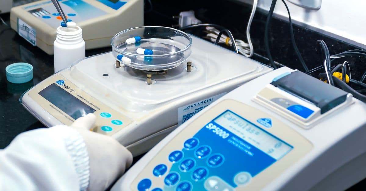
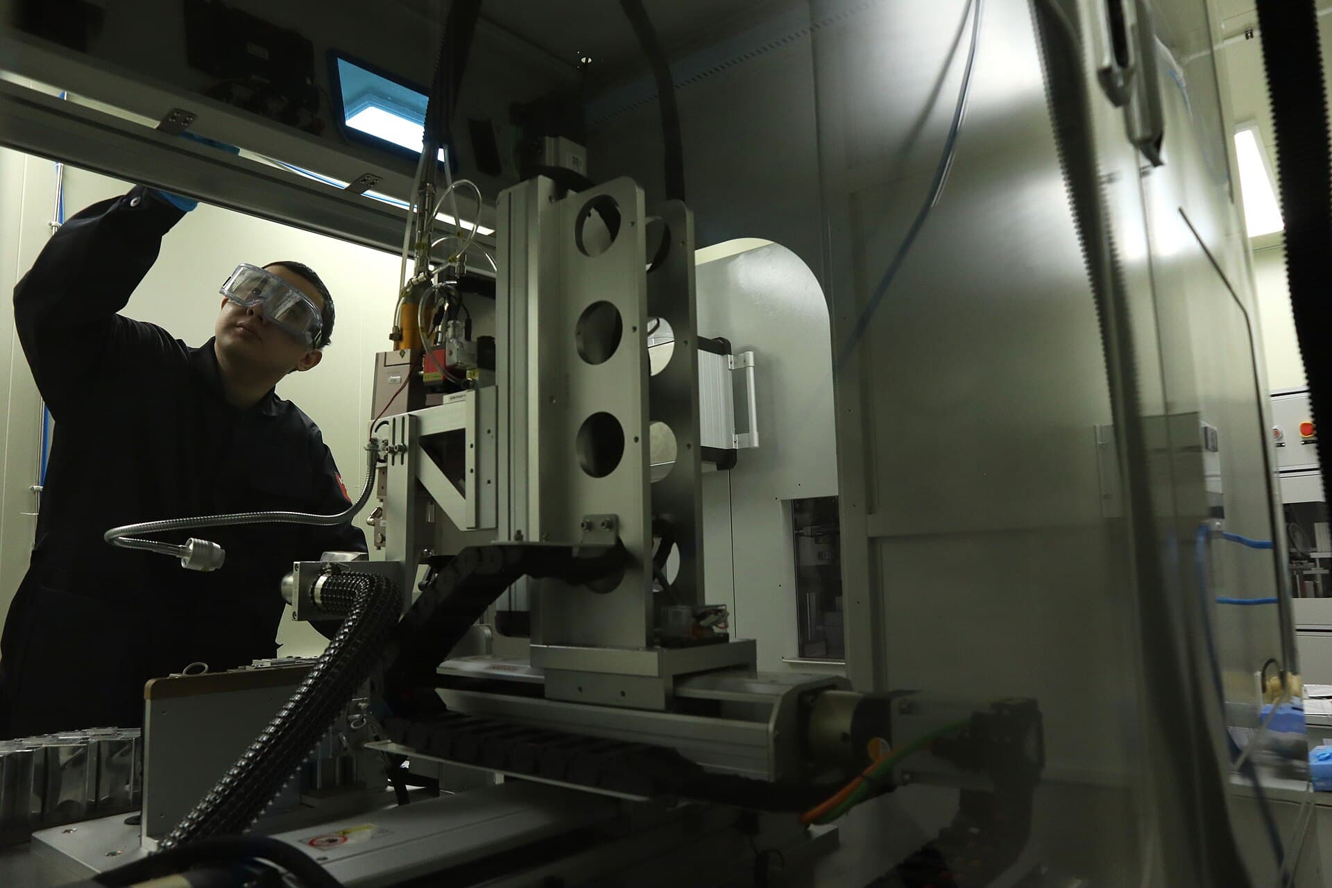
Photo: Pilan Filmes / Pexels
A mass spectrometer can tell a forensic chemist exactly which drug is present in a blood sample, tell an anti-doping lab which banned steroid metabolite shows up in an athlete's urine, and tell NASA's Curiosity rover what gases are locked inside a Martian rock. All three tasks rely on the same basic trick: convert a molecule into charged fragments, weigh those fragments with extreme precision, and let the resulting pattern of weights act as a fingerprint unique to that molecule. The instrument doesn't "see" a structure the way a microscope does. It infers identity from physics, and it has been doing so, in steadily refined forms, for well over a century.
Why molecules need to be charged before they can be weighed
A mass spectrometer cannot act on neutral matter. Its entire sorting mechanism runs on electric and magnetic fields, and fields only push and pull on charged particles. So step one, in every mass spectrometry workflow, is ionization: stripping an electron from a molecule, or adding a proton to it, to give it a net electric charge.
The oldest and still widely used method is electron ionization (EI), which fires a beam of high-energy electrons, typically at 70 electron-volts, through a stream of vaporized sample molecules. The impact knocks a loose electron off each target molecule, but because 70 eV carries far more energy than a typical chemical bond needs to break, the impact usually does more than ionize the molecule. It shatters it. The resulting ion often falls apart into several smaller charged fragments, each one carrying a piece of the original molecule's carbon skeleton with it.
This fragmentation looks destructive at first glance, but it's actually the source of the technique's identifying power. Chemical bonds don't break randomly. A given molecule, under a given set of ionization conditions, breaks at its weakest and most predictable bonds nearly every time, which means the same compound reliably produces the same distribution of fragment sizes, run after run, instrument after instrument. That reproducible pattern, called a fragmentation pattern, functions almost like a barcode. Two completely different molecules with identical overall molecular weight will still, in nearly every practical case, shatter into different-sized pieces, letting the spectrometer tell them apart even when a simple weighing scale could not.
Not every application calls for shattering the molecule, though. Softer ionization methods exist specifically to preserve the intact molecule for weighing, and choosing the right one for the sample type is one of the first decisions an analytical chemist makes.
Ionization methods compared: matching the technique to the sample
Three ionization methods dominate modern mass spectrometry, and each one exists because no single approach works well across gases, liquids, and solids.
| Ionization Method | Typical Sample Type | How It Charges the Molecule | Fragmentation Level |
|---|---|---|---|
| Electron Ionization (EI) | Small, volatile organic compounds analyzed by gas chromatography | High-energy electron beam knocks an electron loose | High — produces rich, library-matchable fragment patterns |
| Electrospray Ionization (ESI) | Large biomolecules, proteins, and drugs in solution, paired with liquid chromatography | High-voltage needle sprays sample into charged droplets that shed solvent, leaving bare ions | Low — usually preserves the intact molecule, often with multiple charges |
| Matrix-Assisted Laser Desorption/Ionization (MALDI) | Solid or crystallized samples, especially large proteins and polymers | Laser pulse vaporizes a sample embedded in a light-absorbing matrix crystal | Very low — gentle enough for intact, fragile macromolecules |
Electron ionization favors small volatile molecules and deliberately fragments them for identification, while electrospray ionization and MALDI are "soft" methods built to keep large, fragile biomolecules intact for accurate mass measurement.
Electrospray ionization (ESI), developed and refined for biomolecule analysis in the late 1980s, is especially important in proteomics, the large-scale study of proteins, because it lets a liquid sample carrying an intact protein enter the spectrometer without tearing the molecule apart. MALDI, similarly gentle, is preferred when a sample can be embedded in a crystalline matrix, a common workflow for identifying microorganisms in clinical microbiology labs, where a bacterial colony's protein profile can be matched against a reference library in minutes.
The mass analyzer: sorting ions by mass-to-charge ratio
Once ionized, charged fragments are accelerated through an electric field into a mass analyzer, the component that physically separates ions according to their mass-to-charge ratio, written as m/z, where m is the ion's mass and z is the number of charges it carries. This ratio, not raw mass alone, is what the instrument actually measures, which is a subtlety that trips up a lot of newcomers to the field.
One of the earliest working designs, the magnetic sector analyzer, bends a stream of ions using a magnetic field. Lighter ions, for a given charge, curve more sharply than heavier ones traveling at the same velocity, in roughly the same way a lighter ball thrown at a given speed curves more easily around an obstacle than a heavier ball thrown with the same force. By placing a detector at a fixed point and varying the magnetic field strength, or by scanning across a range of field strengths, the instrument can register ions arriving at every mass-to-charge value across the sample.
Modern instruments more often use alternative analyzer designs, including quadrupole analyzers, which use oscillating electric fields between four parallel rods to filter ions by m/z, and time-of-flight (TOF) analyzers, which measure how long ions take to travel a fixed distance after acceleration, since lighter ions of the same charge arrive sooner. Each design trades off resolution, speed, and cost differently, but every one of them rests on the identical underlying principle Francis Aston established with his magnetic sector mass spectrograph: mass-to-charge ratio, not mass alone, is the sortable quantity.
Why does mass spectrometry measure mass-to-charge ratio instead of mass directly?
Mass spectrometers use electric and magnetic fields to steer ions, and those fields respond to an ion's charge as much as its mass. A field that would bend a singly charged ion of mass 1,000 a certain amount will bend a doubly charged ion of mass 2,000 by roughly that same amount, because both have the same m/z. That's why instrument software and interpretation always account for charge state, particularly with large biomolecules that frequently pick up multiple charges during electrospray ionization.
A century of precedent: from Thomson's electron to Aston's isotopes
The physics behind mass spectrometry did not appear fully formed. It traces directly back to J.J. Thomson's 1897 cathode-ray tube experiments at the University of Manchester's counterpart, Cambridge's Cavendish Laboratory, in which he deflected cathode rays with electric and magnetic fields and concluded he was observing particles far smaller than atoms, which he calculated to have an unusually large charge-to-mass ratio (Science History Institute). That discovery, the electron, established that charged subatomic particles could be manipulated and measured using exactly the field-deflection approach mass spectrometry still relies on.
Thomson pushed the same idea further in 1913, building what he called a "positive-ray parabola apparatus," an early instrument that channeled a stream of ionized neon gas through combined magnetic and electric fields and captured the deflected paths on a photographic plate. The ions traced parabolic curves whose shape depended on their mass-to-charge ratio. When Thomson examined the resulting photographic plate, he found two distinct parabolas rather than one, evidence that neon gas, thought to be a single uniform element, actually consisted of atoms with two different masses. That finding was among the first direct experimental demonstrations of isotopes, atoms of the same element with different numbers of neutrons and therefore different masses (Discovery of the Isotopes with Z≤10, arXiv).
Francis Aston, who had worked as Thomson's assistant, spent the following years refining that crude parabola apparatus into a genuinely precise instrument. In 1919 he built the first true mass spectrograph, using a magnetic-sector design that focused ions of the same m/z onto a single point on a photographic plate rather than spreading them into a smeared curve, dramatically improving both resolution and precision (Lindau Nobel Laureate Meetings). Using this instrument, Aston went on to identify isotopes across more than 50 of the naturally occurring elements and formulated what became known as the whole number rule, the observation that isotopic masses cluster very close to whole-number multiples of the mass of a hydrogen atom (Nobel Prize official facts page, 1922). The Royal Swedish Academy of Sciences awarded Aston the 1922 Nobel Prize in Chemistry specifically "for his discovery, by means of his mass spectrograph, of isotopes, in a large number of non-radioactive elements, and for his enunciation of the whole-number rule" (Nobel Prize official summary, 1922).
That lineage matters for more than historical trivia. The instrument categories used across pharmaceutical labs, crime labs, and planetary science missions today are direct engineering descendants of Aston's magnetic sector design, even though most modern analyzers have since moved to quadrupole, time-of-flight, or ion-trap technology built on the same mass-to-charge sorting principle he established a century ago.
Reading the spectrum: how a pattern of peaks becomes an identification
The raw output of a mass spectrometer is a mass spectrum, a plot of ion abundance, meaning how many ions were detected, against mass-to-charge ratio. Reading one is less like reading a single measurement and more like reading a barcode made of many measurements at once.
The peak at the highest reliable m/z value, called the molecular ion peak, typically corresponds to the intact ionized molecule, adjusted for whatever mass was gained or lost during ionization, and it gives the compound's molecular weight directly. Smaller peaks scattered at lower m/z values correspond to the characteristic fragments produced when the molecule broke apart during ionization. Because that breakage follows the molecule's actual chemical bond strengths, which are physical properties rather than arbitrary outcomes, the relative heights and positions of those smaller peaks are just as diagnostic as the molecular ion peak itself, sometimes more so.
Analysts rarely interpret a spectrum from first principles for routine identification work. Instead, software compares an unknown compound's experimental spectrum against enormous reference libraries, most notably the National Institute of Standards and Technology (NIST) mass spectral library, which contains reference electron-ionization spectra for several hundred thousand compounds and is maintained and periodically updated by NIST as a public analytical standard (NIST). A match against a library entry, scored by how closely the fragment pattern and relative peak intensities align, is usually sufficient for identification within a reasonable statistical confidence.
Can two different molecules produce the same mass spectrum?
It's rare but not impossible, particularly among structural isomers, molecules built from the same atoms arranged differently, which can sometimes fragment along similar bonds and produce overlapping spectra. Analysts handle this by combining mass spectrometry with a prior separation step, since isomers that produce similar spectra often still separate cleanly by chromatography, and by using higher-resolution instruments that can distinguish extremely close m/z values that lower-resolution instruments would register as identical.
Coupling with separation: why GC-MS and LC-MS dominate real-world analysis
Almost no sample that actually matters, biologically or forensically, arrives as a single pure compound. Blood, urine, soil, and environmental water samples are complex mixtures of thousands of molecules, and feeding a mixture directly into a mass spectrometer produces an unreadable jumble of overlapping fragment patterns from every component at once.
The solution is to physically separate the mixture before it reaches the ion source. Gas chromatography-mass spectrometry (GC-MS) vaporizes a sample and pushes it through a long, narrow, coated column; different compounds interact with the column's coating to different degrees and therefore travel through at different speeds, exiting, or "eluting," at different times. Each eluting compound then enters the mass spectrometer individually, moments apart from its neighbors, so the resulting data is a clean series of individual spectra rather than one chaotic composite. Liquid chromatography-mass spectrometry (LC-MS) works on the same time-separation principle but pushes a liquid sample through a column instead of a gas, making it the preferred choice for larger, less volatile, or heat-sensitive molecules, such as many pharmaceuticals and biological macromolecules, that would break down if vaporized for GC.
| Component | Function |
|---|---|
| Chromatography stage (GC or LC) | Physically separates mixture components in time, before they reach the ion source |
| Ion source | Converts neutral molecules into charged ions, often fragmenting them in predictable patterns |
| Mass analyzer | Sorts ions by mass-to-charge ratio (m/z) using electric or magnetic fields |
| Detector | Counts ions arriving at each m/z value, producing the raw spectrum |
Every mass spectrometry workflow follows this same four-stage pipeline; the specific technology at each stage changes depending on the sample and the required sensitivity.
Why is mass spectrometry usually paired with chromatography rather than used alone?
Real-world samples are almost always mixtures rather than a single compound, and a mass spectrum of a mixture produces overlapping fragment patterns that are extremely difficult to interpret reliably. Chromatography solves this by physically separating the mixture's components in time before each one individually enters the mass spectrometer, which is why GC-MS and LC-MS, not standalone mass spectrometry, are the standard configurations in most forensic, clinical, and pharmaceutical laboratories.
Where mass spectrometry does consequential, high-stakes work
Anti-doping testing is one of the clearest real-world demonstrations of mass spectrometry's identifying power. The World Anti-Doping Agency's accredited laboratories rely heavily on GC-MS and LC-MS to detect banned substances and their metabolic breakdown products in athlete urine and blood samples, often at concentrations of nanograms per milliliter, distinguishing a handful of target molecules from thousands of naturally occurring compounds already present in human biological fluid (WADA Prohibited List and testing framework). The technique's specificity, meaning its ability to correctly identify a target compound without being fooled by chemically similar molecules, is precisely what makes doping violations legally defensible; a spectrum match against a certified reference standard is treated as strong analytical evidence.
Forensic toxicology depends on the same combination of separation and mass identification to determine cause of death, detect drug-facilitated crimes, and identify unknown substances seized in criminal investigations, typically running GC-MS or LC-MS panels against blood, urine, or tissue samples to identify both parent drugs and their metabolites at concentrations far below what simpler colorimetric tests could reliably detect.
Space science has adopted the same core instrument for a very different purpose. NASA's Curiosity rover carries a mass spectrometer as part of its Sample Analysis at Mars (SAM) instrument suite, used to analyze the chemical and isotopic composition of Martian soil, rock, and atmospheric gas samples in search of organic compounds and other evidence relevant to the planet's past or present habitability (NASA Mars Science Laboratory, SAM instrument). Proteomics, the systematic study of the full set of proteins expressed by a cell or organism, similarly depends on high-resolution mass spectrometry, typically paired with electrospray ionization, to identify and quantify thousands of proteins from a single biological sample, work that underpins much of modern biomedical research into disease biomarkers and drug targets.
What people get wrong about mass spectrometry
A common misconception is that mass spectrometry directly "reads" a molecule's structure the way a barcode scanner reads a printed label. It doesn't. The instrument infers identity indirectly, by comparing an experimentally measured fragmentation pattern against known reference spectra in a library, which means a genuinely novel compound with no matching library entry can still produce a perfectly valid, interpretable spectrum, but identifying it may require an expert chemist to manually work out a plausible structure from the fragment masses rather than relying on automatic database matching.
A second, closely related misconception is that mass spectrometry measures atomic or molecular mass the way a kitchen scale measures weight. It actually measures mass-to-charge ratio, and because ions can carry more than one charge, particularly large biomolecules ionized by electrospray, two ions of genuinely different mass but the same ratio of mass to charge can, in principle, land at the same position in a spectrum. This is exactly why instrument resolution and careful charge-state analysis matter for correctly interpreting complex spectra, and why analysts working with large proteins routinely look at a whole envelope of multiply charged peaks rather than a single number.
A third misconception, more relevant to non-scientists reading about drug testing or forensic cases in the news, is that a positive mass spectrometry result is somehow probabilistic or fuzzy in the way a facial recognition match might be. In practice, when a spectrum is matched against a certified reference standard run on the same instrument under the same conditions, the identification is treated by toxicologists and forensic chemists as definitive rather than merely suggestive, which is part of why mass spectrometry results carry substantial weight in both anti-doping arbitration and criminal court proceedings.
The takeaway
Mass spectrometry identifies molecules not by directly reading their structure, but by ionizing them, letting them fragment in chemically predictable ways or preserving them intact depending on the ionization method chosen, and then sorting the resulting charged particles by mass-to-charge ratio to produce a distinctive spectral fingerprint. That fingerprint, built on a lineage of physics running from J.J. Thomson's 1897 discovery of the electron through his 1913 isotope-revealing parabola apparatus to Francis Aston's Nobel Prize-winning 1919 mass spectrograph, now underpins anti-doping enforcement, forensic toxicology, protein science, and the search for organic molecules on Mars, all using essentially the same century-old principle refined into faster, more sensitive instruments.
Read next:
FAQ
Q: What does mass-to-charge ratio (m/z) mean in mass spectrometry?
Mass-to-charge ratio is the value a mass analyzer actually measures: an ion's mass divided by its electric charge. Ions are sorted and detected based on this ratio rather than mass alone, which is why understanding an ion's charge state is essential for correctly interpreting a spectrum, especially for large, multiply charged biomolecules analyzed by electrospray ionization.
Q: Why do molecules need to be ionized before mass spectrometry analysis?
Mass analyzers use electric or magnetic fields to sort particles, and those fields only act on charged particles. Neutral molecules must first be converted into ions, commonly through electron bombardment or gentler methods like electrospray ionization, a step that also often fragments the molecule into smaller, chemically predictable pieces.
Q: Who developed the foundational technology behind mass spectrometry?
J.J. Thomson discovered the electron in 1897 and later built an early instrument that used electric and magnetic field deflection to demonstrate the existence of isotopes in neon gas around 1913. Francis Aston, his former assistant, refined this into a precise mass spectrograph in 1919, identified isotopes across more than 50 elements, and won the 1922 Nobel Prize in Chemistry for that work.
Q: Why is mass spectrometry usually combined with chromatography?
Real samples are typically complex mixtures rather than single pure compounds, and a mass spectrum of an unseparated mixture produces overlapping, unreadable fragment patterns. Chromatography, gas chromatography in GC-MS or liquid chromatography in LC-MS, separates the mixture's components in time before each one enters the mass spectrometer individually, allowing accurate identification of each component.
Q: What is the difference between electron ionization and electrospray ionization?
Electron ionization bombards molecules with a high-energy electron beam, deliberately fragmenting them into a rich, identifiable pattern used mostly for small volatile compounds analyzed by GC-MS. Electrospray ionization is a gentler technique that sprays a liquid sample into charged droplets, usually preserving the intact molecule, and is preferred for large, fragile biomolecules like proteins.
Q: How does mass spectrometry detect banned substances in athletes?
Anti-doping laboratories accredited by the World Anti-Doping Agency use GC-MS and LC-MS to detect target compounds and their metabolites at very low concentrations within complex biological samples like blood or urine, comparing the resulting fragmentation patterns against certified reference spectra for banned substances.
Q: Can mass spectrometry identify a completely unknown compound?
Yes, but not automatically. If a compound has no matching entry in a reference library like the NIST mass spectral library, an expert chemist can still work out a plausible structure by manually interpreting the pattern of fragment masses, though this process is slower and less certain than a direct library match.
Follow new articles
Add SciHub101 to your feed reader to see new science and technology stories.
About the Author
SciHub101 is an independent science and technology publication founded and edited by Buğra Sözeri, a software engineer and app developer based in Türkiye. AI tools assist with drafting and research. Source documentation in the existing archive is uneven, and articles are being reviewed and corrected. The Editorial Team byline identifies editorial responsibility; it does not certify specialist or independent human review. Where we compare products, anything built by our own team is labelled as such — see the editorial guidelines and About page for how corrections and disclosures work.
Related Articles

Machine Learning for Biomarker Discovery: Finding Disease Signals in Biological Noise
AI is combing through genomic, proteomic, and metabolomic data to identify novel biomarkers—measurable biological signals that predict disease, drug response, and patient outcomes.
Quantum Error Correction Explained: Physical Qubits, Logical Qubits and Thresholds
Quantum error correction protects one logical qubit across many noisy physical qubits. Learn how syndromes, surface codes, and thresholds work in practice.
Sodium-Ion Batteries Explained: Chemistry, Advantages, Trade-Offs and Uses
Sodium-ion batteries move sodium instead of lithium ions. See how the cathode, hard-carbon anode and electrolyte work, the numbers, and where they fit.

Structural Battery Composites Explained: When Materials Carry Loads and Store Energy
Structural battery composites let carbon fiber carry loads and store energy. See how they work, what the lab numbers mean, and why products are still rare.