How MRI Works — Magnets and Body Imaging
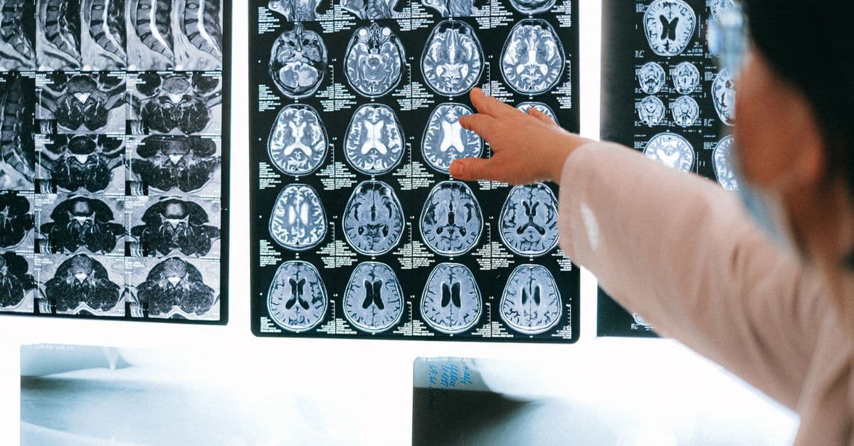
Photo: Anna Shvets / Pexels
An MRI machine is one of the strangest objects in a modern hospital. It is, essentially, a giant superconducting electromagnet — so powerful that loose objects in the room become projectiles, so cold inside that liquid helium is constantly evaporating off — and yet it produces no radiation, makes no incisions, and can map the soft tissue of a living human brain in millimeter detail.
The technology that became Magnetic Resonance Imaging emerged from nuclear physics in the 1940s, was adapted into medical imaging by Paul Lauterbur and Peter Mansfield in 1973, and earned them the Nobel Prize in Physiology or Medicine in 2003. Today there are roughly 50,000 MRI scanners worldwide.
What MRI Actually Measures
MRI doesn't image your tissues directly. It images hydrogen nuclei — single protons — in the water and fat of your body. Since humans are roughly 60% water by mass, every voxel of tissue contains an enormous population of hydrogen protons, each of which acts like a tiny spinning magnet.
The trick of MRI is to manipulate those protons with powerful external fields, then listen as they relax back to their normal state, and finally turn the timing of that relaxation into a picture.
Step by Step: How an MRI Scan Happens
1. The Main Magnet Aligns Your Protons
Clinical MRIs typically run at 1.5 Tesla (T) or 3 T, with research scanners reaching 7 T or higher. To put that in scale: Earth's magnetic field is about 50 microtesla, so a 3T scanner is roughly 60,000 times stronger than the field you're standing in right now. This main field is produced by a superconducting coil cooled to about 4 Kelvin with liquid helium.
When you slide into the bore, the field gently nudges a tiny fraction of your hydrogen protons into alignment with it. Most cancel out in pairs, but a small surplus — perhaps a few protons per million — lines up. That surplus is what MRI exploits.
2. Radiofrequency Pulses Tip the Protons
A radiofrequency (RF) coil pulses electromagnetic waves at the Larmor frequency — the natural precession frequency of hydrogen in the given magnetic field (about 64 MHz at 1.5T, 128 MHz at 3T). This is identical to AM/FM radio physics, just precisely tuned. The pulse tips the aligned protons sideways, like spinning tops nudged off vertical.
3. Protons Relax and Emit a Signal
When the RF pulse stops, protons relax back to alignment. As they do, they release the absorbed energy as a faint radio signal — picked up by the same coils. Two key relaxation times are measured:
- T1 (longitudinal relaxation): How quickly protons re-align with the main field.
- T2 (transverse relaxation): How quickly the protons' coordinated wobble decoheres.
Different tissues have characteristically different T1 and T2 values. Fat, water, gray matter, white matter, tumors, blood — each has its own fingerprint. By weighting scans toward T1 or T2 contrast, radiologists can highlight different anatomy.
4. Gradient Coils Locate the Signal
A bare proton signal would tell you nothing about where in the body it came from. Three sets of gradient coils apply small additional fields that vary across X, Y, and Z, so the Larmor frequency becomes position-dependent. The signal arriving at the antenna now encodes spatial information. (The loud banging you hear during a scan is these gradient coils flipping on and off at high speed against the main magnetic field.)
5. Computer Reconstruction
A two-dimensional Fourier transform — the same math used in audio compression and Wi-Fi — converts the frequency-encoded signal into an image of proton density and relaxation behavior. Modern scanners may also use compressed sensing and AI reconstruction to recover full images from sparse data, cutting scan times by 30–60%.
What MRI Is Good At — And What It Isn't
MRI excels at soft tissue contrast. Brain, muscle, ligaments, cartilage, organs, tumors — anything water-rich and subtly different from its neighbors. It is the modality of choice for neurology, orthopedic injury (torn ACL, slipped disc), and many oncology workups.
MRI is poor at imaging bone cortex (low water content, appears dark) and lung parenchyma (air, no signal). For broken bones you want X-ray or CT; for acute lung disease, CT.
Comparison Table: MRI vs CT vs X-ray vs Ultrasound
| Modality | Physics | Ionizing Radiation | Best For | Limitations | Typical Scan Time |
|---|---|---|---|---|---|
| X-ray | Photons through tissue | Yes (low dose) | Broken bones, chest, dental | 2D only, poor soft tissue | Seconds |
| CT | Rotating X-rays + reconstruction | Yes (higher dose) | Trauma, lung, acute bleeding, bone | Less soft tissue detail than MRI, radiation | 1–10 min |
| MRI | Magnets + RF waves | No | Brain, joints, soft tissue tumors, spine | Slow, noisy, expensive, claustrophobic, no metal | 20–60 min |
| Ultrasound | Sound waves + echo | No | Pregnancy, abdomen, vessels, heart | Operator-dependent, can't penetrate bone/air | 10–30 min |
Contrast Agents and Gadolinium
To enhance certain features — blood vessels, lesions, inflammation — radiologists inject gadolinium-based contrast agents (GBCAs). Gadolinium is paramagnetic, dramatically shortening T1 in nearby tissue and brightening it on T1-weighted scans.
GBCAs are generally safe but carry two known concerns: nephrogenic systemic fibrosis in patients with severe kidney disease (now rare due to careful screening), and gadolinium retention in brain tissue from repeated scans, whose long-term significance is still under study. Macrocyclic agents (like gadoteridol) retain less than older linear agents and are now preferred.
Functional MRI (fMRI)
In 1990, Seiji Ogawa discovered that oxygenated and deoxygenated blood have different magnetic properties. Active brain regions consume more oxygen, which triggers a slightly delayed surge of oxygen-rich blood — and that blood has a measurably different MRI signal. This is the BOLD (blood-oxygen-level-dependent) signal, the foundation of functional MRI.
fMRI has become the workhorse of cognitive neuroscience: mapping language areas before brain surgery, studying decision-making, attention, memory, and even cross-correlating patterns of activation with subjective experience. Caveats apply — fMRI measures blood flow, not neurons directly, and the temporal resolution (seconds) is coarse compared to neural firing (milliseconds). See our piece on memory and the brain for how fMRI has changed our understanding of cognition.
Safety: The Magnet Is Always On
The main magnet is never turned off between patients — it's a superconducting coil holding a persistent current. This is why MRI safety is unforgiving:
- No ferromagnetic objects in the room. Oxygen tanks, scissors, and chairs have become deadly projectiles in documented accidents.
- Pacemakers and implanted devices require careful screening. Many modern devices are MR-conditional, but older ones are absolute contraindications.
- Tattoos with metallic pigment can heat slightly during scanning.
- Claustrophobia affects up to 15% of patients in conventional closed bores; open MRI or wide-bore scanners help.
- Noise during gradient switching can exceed 100 dB. Earplugs and headphones are standard.
If a magnet must be shut down emergently — say, a person is pinned against it by a steel object — engineers initiate a quench, venting the helium and collapsing the superconductivity. This destroys the magnet and costs hundreds of thousands of dollars to refill, but it saves lives.
Recent Advances
- 7T clinical scanners (FDA-approved for clinical use since 2017) reveal anatomy invisible at 3T, particularly for epilepsy and multiple sclerosis lesions.
- Compressed sensing acquires only a fraction of the usual data and reconstructs the full image — slashing scan times.
- AI-based denoising and reconstruction (Subtle Medical, GE AIR Recon DL) improves image quality from shorter scans.
- MR-Linac systems combine MRI with linear accelerators for image-guided radiation therapy in real time.
- Portable MRI (Hyperfine Swoop, 0.064T) brings imaging to ICUs and ambulances at a fraction of the cost. Lower resolution, but a transformational reach.
See our related explainer on magnetism in physics for the underlying field theory.
FAQs
Q: Why is MRI so loud? A: The gradient coils experience strong forces from the main magnet when current flows through them, causing them to physically vibrate against their mounts at audio frequencies. Modern "silent" sequences using clever pulse shapes can cut noise significantly.
Q: Why does an MRI take so long? A: Each image slice requires many RF pulses and gradient encodings to build up enough signal. A full brain MRI may need 10–15 different sequences (T1, T2, FLAIR, diffusion, etc.), each taking 3–6 minutes. Newer scanners with parallel imaging and AI reconstruction are cutting this dramatically.
Q: Can I get an MRI with a tattoo or piercing? A: Most modern tattoos are safe. Remove piercings if possible. Inform the technologist of any tattoos, especially older ones, which may briefly warm during scanning.
Q: Is MRI safer than CT? A: For radiation, yes — MRI uses none. But MRI has its own risks: magnet hazards, contrast reactions, and longer scan times that make it unsuitable for unstable patients. The right choice depends on the clinical question.
Q: Can MRI read minds? A: No, despite headlines. fMRI can identify which broad brain regions are active during specific tasks and, in research settings, sometimes decode coarse categories of what a subject is seeing or imagining. It cannot read thoughts in any meaningful sense, and even sophisticated decoding requires extensive per-subject training.
Q: What's the strongest MRI ever built? A: The Iseult scanner at CEA in France operates at 11.7 Tesla for human imaging and produced its first images of a living human brain in 2024. Animal-research scanners exceed 21T.
Sources
- Lauterbur, P. C. (1973). "Image Formation by Induced Local Interactions." Nature.
- Ogawa, S. et al. (1990). "Brain magnetic resonance imaging with contrast dependent on blood oxygenation." PNAS.
- American College of Radiology MRI Safety Guidelines (2024).
- FDA — Magnetic Resonance Imaging device safety communications.
- Nobel Prize 2003 — Lauterbur and Mansfield.
- Iseult Project, CEA Paris-Saclay (2024 first-image announcement).
Weekly digest
Stay ahead of the curve
Get the best science, AI, and tech stories delivered to your inbox every week. No spam — unsubscribe any time.
About the Author
SciHub101 is an independent science and technology publication founded and edited by Buğra Sözeri, a software engineer and app developer based in Türkiye. Articles are drafted against peer-reviewed literature and primary sources, fact-checked, and edited by a human before publication. Where we compare products, anything built by our own team is labelled as such — see the editorial guidelines and About page for how corrections and disclosures work.
Related Articles
What Is Light? Photons and Wave-Particle Duality
Light is both wave and particle. Explore photons, the electromagnetic spectrum, polarization, lasers, and quantum entanglement explained simply.

Climate Change Explained — The Greenhouse Effect
How the greenhouse effect works, which gases drive it, and the evidence that today's warming is anthropogenic — explained clearly.
How MRI Machines Work: Magnetic Resonance Imaging Explained
MRI sees inside your body without radiation by making hydrogen atoms in your tissues sing at a specific radio frequency.
How Magnetism Works — From Atoms to Earth
How magnetism works: electron spin, magnetic domains, ferromagnetism, Earth's geodynamo, animal navigation, and MRI, explained mechanism by mechanism.

How Pain Works in the Brain: It's Not What You Think
Pain is not a signal your body sends to your brain. It is a decision your brain makes — and that distinction has transformed medicine.