How MRI Machines Work: Magnetic Resonance Imaging Explained
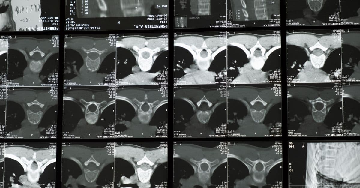
Photo: cottonbro studio / Pexels
An MRI machine contains enough liquid helium to fill a small swimming pool, cooled to -269°C (barely above absolute zero), generating a magnetic field 60,000 times stronger than Earth's. Yet the images it produces come not from radiation or cameras but from radio waves emitted by hydrogen atoms in your body, nudged into quantum excitement and captured as they relax. For over four decades, MRI has become the gold standard for soft-tissue imaging — revealing brain tumours, torn knee ligaments, and organ damage with stunning clarity. Every scan is a quantum mechanics experiment conducted on a living person, translating the language of nuclear magnetism into images that can save lives. For the biological side of the technology, see MRI technology and magnetic resonance imaging explained.
The quantum foundation: proton spin
You don't need quantum mechanics to understand MRI at a practical level, but you need a little. Protons — the nuclei of hydrogen atoms — have a quantum property called spin. Spin makes protons behave like tiny bar magnets with a north and south pole. Another everyday device built on electromagnetic physics is the touchscreen, explained in how touchscreens work.
In the absence of an external magnetic field, the spins of all protons in your body point in random directions. Their tiny magnetic moments cancel out and you're magnetically invisible.
Place your body inside a powerful MRI magnet (typically 1.5 Tesla or 3 Tesla for clinical use — the magnet in your phone's compass generates about 0.00005 Tesla by comparison), and something interesting happens. Protons tend to align with the strong external field — either parallel (with the field, lower energy) or anti-parallel (against the field, higher energy). A slight excess aligns parallel, producing a net magnetization in the patient along the magnet's field direction (called the longitudinal direction).
The protons don't just align statically — they precess, wobbling around the magnetic field axis like a spinning top wobbles around the direction of gravity. The frequency of this precession is called the Larmor frequency, and it's directly proportional to the magnetic field strength. At 1.5 Tesla, hydrogen protons precess at about 63.9 MHz — in the radio frequency range.
The radiofrequency pulse: disturbing the equilibrium
Here's where MRI becomes clever. To detect the proton magnetization, you need to disturb it. An RF pulse at exactly the Larmor frequency is transmitted into the patient. This resonant pulse (hence "magnetic resonance") tips the net magnetization away from the longitudinal direction — a 90-degree pulse tips it into the transverse plane.
Now the magnetization is precessing in the transverse plane, perpendicular to the main field. This rotating magnetization induces a voltage in receiver coils placed around the patient — Faraday induction, the same principle as an electrical generator. This induced voltage is the MRI signal.
After the RF pulse, protons relax back to their equilibrium state. This relaxation happens through two independent processes:
T1 (spin-lattice relaxation): Protons return their energy to surrounding molecules, restoring the longitudinal magnetization. Different tissues have different T1 values: fat relaxes quickly (~250ms), brain gray matter more slowly (~1,000ms), cerebrospinal fluid very slowly (~4,000ms).
T2 (spin-spin relaxation): Protons interact with each other, causing their precessions to fall out of sync (dephase), reducing the transverse signal. Fluids have long T2 values (water appears bright); rigid structures like cortical bone have very short T2 (appear dark).
By choosing different timing parameters (echo time TE, repetition time TR), radiologists can create images that emphasize T1 or T2 differences between tissues. T2-weighted images are particularly useful for brain imaging — lesions, edema, and tumors often appear very bright because they contain more water than surrounding tissue.
Gradient coils: encoding position
A uniform magnetic field would make every hydrogen proton in your body resonate at the same frequency — the signal from your brain would be indistinguishable from your foot. To create spatial images, MRI uses gradient coils.
Gradient coils are large electromagnets that superimpose a controlled, spatially varying magnetic field on top of the main field. If the magnetic field varies linearly along, say, the head-to-foot direction, then the Larmor frequency varies along the same direction. Protons in your head precess at a slightly different frequency than protons in your chest.
By acquiring data while sweeping through different gradient configurations — slice selection gradients, frequency-encoding gradients, and phase-encoding gradients — the scanner builds up a dataset in "k-space" (spatial frequency domain). A mathematical operation called the 2D Fourier transform converts this k-space data into the final image.
Modern MRI sequences use complex patterns of RF pulses and gradient switching to efficiently fill k-space and create different image contrasts. A standard brain MRI scan might acquire 200 slices using sequences that take several minutes each; a cardiac MRI must acquire data gated to the heartbeat to avoid motion artifacts.
The engineering inside an MRI scanner
The hardware of an MRI scanner is remarkable:
Superconducting magnet: The main field is generated by niobium-titanium superconducting wire carrying thousands of amperes of current with zero resistance. The wire must be cooled to 4K using liquid helium. A standard 1.5T clinical magnet requires about 1,500 liters of liquid helium and might weigh 5–8 tonnes. The magnet field is always on — you cannot "turn off" an MRI magnet during normal operation.
Shimming: The field must be extremely uniform (within a few parts per million) across the imaging volume. Passive shims (small iron pieces) and active shim coils correct for manufacturing imperfections and variations caused by the patient's presence.
Gradient coils: Three sets of coils (X, Y, Z) create precisely controlled field gradients, switched at frequencies up to several kHz. This switching is what makes the loud banging and knocking sounds during an MRI scan — the Lorentz forces on the current-carrying coils cause vibration.
RF system: A high-power RF amplifier (up to 35 kW for 3T systems) drives the transmit coil. Sensitive receive coils (often flexible surface coils positioned close to the anatomy of interest) detect the picoVolt-level signals from tissue.
MRI variants: from fMRI to MRS
Beyond anatomical imaging, MRI has remarkable functional and spectroscopic capabilities:
fMRI (functional MRI): Detects brain activity by measuring blood oxygenation (the BOLD — Blood Oxygenation Level Dependent — signal). Oxygenated and deoxygenated hemoglobin have different magnetic properties; active brain regions receive more oxygenated blood, creating a detectable signal change. fMRI has revolutionized neuroscience, mapping language, memory, and motor function.
MRS (MR Spectroscopy): Instead of imaging, measures the concentration of specific molecules in tissue — distinguishing tumor metabolites from normal tissue, for instance.
Diffusion tensor imaging (DTI): Maps the diffusion of water molecules in brain tissue, revealing white matter tract architecture — the "wiring diagram" of the brain.
Safety and Contraindications
While MRI is widely considered safe, the physics of strong magnetic fields and radiofrequency energy introduce genuine risks that must be carefully managed. Ferromagnetic objects become projectiles in the magnetic field — cases are documented of oxygen tanks, scissors, and surgical instruments accelerating into the magnet with lethal force.
Metal implants present three concerns:
- Ferromagnetism: Iron-based implants are attracted and may shift or twist inside the body.
- Induced currents: The changing gradient fields induce electrical currents in conductive materials, potentially causing heating (implant heating).
- Malfunction: Electrical devices (pacemakers, cochlear implants, insulin pumps) may malfunction or have their programming altered.
Most modern implants are labelled "MRI-safe" (safe for patients under specified conditions) or "MRI-conditional" (safe only at certain field strengths and under specific protocols). Titanium, non-ferromagnetic stainless steel, and ceramic materials are widely used in orthopedic implants precisely because they are MRI-compatible.
Heating and tissue damage is a concern for patients with certain conditions or implants. The radiofrequency energy absorbed by tissue (measured as Specific Absorption Rate, SAR) is strictly regulated by scanning protocols. For most patients, the energy deposition is trivial — less than a minor fever. However, patients with implanted wires (like temporary pacemaker leads) or certain metallic foreign bodies may experience significant localized heating.
Gadolinium contrast agents are used to enhance image contrast by accelerating T1 relaxation in nearby tissue. Gadolinium is highly toxic as a free ion, so it's administered in chelated complexes that prevent the gadolinium from entering the body. In patients with renal failure, chelation may fail and gadolinium can accumulate, causing gadolinium deposition disease — a potentially serious condition causing joint pain, muscle weakness, and burning sensations.
Source: FDA (2023). Guidance on Gadolinium-Based Contrast Agents: Assessment of Bioaccumulation and Renal Safety. — Regulatory review of gadolinium safety concerns.
Comparison Table: Imaging Modalities
Different imaging techniques have distinct strengths and limitations:
| Modality | Radiation | Soft Tissue Contrast | Speed | Cost | Best For |
|---|---|---|---|---|---|
| X-ray | Yes (ionizing) | Poor | Very fast | Low | Bones, pneumonia, chest |
| CT | Yes (ionizing) | Moderate | Fast (seconds) | Moderate | Trauma, lung detail, 3D |
| MRI | No | Excellent | Slow (minutes) | High | Brain, spinal cord, joints |
| Ultrasound | No | Poor-moderate | Fast | Very low | Pregnancy, real-time organs |
| PET | Yes (radioactive) | Poor (structural) | Moderate | Very high | Metabolic activity, cancer staging |
| fMRI | No | Good | Moderate | High | Brain activation mapping |
MRI's lack of radiation and superior soft-tissue contrast make it invaluable for brain and neurological imaging, despite its cost and time requirements.
The Evolution of MRI Technology
1970s: Nuclear Magnetic Resonance becomes Medical Paul Lauterbur and Peter Mansfield independently developed MRI imaging. Lauterbur used gradient coils (1973) to spatially encode NMR signals. Mansfield developed fast imaging sequences (1977) to speed up data acquisition. Both were awarded the Nobel Prize in Physiology or Medicine in 2003. The first human MRI scan was performed in 1977; the first clinical MRI scanner was installed in 1980.
1990s: Functional MRI and Clinical Expansion The discovery of the BOLD (Blood Oxygenation Level Dependent) signal by Seiji Ogawa (1990) revolutionized neuroscience. fMRI could now map active brain regions non-invasively, winning Ogawa a Lasker Award in 2003. Clinical MRI became standard for neuroimaging, spinal imaging, and cardiac applications.
2000s–2010s: Parallel Imaging and Acceleration Parallel imaging techniques (GRAPPA, SENSE) used multiple receiver coils simultaneously, reducing acquisition time by 2–4 fold. Diffusion Tensor Imaging (DTI) revealed white matter architecture. Perfusion imaging measured tissue blood flow. These advances enabled clinical brain mapping for surgical planning.
2020s–Present: Ultra-High Field and AI 7 Tesla and 11.7 Tesla research MRI scanners now provide extraordinary resolution. Artificial intelligence is being deployed to accelerate image acquisition (reducing scan time from 45 minutes to 10 minutes) through compressed sensing and deep learning reconstruction. Clinical adoption of AI-enhanced MRI is accelerating.
Source: Geva, T. (2006). Magnetic Resonance Imaging: Historical Perspective. Journal of Cardiovascular Magnetic Resonance, 8(4). — Historical review of MRI development and clinical adoption.
Current Applications and Emerging Directions
Clinical applications: MRI dominates diagnosis of neurological disorders (stroke, dementia, multiple sclerosis, epilepsy), musculoskeletal injuries (torn ligaments, cartilage damage), spinal disorders, cardiac imaging (heart wall thickness, viability), liver and pancreatic disease, and malignancy detection (particularly in soft tissue and the brain).
Emerging directions:
- Real-time interventional MRI: Using MRI to guide minimally invasive procedures (biopsies, ablations, endoscopy) in real time.
- Hyperpolarization techniques: Creating non-equilibrium states in nuclei (like xenon-129 or carbon-13) to enhance signal by 10,000-fold, enabling metabolic imaging.
- PET-MRI hybrid scanners: Combining functional PET imaging (metabolic) with structural MRI (anatomical) in one device.
- Portable MRI: Development of lower-field (0.5T) systems that are smaller and cheaper for point-of-care use in clinics and rural hospitals.
The bottleneck for MRI adoption in resource-limited settings is cost (2–3 million US dollars per scanner) and expertise (radiologists and physicists required). Portable MRI and AI interpretation hold promise for democratizing access.
Clinical Workflow and Patient Experience
An MRI scan is a carefully choreographed sequence of events designed to protect safety and maximize image quality. Understanding this workflow illustrates the complexity hidden behind a simple "get an MRI."
Pre-scan screening is critical. Patients complete detailed questionnaires about metal implants, pacemakers, metallic tattoos, medical history, and claustrophobia. An MRI technologist reviews answers and explains the procedure. Some answers (certain pacemakers, recent metallic foreign bodies, extreme anxiety) may warrant cancellation or rescheduling.
The scan itself involves positioning the patient on a sliding table, placing receiver coils around the anatomy of interest (a knee coil for knee imaging, a head coil for brain), and instructing the patient to remain motionless. Any movement corrupts images — a brief twitch can ruin a 5-minute acquisition. The scanner automatically positions the table into the bore and begins pulses.
The patient experiences:
- Loud knocking and banging (the gradient coil vibration) — hearing protection is essential
- Radio pulses (imperceptible)
- Vibration from the scanning bed
- Instructions ("hold your breath", "don't move") delivered via speaker
- A call button for emergency exit
The entire process (setup + imaging) typically takes 30-60 minutes. For anxiety-prone patients, this is brutally long. Pediatric patients often require sedation.
Post-scan, the radiologist (a physician specialized in interpreting images) reviews the images, writes a report within hours, and the referring physician discusses results with the patient.
Total turnaround time from order to report can be 48-72 hours in busy hospitals — a bottleneck that's partly why MRI capacity is often saturated.
Source: American College of Radiology (2024) ACR Appropriateness Criteria — Clinical workflow and scheduling guidelines.
Portable and Low-Field MRI: Democratizing Access
Traditional MRI scanners cost $2–3 million, require specialized facilities, and occupy 400+ square feet. This limits availability, especially in low-income countries and rural areas.
Portable MRI systems operating at 0.5T or lower are being developed to change this. Companies like Hyperfine (acquired by GE Healthcare in 2021) and others are building refrigerator-sized MRI devices that:
- Cost $300,000–$500,000 (10x cheaper)
- Weigh <1 ton (can be moved between rooms)
- Require less cooling infrastructure
- Produce lower-quality images than 3T systems but adequate for many clinical questions
Trade-offs: 0.5T MRI has lower signal-to-noise ratio — images are noisier, resolution is lower, and scanning takes longer. A 0.5T brain MRI might take 90 minutes where a 3T takes 30 minutes. Certain sequences (fMRI, diffusion-weighted imaging) become impractical.
Use cases: Portable MRI is ideal for:
- Point-of-care assessment in emergency departments (stroke evaluation)
- Underserved rural clinics
- Bedside imaging for hospitalized patients (avoiding transport)
- Global health initiatives in low-resource settings
A patient with a suspected stroke in a rural area can now receive an MRI bedside, dramatically improving care quality. The trade-off (slightly lower quality) is worthwhile when the alternative is no imaging at all or hours of transportation to a distant hospital.
Source: Cooley et al. (2023). "Low-Field MRI: Clinical Applications and Technical Innovations." Magnetic Resonance in Medicine, 89(3). — Review of portable MRI capabilities and limitations.
Advanced Quantitative MRI
Traditional MRI produces qualitative images — "there's a lesion on the brain." Quantitative MRI measures specific tissue properties numerically, enabling precise comparisons over time and across patients.
T1 and T2 mapping: Instead of T1-weighted or T2-weighted images (which mix T1/T2 contrast with other factors), quantitative mapping measures actual T1 and T2 values in milliseconds. This is reproducible — two scans in different hospitals should produce nearly identical T1 values for the same tissue.
Applications:
- Cardiac fibrosis: Elevated T1 detects scar tissue in the heart, predicting arrhythmia risk
- Liver iron content: T2* relaxation quantifies iron deposits, useful in hereditary hemochromatosis or transfusion-dependent patients
- Brain tissue integrity: T2 changes in white matter detect degeneration in multiple sclerosis before visible lesions form
Diffusion tensor imaging (DTI) quantifies water diffusion in different directions. White matter (axons) restricts diffusion along the axon direction (large fractional anisotropy), while gray matter allows diffusion equally in all directions. DTI maps the brain's structural connectivity — the wiring diagram of neural pathways.
These quantitative measures are more reproducible and sensitive to early pathology than qualitative imaging. However, they require specialized sequences, longer acquisition times, and expertise to interpret. Clinical adoption is growing but remains concentrated in academic centers.
Source: Kaur et al. (2023). "Quantitative MRI Biomarkers for Tissue Characterization." Nature Reviews Biomedical Engineering, 6(2). — Review of quantitative MRI applications.
Motion Correction and Artifact Management
Motion is the sworn enemy of MRI. A patient cough, swallow, or heartbeat during data acquisition corrupts images. MRI technologists spend enormous effort minimizing motion:
Physiological gating synchronizes data acquisition with the patient's heartbeat or respiration. A cardiac MRI acquires data only during specific phases of the heartbeat, avoiding motion blur. Respiratory gating does the same for chest/abdominal imaging.
Parallel imaging reduces scan time (allowing shorter breath-holds), thus reducing motion opportunity.
Newer techniques like compressed sensing reconstruct high-quality images from undersampled k-space data, reducing acquisition time further.
Retrospective motion correction uses software to detect and correct subtle motion in already-acquired data — a post-processing step that improves quality without longer scans.
These are the reasons MRI scan protocols are so complex; each sequence is carefully designed to balance image quality, acquisition time, and patient motion tolerance.
MRI in Research and Drug Development
Beyond clinical imaging, MRI is central to neuroscience research and pharmaceutical development:
Preclinical models: Researchers MRI mouse brains (at ultra-high field, 9.4T) to study neurodegeneration, cancer, or stroke models. Small-animal MRI reveals brain pathology in disease models, guiding drug development.
Clinical trial imaging: Pharmaceutical companies use MRI to measure drug efficacy — does a treatment for Alzheimer's disease reduce brain atrophy? Does a cancer drug shrink tumors? MRI provides objective endpoints beyond subjective symptom reporting.
Connectomics: Mapping the brain's structural and functional connectivity requires high-resolution fMRI and DTI. Large collaborative projects (Human Connectome Project, Allen Institute) use MRI to build detailed wiring diagrams of the brain.
Precision medicine: Individualized treatment planning relies on MRI. A surgeon planning to remove a brain tumor performs MRI to identify eloquent cortex (language, motor areas) and plan a trajectory that maximizes tumor removal while minimizing neurological damage.
Frequently Asked Questions
Q: Is MRI safe for everyone?
A: MRI is generally safe for most patients, but ferromagnetic objects (iron, steel) become projectiles in the strong magnetic field. Devices with electronics (pacemakers, older cochlear implants) may malfunction. Most modern implants are MRI-safe or MRI-conditional. Gadolinium contrast agents carry a small risk of deposition disease in patients with severe kidney failure.
Q: Why are MRI machines so loud?
A: Gradient coils carrying large currents experience Lorentz forces when current direction changes rapidly. This makes the coils flex and vibrate at frequencies up to several kHz, creating the characteristic knocking, buzzing, and banging. Noise levels can reach 100–140 dB, comparable to a jet engine, making hearing protection essential.
Q: What's the difference between 1.5T and 3T MRI?
A: Higher field strength (Tesla) increases signal-to-noise ratio, allowing higher resolution images or faster scanning. 3T scanners produce better brain and musculoskeletal images than 1.5T but cost more and pose stronger safety concerns for metallic objects. 7T and 11.7T research scanners provide extraordinary spatial resolution for neuroscience.
Q: Why does MRI take so long compared to CT scan?
A: CT X-rays sweep quickly and acquire data in seconds. MRI must acquire hundreds of different gradient configurations to fill k-space (spatial frequency domain). A brain MRI might require 45 minutes, whereas a chest CT takes 10 seconds. Newer techniques like parallel imaging and compressed sensing are reducing acquisition times significantly.
Q: Can claustrophobic patients use MRI?
A: Older closed-bore MRI machines can be claustrophobic — the patient's head is inside a narrow tunnel. Open-bore systems address this, but have lower field strength and image quality. Wide-bore systems (70 cm instead of 60 cm bore) reduce claustrophobia while maintaining image quality. Sedation is an option for severely anxious patients.
Q: How is MRI different from X-ray and CT?
A: X-rays use ionizing radiation (cancer risk, accumulation over lifetime). CT uses more radiation but produces 3D images. MRI uses no radiation, just magnetic fields and radiofrequency energy, making it ideal for repeated imaging or populations at risk (children, pregnant women). MRI has superior soft-tissue contrast but is slower and more expensive.
Read next:
Follow new articles
Add SciHub101 to your feed reader to see new science and technology stories.
About the Author
SciHub101 is an independent science and technology publication founded and edited by Buğra Sözeri, a software engineer and app developer based in Türkiye. AI tools assist with drafting and research. Source documentation in the existing archive is uneven, and articles are being reviewed and corrected. The Editorial Team byline identifies editorial responsibility; it does not certify specialist or independent human review. Where we compare products, anything built by our own team is labelled as such — see the editorial guidelines and About page for how corrections and disclosures work.
Related Articles
How Skyscrapers Are Built: The Engineering of Vertical Cities
Building a skyscraper means solving physics problems at enormous scale — wind, weight, sway, and the challenge of going higher.
How Google DeepMind Uses AI to Cut Data Center Energy Consumption
DeepMind's reinforcement learning system cut Google's data center cooling energy by 40%—a case study in applying AI to climate-critical infrastructure.
How Satellites Orbit Earth: The Physics and Engineering Behind Eyes in the Sky
There are over 8,000 active satellites orbiting Earth right now. Here's how they stay up and what they do.
History of Electricity: Faraday, Tesla, Edison, and the Force That Powers the World
Electricity was a scientific curiosity in 1800. Within 100 years it was lighting cities and running factories.

How Jet Engines Work: The Physics of Flight Power
A single GE90-115B turbofan is rated at 115,300 pounds-force of thrust. The physics behind it is a short chain of ideas: squeeze air, burn fuel in it, and let the hot gas do work.
How Fiber Optic Cables Transmit Data
A strand of glass thinner than a human hair can carry more information than all the copper wires ever made. Here's the physics behind it.