MRI Technology: Magnetic Resonance Imaging Explained
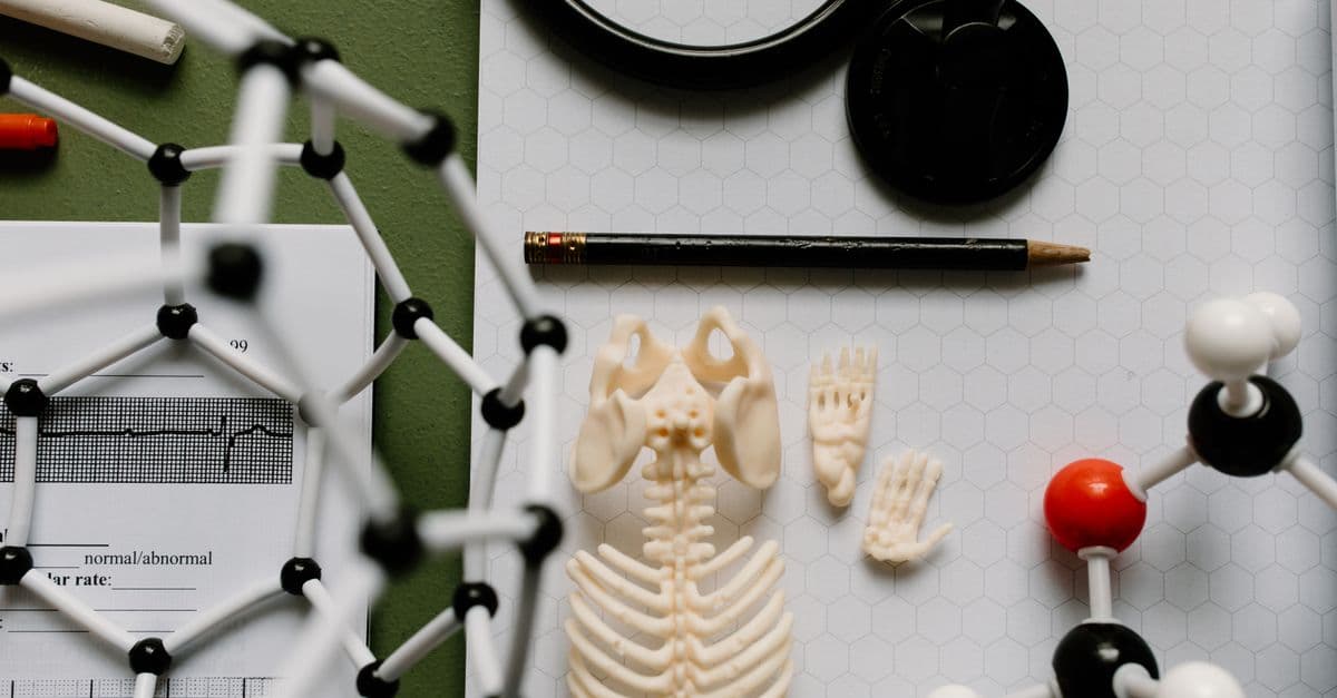
Photo: Tara Winstead / Pexels
Magnetic Resonance Imaging (MRI) represents a revolutionary medical imaging modality, enabling visualization of soft tissues with extraordinary detail without ionizing radiation. Developed from nuclear magnetic resonance principles discovered in physics, MRI technology translated scientific concepts into medical imaging tools. Paul Lauterbur demonstrated in 1973 that magnetic field gradients could localize signals for imaging. Peter Mansfield developed rapid imaging techniques enabling clinical application. Their work earned the Nobel Prize and transformed medical imaging. Today, MRI enables diagnosing brain tumors, spinal cord injuries, cartilage damage, and countless other conditions invisible to other imaging modalities. MRI's lack of radiation, superior soft tissue contrast, and multidirectional imaging capabilities make it invaluable in modern medicine.
Educational content, not medical advice. Talk to your doctor before changing any medication, treatment, or routine. Information here reflects current research but every person's situation differs.
Key Takeaways
- MRI uses magnetic fields and radio waves to generate images without ionizing radiation
- Hydrogen nuclei in water and fat align in magnetic fields and emit signals when excited
- Gradient magnetic fields localize signals enabling reconstruction of detailed images
- MRI excels at soft tissue imaging—brain, spinal cord, cartilage, organs
- Contraindications include metallic implants and claustrophobia concerns
Background
Nuclear magnetic resonance emerged from physics research in the 1930s-1940s. Scientists discovered that atomic nuclei in magnetic fields absorbed and re-emitted radiofrequency energy. Isidor Rabi won the Nobel Prize for this discovery. Bloch and Purcell developed techniques measuring these nuclear magnetic resonance properties, earning their own Nobel Prize.
These physics discoveries remained laboratory curiosities until researchers considered medical applications. Could NMR principles enable imaging internal anatomy? The challenge was creating spatial information from signals—distinguishing which tissue produced specific signals.
The Experiment/Discovery
Signal Localization (1973): Paul Lauterbur's breakthrough involved applying magnetic field gradients to standard NMR equipment. Rather than a uniform magnetic field, he applied gradients—different field strengths at different locations. This enabled determining where signals originated through frequency analysis. Different field strengths caused nuclear spins to precess at different frequencies; analyzing signal frequencies revealed spatial location.
Lauterbur demonstrated his technique on a small water sample containing cancer cells. The resulting image showed the sample's structure—the first NMR image. The concept was straightforward but revolutionary.
Rapid Imaging (1974-1980s): Peter Mansfield developed mathematical approaches enabling rapid MRI acquisition. Sequential pulse application and sophisticated signal processing enabled reconstructing images quickly. This translated MRI from research novelty to potential clinical tool.
Clinical Implementation (1980s-present): Early clinical MRI systems were clunky and slow, but by the 1980s, MRI became established in hospitals. Constant technological improvements—stronger magnets, faster gradient coils, advanced software—continuously enhanced image quality and scanning speed.
The Results
MRI revealed soft tissue architecture with unprecedented clarity. Brain tumors, previously requiring invasive procedures for diagnosis, became visible on MRI. Spinal cord injury patterns became apparent, guiding treatment. Cartilage damage in knee injuries was visualized, enabling precise surgical planning.
MRI uniquely excelled at certain tissue characterization. Different tissues' water and fat content varied, producing characteristic MRI signals. This enabled distinguishing tissue types—gray matter from white matter, benign from malignant lesions. This tissue characterization capability was unique to MRI.
The lack of ionizing radiation represented a major advantage. Patients could receive repeated MRI without radiation risk. This enabled monitoring disease progression and treatment response safely. Pediatric imaging, previously limiting X-ray use to essential cases, could be performed liberally with MRI.
MRI's multidirectional imaging capacity enabled evaluating anatomy from any angle—sagittal, coronal, axial, and oblique planes. This comprehensive anatomical visualization improved diagnostic accuracy compared to single-plane imaging.
Impact on Medicine
Neuroimaging: Brain tumor diagnosis, stroke imaging, and dementia evaluation are revolutionized by MRI. Neurologists now routinely order MRI for evaluating headaches, cognitive changes, and suspected neurological disease. Early stroke detection and characterization guide thrombolytic therapy decisions.
Spinal Imaging: Spinal cord injury, compression, and degeneration visualization enables surgical planning. MRI detects spinal canal narrowing causing myelopathy, disc herniation causing radiculopathy, and cord injury severity.
Orthopedic Surgery: Cartilage and ligament tears become visible on MRI, enabling precise diagnosis and surgical planning. Anterior cruciate ligament injury, meniscal tears, and rotator cuff injuries are diagnosed definitively through MRI.
Cardiac Imaging: Cardiac MRI evaluates myocardial infarction, cardiomyopathy, and valvular disease. Unlike echocardiography depending on imaging windows, MRI provides complete anatomical visualization.
Oncology: Tumor characterization and staging rely increasingly on MRI. Breast cancer screening, liver lesion characterization, and tumor surveillance all employ MRI. Superior soft tissue contrast enables detecting small lesions missed by other modalities.
Current Applications
Brain Imaging: MRI remains gold standard for intracranial pathology. Brain tumors, stroke, dementia, multiple sclerosis, and seizure evaluation all employ MRI. Functional MRI enables mapping brain activity—revolutionizing neuroscience and neurosurgery.
Spine Imaging: Spinal cord pathology diagnosis relies on MRI. Degenerative disc disease, syrinx (fluid within spinal cord), and spinal cord compression are evaluated through MRI.
Abdominal Imaging: Liver, pancreatic, and biliary pathology evaluation increasingly uses MRI. Diffusion-weighted imaging and magnetic resonance cholangiopancreatography provide organ-specific diagnostic information.
Vascular Imaging: Magnetic resonance angiography and venography visualize blood vessels without ionizing radiation or contrast. This enables assessing vascular disease and evaluating vascular malformations.
Musculoskeletal Imaging: Cartilage, ligament, tendon, and muscle pathology are visualized with exquisite detail through MRI. Sports medicine relies heavily on MRI for injury assessment.
Future Directions
Stronger Magnets: Higher magnetic field strengths enable improved image resolution and speed. 7.0-Tesla magnets (compared to standard 1.5-3.0T) provide superior image quality. Ultrahigh field systems (10.5T and beyond) are under development.
Faster Acquisition: Emerging techniques—parallel imaging, compressed sensing—enable dramatically faster acquisitions. Future MRI may complete brain imaging in seconds rather than minutes.
Molecular MRI: Using targeted contrast agents, MRI could visualize specific molecules or biological processes. This promises detecting cancer earlier and monitoring molecular changes during treatment.
Hybrid Imaging: Combining MRI with PET (positron emission tomography) creates hybrid systems enabling simultaneous anatomical and metabolic imaging. This promises improved disease characterization.
Artificial Intelligence: Machine learning algorithms enable reconstructing high-quality images from reduced data, decreasing scan time. AI may also improve diagnostic accuracy through automated analysis.
The Physics Behind the Machine: Engineering Brilliance
Modern MRI machines represent an extraordinary engineering achievement combining quantum physics, supercooled magnets, and precision electronics. The core is a superconducting magnet cooled to near absolute zero (4 Kelvin) with liquid helium. At this temperature, electrical resistance drops to zero, allowing current to circulate indefinitely without power loss, maintaining magnetic fields of 1.5 to 3 Tesla (Earth's field is roughly 50 microtesla — 30,000–60,000 times weaker).
The radiofrequency (RF) system transmits precisely tuned pulses at the Larmor frequency (the resonance frequency of hydrogen nuclei in the magnetic field). When the RF pulse turns off, excited nuclei relax, emitting signals detected by receiver coils surrounding the body part being scanned. Gradient magnetic fields (coils creating variable field strength across space) are crucial for localization — by applying gradients in three perpendicular directions, different spatial locations experience different field strengths, causing nuclei to emit at different frequencies. Fourier mathematics reconstructs these signal patterns into detailed images.
The entire imaging process is remarkably sophisticated: modern pulse sequences acquire complex images in seconds to minutes, whereas early machines required 10+ minutes per image. Speed advances come from clever mathematics (compressed sensing, parallel imaging using multiple receiver coils simultaneously) and faster electronics, not necessarily stronger magnets.
MRI Contrast Agents: Enhancing Images
While MRI works well without contrast agents for many applications, contrast agents dramatically improve diagnostic capability by changing how tissues appear on images. The most common contrast agents are gadolinium-based compounds, paramagnetic metals that alter the relaxation properties of nearby water molecules, creating brighter signal in specific tissues.
When gadolinium enters tissues — particularly tumors or inflamed regions with increased blood flow — water molecules interact differently with the magnetic field. On T1-weighted images, gadolinium appears bright (hyperenhancing). By comparing pre- and post-contrast images, radiologists detect tumors, assess blood-brain barrier disruption, evaluate inflammation, and characterize tissue vascularity.
Other specialized contrast agents target specific tissues. Dysprosium-based agents are used for cardiac imaging. Iron oxide nanoparticles accumulate in specific cell types. Researchers are developing targeted contrast agents that bind specifically to cancer cells or inflammatory markers, promising molecular imaging — using MRI to visualize disease at the cellular and molecular level, not just anatomical changes.
Safety considerations apply: gadolinium must be eliminated by kidneys. Patients with severe renal impairment (eGFR <30 mL/min/1.73m²) risk nephrogenic systemic fibrosis (NSF), a rare but serious condition. Pre-scan renal function assessment is standard practice. Pregnancy-related caution applies; gadolinium crosses the placenta and is avoided unless clinically essential.
Source: Tweedle, M.F. (2020). "Gadolinium-Enhanced MRI: Principles and Applications." Radiology, 297(2): 344–356.
Common MRI Sequences Explained
MRI's versatility comes from pulse sequences — specific patterns of radiofrequency pulses and gradient magnetic fields that produce images with different tissue characterization. Different sequences highlight different tissue properties:
T1-weighted imaging shows anatomical detail with excellent gray-white matter contrast. Fat appears bright (white), water dark (black). T1 sequences are rapid and form the backbone of MRI protocols.
T2-weighted imaging reverses contrast — water appears bright, fat dark. T2 sequences excel at detecting pathology (tumors, edema, infections appear bright because they contain excess water).
FLAIR (Fluid-Attenuated Inversion Recovery) suppresses CSF (cerebrospinal fluid) signal, making subtle brain lesions more visible. FLAIR is particularly valuable for detecting demyelinating disease (multiple sclerosis) and subtle stroke.
Diffusion-Weighted Imaging (DWI) measures water diffusion. In acute stroke, damaged tissue restricts water diffusion, appearing bright on DWI within minutes — before structural changes appear on conventional MRI. This capability makes DWI essential for acute stroke diagnosis.
Perfusion-Weighted Imaging uses rapid imaging after bolus contrast injection to measure tissue blood flow. Brain tumors, stroke territory, and dementia show characteristic perfusion patterns that aid diagnosis.
MR Angiography visualizes blood vessels without catheterization. Time-of-flight techniques or contrast-enhanced angiography reveal vascular anatomy and detect stenosis, aneurysms, and dissections.
Each sequence takes 2–5 minutes. Modern protocols combine 5–15 sequences, totaling 20–60 minutes per study. Sequence selection depends on clinical question — brain tumor imaging differs from knee cartilage imaging.
Source: Huettel, S. A., Song, A. W., & McCarthy, G. (2014). Functional Magnetic Resonance Imaging. Sinauer Associates.
Clinical Decision-Making: When MRI Is Appropriate
MRI excels at certain diagnoses but is unnecessary — even harmful — for others. Understanding appropriate use improves care and reduces unnecessary testing.
MRI clearly indicated: brain tumor evaluation, stroke within 48 hours, spinal cord pathology, dementia evaluation, multiple sclerosis, cartilage injury, anterior cruciate ligament tear, cardiac viability assessment, liver lesion characterization, prostate cancer staging.
MRI not indicated: acute fractures (CT or X-ray superior), acute head injury with likely skull fracture (CT is faster and equally sensitive), metallic foreign bodies (MRI contraindicated; requires imaging methods that don't interact with metal).
MRI contextual: chest imaging for lung pathology (CT generally superior due to speed), abdominal imaging (ultrasound or CT often faster for acute evaluation), cancer screening in high-risk populations (increasingly used but cost-benefit debated).
Appropriate use imaging guidelines exist to help clinicians select optimal modalities. Overuse wastes resources and may delay diagnosis by causing unnecessary scanner time allocation. Underuse misses treatable pathology. Experienced radiologists and clinicians understand these nuances and select modalities appropriately.
Source: American College of Radiology (2025). "ACR Appropriateness Criteria." American College of Radiology.
The History of MRI Development and Evolution
1930s-1940s: Physics foundations. Isidor Rabi discovers nuclear magnetic resonance phenomenon. Edward Purcell and Felix Bloch develop NMR measurement techniques, earning Nobel Prize. NMR remains laboratory physics tool.
1970: Paul Lauterbur applies magnetic field gradients to NMR, enabling spatial localization. Demonstrates imaging capability on small samples. Concept is revolutionary but impractical at first.
1974-1980s: Peter Mansfield develops rapid imaging mathematics (Fourier transformation approaches). Enables faster acquisition and clinical feasibility. Both Lauterbur and Mansfield eventually earn Nobel Prize.
1982-1984: First clinical MRI systems installed. Early machines produce crude images slowly (15+ minutes per image). Space requirements enormous; machines weigh 40+ tons. Cost prohibitive for most institutions.
1985-1995: Technological refinement. Faster computers, better algorithms, gradient coil improvements. Clinical utility expands dramatically. Brain imaging becomes routine. Functional MRI emerges as research tool.
1996-2010: Widespread adoption. MRI becomes standard hospital equipment for neuroimaging, orthopedic imaging, and oncology. Contrast agents developed and refined. Parallel imaging enables faster scanning. High-field magnets (3.0T) become common.
2010-present: Advanced applications. Functional MRI becomes routine neuroscience tool. Molecular imaging agents under development. AI integration improves image reconstruction and interpretation. 7.0T and higher field magnets clinical research tools.
MRI's trajectory parallels information technology advancement — as computers became faster and algorithms more sophisticated, MRI became faster and more capable. Further advances will likely emphasize speed, image quality, and AI-assisted interpretation.
Source: Geva, T. (2006). "Magnetic Resonance Imaging: Historical Perspective." Journal of Cardiovascular Magnetic Resonance, 8(4): 573–580.
Frequently Asked Questions
Q: How does MRI create images without using radiation?
A: MRI uses magnetic fields and radiofrequency waves instead of ionizing radiation. Hydrogen nuclei (primarily in water molecules) align in a strong magnetic field. Radiofrequency pulses excite these nuclei to higher energy states; when the pulse ends, they relax and emit signals. Magnetic field gradients encode spatial location into signal frequency. Computers use Fourier transformation to convert signal patterns into detailed images.
Q: Is MRI safe, and what are the actual risks?
A: MRI has no known biological hazards from magnetic fields or radiofrequency at clinical field strengths. Unlike X-rays, there's no ionizing radiation exposure. Primary risks involve ferromagnetic implants (pacemakers, old aneurysm clips) that can malfunction in strong fields. Claustrophobia and acoustic noise (MRI produces loud clicking sounds) can be uncomfortable. Standard pre-MRI screening eliminates most implant-related risks.
Q: Why does MRI take longer than CT scanning?
A: CT acquires images in seconds using rapid X-ray rotation. MRI requires sequential excitation and signal acquisition of tissues — a fundamentally slower process. However, newer fast techniques (parallel imaging, compressed sensing) have dramatically reduced MRI times. Brain imaging that once took 20 minutes now takes 5 minutes; future advances promise even faster imaging.
Q: Can patients with metal implants safely receive MRI?
A: It depends on the implant type. Ferromagnetic metals (pacemakers, ferromagnetic aneurysm clips, some cochlear implants) are incompatible with MRI and risk malfunction or heating. However, many implants (modern cardiac stents, titanium orthopedic hardware, non-ferromagnetic clips) are MRI-safe. Detailed pre-MRI screening with specific implant information is standard practice.
Q: Why is MRI so much more expensive than X-rays or ultrasound?
A: MRI equipment costs $1–3 million, requires specialized installation (RF-shielded room, cryogenic systems), and demands expensive ongoing maintenance. Operational costs include liquid helium (~$3,000/month), staffing, and electricity. Despite high costs, MRI's superior soft-tissue diagnostic capability justifies expense for appropriate clinical questions.
Q: Is MRI safe during pregnancy?
A: Yes, particularly after the first trimester. MRI has no ionizing radiation and no documented adverse effects on the fetus at clinical field strengths. Gadolinium-based contrast agents are avoided unless clinically necessary (they cross the placenta). MRI is increasingly the preferred imaging modality for pregnant patients requiring diagnostic imaging.
The Patient Experience: What Actually Happens During an MRI Scan
Understanding the patient journey through an MRI provides insight into why the machine is so effective—and what its limitations are. Patients enter the scanner room and change into hospital gowns (to eliminate all metallic buttons and zippers). A technologist performs pre-scan screening: any metal implants, tattoos with metallic ink, or contraindicated medical devices? Any claustrophobia or motion disorders? The machine's strong magnetic field is always on, even when not scanning—it doesn't activate and deactivate, so safety concerns apply the entire time.
As the patient lies on the scanning table, they're moved into the bore (the tube). The bore is tight—typically 60 centimeters (24 inches) in diameter. Some patients experience claustrophobia even if they're not typically claustrophobic; the enclosed space, loud noise, and loss of control create anxiety. Open-bore magnets (weaker fields, wider openings) exist but produce lower-quality images. The technologist remains at a control console outside the scanner, communicating via intercom. The patient holds an emergency call button in case of distress.
Once scanning begins, the machine produces rhythmic banging, clanging, and high-pitched sounds—gradient coils switching rapidly, RF coils transmitting and receiving. Earplugs or headphones are provided. A typical brain scan requires 10–20 minutes of continuous noise and immobility. Any patient motion (swallowing, fidgeting, even breathing) degrades image quality, requiring restarts. This is why patients with dementia, severe anxiety, or restlessness often receive sedation (propofol or midazolam) for MRI.
Some sequences produce specific sound patterns: the echoplanar imaging (EPI) used in functional MRI creates a distinctive high-pitched chirping. Diffusion-weighted imaging (used for acute stroke) produces sustained humming. The variety of sounds is auditory feedback that different parts of the scanner are active—but to the patient, it's just overwhelming noise.
Contrast injection, if used, is intravenous—a technician inserts an IV line before scanning. The gadolinium contrast is a room-temperature liquid that, when injected, feels warm as it travels through veins. Some patients describe a metallic taste; others feel mild vertigo. These sensations are expected and temporary. The warmth dissipates in seconds.
Source: Weinreb, J. C., & Rodgers, G. M. (2019). "Patient Safety in MRI: Claustrophobia, Anxiety, and Motion During Scanning." Journal of Magnetic Resonance Imaging, 50(4): 1088–1098.
MRI Artifacts: Why Radiologists See Stripes, Blurs, and Ghosts
Real MRI images contain artifacts—visual distortions from physics, patient motion, metallic interference, and signal processing limitations. Radiologists are trained to recognize artifacts, distinguish them from true pathology, and compensate during interpretation. Understanding artifacts is crucial because they can mimic disease or hide disease.
Metal artifacts are the most visible. A metal implant (even titanium, which is slightly ferromagnetic) distorts the magnetic field locally, causing severe signal loss and geometrical distortion. A hip replacement creates a dark, star-patterned void across multiple slices. This is why CT is preferred for orthopedic imaging of metal implants; X-ray isn't affected by metal, and CT artifacts are less severe. Advanced MRI sequences (metal-artifact-reduction, MAR) can partially recover signal but aren't perfect.
Motion artifacts appear when patients move during scanning. Head motion causes "ghosting"—repeated faint copies of structures appearing perpendicular to the direction of motion. A patient who swallows mid-scan shows a ghost swallow trail. Even subtle motion (eye movement, cardiac pulsation) creates artifacts. High-speed EPI sequences are particularly susceptible. Modern sequences use motion-correction algorithms (post-processing software that realigns slices), but severe motion requires rescanning.
Susceptibility artifacts occur at interfaces between tissues of very different magnetic properties. The most notorious is at air-tissue boundaries: sinuses, lungs, and ear canals (which contain air) distort the field. This is why brain MRI is excellent but lung MRI is poor (lungs are mostly air). The field distortion blurs and distorts the adjacent tissue, creating false signals or signal voids. This is why CT is standard for lung cancer screening.
Aliasing artifacts (spatial folding) occur when the field-of-view (FOV) is too small. Structures outside the FOV fold back into the image, creating ghosts or duplicates. Modern scanners automatically set appropriate FOV, but this can happen if scanning a small region or using sequences with limited coverage.
Ringing artifacts (Gibbs ringing) appear as oscillating bands near sharp edges—between the brain and CSF, for example. They result from incomplete Fourier data collection; the image reconstruction assumes data extends beyond its actual boundaries. Filters reduce ringing but also blur the image slightly.
Herringbone or zipper artifacts indicate RF noise from external sources (nearby electronics, cell phones, pagers). The RF-shielded room contains the scanner, but if the shielding is compromised, external RF signals leak in and corrupt the image.
Recognizing these artifacts is essential because they can be mistaken for stroke, tumor, or other pathology. A board-certified neuroradiologist will note "motion artifact limits evaluation" or "metal artifact from prior surgery" in the report, contextualizing the image limitations.
Source: Kramer, S. P., & Schears, G. J. (2020). "MRI Artifacts and Quality Assessment." Radiographics, 40(1): 74–87.
MRI vs CT vs Ultrasound: Detailed Clinical Comparison
| Characteristic | MRI | CT | Ultrasound |
|---|---|---|---|
| Radiation | None (magnetic fields) | Ionizing X-rays (~10mSv for chest CT) | None (sound waves) |
| Soft tissue contrast | Excellent (gray-white matter, edema, tumor) | Good (but less than MRI) | Good (but operator-dependent) |
| Speed | Slow (5–60 min/study) | Fast (5–10 sec) | Fast (5–15 min) |
| Metal sensitivity | Yes (contraindication for ferromagnetic implants) | No (metal causes artifacts but safe) | No metal issues |
| Bone imaging | Fair (cortical bone appears dark) | Excellent (dense bone visible) | Fair (bone surface only) |
| Real-time capability | No (rigid sequences, no live video) | No (static images only) | Yes (dynamic real-time video) |
| Cost | High ($800–2,500/scan) | Moderate ($300–1,000/scan) | Low ($100–300/scan) |
| Claustrophobia risk | High (narrow bore) | Low (wide aperture) | None |
| Availability | Limited (major medical centers) | Common (most hospitals) | Very common (most facilities) |
| Brain stroke imaging | Excellent (DWI detects acute stroke within minutes) | Good (rules out hemorrhage but misses early ischemia) | Poor (cannot penetrate skull) |
| Abdominal imaging | Good (liver, pancreas, adrenal) | Excellent (standard for CT abdomen) | Good for real-time (fluid assessment) |
| Liver lesion characterization | Excellent (multiphasic dynamic imaging) | Excellent (dynamic contrast imaging) | Good (depends on body habitus) |
| Vascular imaging | Excellent (MR angiography without catheter) | Excellent (CTA standard for aneurysm) | Good (carotid ultrasound routine) |
| Musculoskeletal | Excellent (cartilage, ligament, meniscus) | Poor (soft tissue not visible) | Good (rotator cuff, ACL visualization) |
MRI excels at soft tissue; CT excels at bone and speed; ultrasound excels at real-time and cost.
MRI Safety Screening: Detailed Contraindications and Precautions
Absolute contraindications—situations where MRI is dangerous—are rare in modern practice because most implants are now MRI-compatible.
Ferromagnetic implants are the primary danger. Old pacemakers (pre-2000) contained ferromagnetic materials and would malfunction in strong fields, potentially stopping heartbeat. Modern pacemakers (post-2010) are MRI-conditional: they work safely in MRI at 1.5T or 3T, but the patient's device card must be verified pre-scan. Some older ferromagnetic aneurysm clips (pre-1990s) could shift or heat in the magnetic field, causing neurological injury. Modern clips are titanium and MRI-safe.
Metallic foreign bodies pose risk. A ferromagnetic object (old shrapnel, metal filing in eye) can move in the magnetic field, causing tissue damage. Pre-scan screening specifically asks about metallic particles. Eye injuries from metal fragments warrant CT imaging instead.
Severe renal disease (eGFR <30 mL/min/1.73m²) contraindicates gadolinium contrast. Gadolinium-based contrast agents carry a small risk of nephrogenic systemic fibrosis (NSF), a rare but serious condition causing skin fibrosis, muscle pain, and organ involvement. The risk is low with modern linear chelating agents (gadoterate, gadopentetate), but high-risk patients are screened with renal function tests before contrast administration.
Conditional contraindications require risk-benefit discussion:
- Severe claustrophobia — sedation or open-bore MRI if diagnosis is necessary
- Unstable cardiac disease — patient monitoring required; 3T magnets may be unsafe for severe arrhythmia patients
- Gadolinium allergy — documented rare; alternative imaging or pre-medication if benefit outweighs risk
- Metallic tattoos — some inks contain ferromagnetic particles causing localized heating or artifact; patient tolerance often varies
Pre-scan screening forms ask detailed questions about all implants, occupational metallic exposure, and medical history. Patients remove all jewelry, watches, and piercings. A radiologist reviews the screening form before scanning proceeds. This systematic approach has made serious adverse events from MRI extremely rare.
Source: Shellock, F. G., & Spinazzi, A. (2018). "MRI Safety Update 2018: Critical Review and Guidance for Use of Gadolinium-Based Contrast Agents and Procedural Monitoring in Patients with Cumulative Renal Impairment." Journal of Magnetic Resonance Imaging, 47(3): 515–521.
Novel MRI Applications in 2026: Emerging Techniques Beyond Standard Imaging
While conventional MRI has reached clinical maturity, emerging applications promise new diagnostic and therapeutic capabilities.
Hybrid PET-MRI combines positron emission tomography (which shows molecular/metabolic activity) with MRI (which shows anatomy). A 2025 study found that simultaneous PET-MRI improved cancer staging accuracy by 20–30% compared to sequential CT-PET, because anatomical and metabolic information are perfectly aligned in space and time. These systems cost $5–8 million but are increasingly installed in major cancer centers.
Synthetic MRI uses machine learning to generate multiple conventional sequences (T1, T2, FLAIR) from a single, rapid acquisition. Instead of scanning 20+ minutes, a synthetic MRI protocol acquires data in 5 minutes, and AI reconstructs the standard sequences post-hoc. A 2024 clinical trial found synthetic MRI achieved 95% diagnostic accuracy vs conventional sequences for brain tumor assessment, with 75% time savings. This innovation could democratize MRI—shorter scanning times reduce motion artifacts, improve patient throughput, and lower operational costs.
Real-time MRI for interventional procedures—guiding surgery, biopsy, or catheter placement without fluoroscopy. Surgeons see real-time MRI video during brain tumor resection, allowing intraoperative assessment of tumor extent without stopping to obtain conventional MRI. This requires specialized sequences and careful synchronization of surgical instruments with field-free zones, but clinical applications are expanding.
Molecular MRI using targeted contrast agents that bind specific molecules. Iron oxide nanoparticles conjugated to antibodies targeting cancer cells accumulate in tumors, making small lesions visible. A 2026 early-phase trial found molecular MRI detected prostate cancer foci (5–10 mm) that conventional imaging missed, potentially improving early detection. Clinical translation is 1–3 years away.
Diffusion tensor imaging (DTI) maps white matter tracts—the neural "wiring" connecting brain regions. DTI reveals disrupted connections in stroke, dementia, and developmental disorders. Beyond diagnosis, neurosurgeons use DTI preoperatively to plan tumor resection, avoiding critical pathways. Integration with neuronavigation systems during surgery ensures surgeons respect functional anatomy.
These emerging techniques demonstrate MRI's continuing evolution. As magnet strengths increase, AI image reconstruction improves, and novel contrast agents are developed, MRI will expand into domains currently dominated by other modalities.
The takeaway
MRI is a triumph of applied physics translating Lauterbur and Mansfield's innovations into an indispensable medical tool. It provides soft-tissue imaging detail impossible with other modalities, without radiation exposure. The technology continues evolving: stronger magnets, faster sequences, and novel contrast agents expand clinical applications yearly.
From neuroimaging to oncology to sports medicine, MRI has become essential infrastructure in modern healthcare. The machine's sophistication — supercooled magnets, RF physics, parallel signal processing — is invisible to patients and clinicians focused on the images. Yet every MRI scan represents decades of physics, engineering, and clinical refinement. Understanding what happens inside the scanner, recognizing imaging artifacts, and knowing when MRI is appropriate versus other modalities makes clinicians and patients alike more informed consumers of this remarkable technology.
Read next:
Follow new articles
Add SciHub101 to your feed reader to see new science and technology stories.
About the Author
SciHub101 is an independent science and technology publication founded and edited by Buğra Sözeri, a software engineer and app developer based in Türkiye. AI tools assist with drafting and research. Source documentation in the existing archive is uneven, and articles are being reviewed and corrected. The Editorial Team byline identifies editorial responsibility; it does not certify specialist or independent human review. Where we compare products, anything built by our own team is labelled as such — see the editorial guidelines and About page for how corrections and disclosures work.
Related Articles

mRNA Vaccines: COVID-19 Breakthrough Explained
How modified mRNA and lipid nanoparticles enabled rapid COVID-19 vaccine development, building on 30 years of foundational research by Katalin Karikó and others.
Microbiome Research: Bacteria in Human Health Explained
How understanding the microbiome revealed that trillions of bacteria impact health and disease
X-ray Discovery: Seeing Inside the Body Explained
How Wilhelm Röntgen's accidental discovery of X-rays revolutionized medical imaging and diagnosis
How MRI Machines Work: Magnetic Resonance Imaging Explained
MRI sees inside your body without radiation by making hydrogen atoms in your tissues sing at a specific radio frequency.

Countries with the Most Doctors Per Capita (Physicians Per 1,000 People Ranked)
Cuba has 9.54 physicians per 1,000 people — the highest doctor-to-population ratio of any large nation on earth. That is nearly three times the OECD average of 3.7, and more than six times the WHO's minimum recommended threshold of 1.5. Behind Cuba sit Monaco, Greece, Belgium, and Lithuania in a top five dominated by an unexpected mix of a Caribbean socialist state and small European nations.

AI in Organ Transplantation: Matching, Allocation, and Saving More Lives
Machine learning is being tested on organ matching, graft-survival prediction, and kidney nonuse. Here is what the evidence shows and where it stops.