How the Human Heart Pumps Blood: A Complete Guide
Photo: Monstera Production / Pexels
Your heart will beat approximately 2.5 billion times over the course of your life — and it started before you were even born, pumping blood when you were a tiny embryo just a few weeks old. It never takes a vacation, never truly rests, and somehow manages to pump around 2,000 gallons of blood every single day. The engineering behind this fist-sized muscle is more elegant than almost anything humans have ever built.
Educational content, not medical advice. Talk to your doctor before changing any medication, treatment, or routine. Information here reflects current research but every person's situation differs.
Key takeaways
- The heart is a double pump: the right side sends blood to the lungs, the left side sends it to the rest of the body.
- Four valves act as one-way doors, preventing backflow of blood.
- The heart generates its own electrical signals through the sinoatrial (SA) node — your natural pacemaker.
- A single red blood cell completes a full circuit of the body in about 60 seconds.
- Heart disease remains the world's leading cause of death, yet many risk factors are modifiable.
The anatomy of the heart
The heart sits slightly left of center in the chest, nestled between the lungs in a space called the mediastinum. It's roughly the size of your fist and weighs between 8 and 12 ounces in most adults. Surrounding it is the pericardium — a tough, double-layered sac filled with a small amount of lubricating fluid that lets the heart slide freely as it beats.
Inside, the heart is divided into four chambers. The upper chambers are called the atria (singular: atrium), and the lower chambers are the ventricles. A wall called the septum divides the heart down the middle, creating a right side and a left side that, while adjacent, never directly share blood.
The right atrium receives oxygen-depleted blood returning from the body via two large veins: the superior vena cava (draining the upper body) and the inferior vena cava (draining the lower body). The left atrium receives oxygen-rich blood arriving from the lungs via four pulmonary veins.
Between each atrium and ventricle sits a valve. On the right side is the tricuspid valve (three leaflets); on the left is the mitral valve (two leaflets, also called the bicuspid valve). Two more valves guard the exits: the pulmonary valve leads from the right ventricle to the pulmonary artery, and the aortic valve leads from the left ventricle to the aorta. Together these four valves act as one-way doors, ensuring blood always moves in the right direction.
How the pumping cycle works
Every heartbeat is actually a coordinated two-stage squeeze called the cardiac cycle. It has two phases: diastole (relaxation, when the chambers fill) and systole (contraction, when the chambers pump).
Picture it this way: the heart is really two pumps sharing a wall. The right pump handles the pulmonary circuit — it collects used blood from the body and sends it to the lungs to pick up oxygen and drop off carbon dioxide. The left pump handles the systemic circuit — it receives the freshly oxygenated blood from the lungs and drives it out through the aorta to every organ, muscle, and cell in the body.
The sequence of a single beat goes like this:
- Both atria fill with blood and contract simultaneously, pushing blood down through the open tricuspid and mitral valves into the ventricles.
- The ventricles fill, then contract powerfully. Pressure snaps the mitral and tricuspid valves shut (this is the "lub" in the classic "lub-dub" sound a stethoscope picks up).
- The rising pressure forces open the pulmonary and aortic valves. Blood rushes out into the pulmonary artery and the aorta.
- As the ventricles relax, pressure drops, the pulmonary and aortic valves slam shut (the "dub"), and the cycle begins again.
The left ventricle has noticeably thicker walls than the right — it needs to generate much higher pressure to push blood all the way to your toes and back, while the right ventricle only needs to reach the nearby lungs.
The heart's electrical system
The heart doesn't wait for your brain to tell it to beat. It generates its own spark through a built-in electrical system. The sinoatrial (SA) node, a small cluster of cells in the upper right atrium, fires an electrical impulse roughly 60–100 times per minute at rest — earning it the nickname "the natural pacemaker."
That impulse sweeps across both atria, causing them to contract. It then hits the atrioventricular (AV) node, which acts as a gate, briefly slowing the signal so the ventricles have time to fully fill before they're told to fire. From there, the signal travels down the Bundle of His and branches out through the Purkinje fibers, which spread the signal rapidly across the ventricular walls, triggering a synchronized, efficient squeeze.
This is exactly what an electrocardiogram (ECG) measures — the electrical waves produced at each stage of this cycle. The classic P-QRS-T pattern on an ECG corresponds precisely to atrial contraction, ventricular contraction, and ventricular recovery.
What can go wrong
The heart is resilient, but its very relentlessness makes it vulnerable to wear and tear.
Coronary artery disease is the most common heart condition. The coronary arteries are the vessels that supply the heart muscle itself with blood. When fatty plaques build up inside them (a process called atherosclerosis), blood flow narrows. If a plaque ruptures and a clot forms, blood supply is suddenly cut off — that's a heart attack (myocardial infarction). Within minutes, heart muscle cells begin to die from oxygen starvation.
Heart failure doesn't mean the heart has stopped; it means the heart can no longer pump efficiently enough to meet the body's needs. It can result from years of high blood pressure, a previous heart attack, or damaged valves.
Arrhythmias are disruptions to the electrical system. Atrial fibrillation (AFib), where the atria quiver chaotically instead of contracting cleanly, is the most common sustained arrhythmia, affecting tens of millions worldwide and dramatically raising stroke risk. Ventricular fibrillation is immediately life-threatening — it's what causes cardiac arrest.
Valve disease occurs when valves become stiff (stenosis) or leaky (regurgitation), forcing the heart to work harder. Some valve defects are congenital; others develop over a lifetime.
Modern medicine and the heart
Cardiology has made extraordinary strides. Coronary angioplasty — threading a tiny balloon into blocked arteries and expanding it — can reopen a blocked vessel within minutes during a heart attack. Stents, small metal scaffolds left in place, keep the artery open. Bypass surgery reroutes blood flow around blockages using vessels harvested from elsewhere in the body.
Implantable cardioverter-defibrillators (ICDs) continuously monitor heart rhythm and deliver a shock if lethal arrhythmia is detected. Artificial pacemakers take over from a failing SA node. And in the most extreme cases, a heart transplant — or increasingly, a mechanical heart (ventricular assist device) — can sustain life when nothing else will.
Perhaps most remarkable is that prevention remains powerful: not smoking, maintaining healthy blood pressure, exercising regularly, and managing cholesterol and blood sugar can collectively reduce heart disease risk by up to 80 percent.
Autonomic Control: How Your Brain Talks to Your Heart
Although the heart generates its own rhythm via the SA node, the brain has constant input through the autonomic nervous system, which operates below conscious control. The sympathetic branch (fight-or-flight) accelerates heart rate and increases contractility; the parasympathetic branch (rest-and-digest) does the opposite via the vagus nerve.
This is why your heart rate jumps when you're startled, slows when you're calm, and adjusts moment-by-moment based on oxygen demand. Baroreceptors (pressure sensors in artery walls) detect blood pressure and reflexively adjust the heart's output to maintain homeostasis. A sudden drop in blood pressure (standing up, blood loss) triggers immediate acceleration; prolonged high pressure triggers compensation that eventually leads to hypertension-related damage if not addressed.
Source: Thayer, J. F., et al. "The Neurovisceral Integration Model of Autonomic Regulation," Neuroscience & Biobehavioral Reviews (2012) — How the vagus nerve and autonomic nervous system regulate heart function.
The link between chronic stress and heart disease partly flows through this pathway: sustained sympathetic activation wears down heart tissue, accelerates atherosclerosis, and increases arrhythmia risk.
Coronary Blood Flow and the Critical Role of Oxygen
Unlike other muscles that rest during the cardiac cycle, the heart is constantly working and constantly demanding oxygen. The heart represents ~0.5% of body weight but consumes ~5% of resting oxygen delivery.
Coronary arteries are the heart's blood supply lines. The left main coronary artery splits into the left anterior descending and left circumflex; the right coronary artery supplies the right side. These vessels branch recursively into capillaries that blanket the myocardium (heart muscle). Remarkably, most coronary blood flow occurs during diastole (relaxation), when the contracting ventricle isn't squeezing the arteries shut.
When atherosclerotic plaque narrows a coronary artery to less than ~70% of original diameter, the heart can still function at rest but cannot increase blood flow enough during exercise — the patient experiences chest pain (angina). Complete blockage causes myocardial infarction. Some individuals develop collateral circulation — spontaneous growth of tiny backup vessels that can partially compensate if main vessels are blocked. This natural workaround is more common in some individuals than others and is being studied as a potential therapeutic target.
Source: Fuster, V., et al. "Atherothrombosis and High-Risk Plaque," Journal of the American College of Cardiology (2005) — Mechanisms of coronary artery disease and thrombotic events.
Heart Rate Variability and Cardiac Health
Heart rate variability (HRV) — the variation in time between consecutive heartbeats — is a surprisingly good marker of cardiovascular health and resilience. A healthy heart doesn't beat at a perfectly regular 60, 70, or 80 times per minute; instead, intervals vary by tens or hundreds of milliseconds in response to breathing, posture changes, and autonomic tone.
Higher HRV generally indicates better health: the heart is responsive to changing demands. Low HRV correlates with increased risk of sudden cardiac death, heart attack, and all-cause mortality. A very regular heartbeat (almost robotic) is a red flag.
This measurement is now available via smartwatches and fitness trackers. While interpretation requires care (normal ranges vary by age and sex), tracking HRV trends can provide early warning of overtraining, illness, or stress accumulation in athletes and health-conscious individuals.
Development and Congenital Heart Disease
The heart begins forming just 3 weeks into human development—earlier than the brain, lungs, or kidneys. During weeks 4–8, it undergoes a complex series of foldings and septations to form chambers, valves, and outflow tracts. These developmental windows are exquisitely sensitive: maternal infections (rubella), medication exposure (thalidomide, retinoic acid), or genetic mutations can cause defects.
Congenital heart defects affect roughly 1 in 100 live births. They range from trivial (patent foramen ovale, a tiny hole between atria present in ~25% of people) to life-threatening (hypoplastic left heart, where the left pump fails to form). Modern imaging and surgery have made even severe defects manageable — many people with congenital heart disease now reach adulthood and lifespan.
Source: Hoffman, J. I., & Kaplan, S. "The Incidence of Congenital Heart Disease," Journal of the American College of Cardiology (2002) — Epidemiology and natural history of congenital cardiac defects.
The Remarkable Recovery Capacity of the Circulatory System
Despite the heart's strict demands, the cardiovascular system has surprising plasticity. Aerobic exercise triggers remodeling: the heart wall thickens proportionally (physiological hypertrophy), cardiac output increases, and efficiency improves. An athlete's heart may pump 30-50% more blood per beat than a sedentary person's, lowering resting heart rate.
Angiogenesis — the growth of new blood vessels — occurs in response to sustained tissue oxygen demand. Regular exercise stimulates capillary growth, improving oxygen delivery to muscles. This is why exercise is simultaneously protective against and therapeutic for cardiovascular disease.
Conversely, deconditioning (prolonged inactivity) reverses these gains within weeks to months. Bed rest and sedentary lifestyle accelerate atherosclerosis, reduce cardiac output, and increase arrhythmia risk. This is partly why hospitalization and recovery are so physically demanding—the cardiovascular system rapidly loses adaptation.
Frequently Asked Questions
Q: How fast does a normal heart beat?
A: At rest, most adults have a resting heart rate of 60-100 beats per minute. Trained athletes often have rates in the 40s because their stronger hearts pump more blood per beat. During intense exercise, heart rate can reach 180-200 beats per minute in healthy individuals.
Q: Does your heart stop when you sneeze?
A: No—this is a myth. Your heart rhythm may briefly change due to pressure changes in the chest cavity, but it never actually stops. The vagus nerve is temporarily stimulated during a sneeze, which may momentarily slow the heart, but it never pauses.
Q: Why is the left ventricle so much thicker than the right?
A: The left ventricle must generate pressure to push blood through the entire systemic circulation—thousands of kilometers of blood vessels reaching every organ and tissue. The right ventricle only needs to push blood to the nearby lungs at much lower pressure. The extra muscle mass of the left ventricle generates that necessary force.
Q: What is the difference between a heart attack and cardiac arrest?
A: A heart attack is a circulation problem—a blocked artery cuts off blood supply to heart muscle, causing tissue death. Cardiac arrest is an electrical problem—the heart's rhythm becomes chaotic, stopping effective pumping. A heart attack can trigger cardiac arrest, but they are distinct medical emergencies.
Q: Can the heart repair itself after a heart attack?
A: Only partially. Unlike some tissues, adult heart muscle cells have very limited regenerative capacity. Damaged cells are replaced by scar tissue, which doesn't contract, reducing the heart's pumping efficiency. Prevention through lifestyle and medication is far more valuable than recovery after infarction.
Fetal Circulation: The Heart Before Birth
Before a baby draws its first breath, the heart faces a unique problem: the lungs are filled with fluid, not air, so they cannot oxygenate blood. Instead, the fetus relies on placental oxygenation — the mother's blood exchanges gases with fetal blood across the placental barrier. The fetal heart must route blood to the placenta efficiently while minimizing flow to the non-functional lungs.
The fetal circulation achieves this through three special shunts:
The foramen ovale: A hole in the atrial septum (wall between the atria) allows blood from the right atrium to flow directly into the left atrium, bypassing the right ventricle. This keeps oxygenated blood (from the placenta) separated from deoxygenated blood destined for the lungs.
The ductus venosus: A vessel that shunts blood from the umbilical vein (carrying oxygenated blood from the placenta) directly into the inferior vena cava, bypassing the liver.
The ductus arteriosus: A vessel connecting the pulmonary artery directly to the aorta, allowing most blood from the right ventricle to bypass the fluid-filled lungs and go directly to the body.
At birth, when the baby takes its first breath, the lungs expand and pulmonary resistance drops dramatically. Increased blood flow to the lungs causes pressure changes in the atria that slam shut the foramen ovale. The ductus venosus and ductus arteriosus constrict as oxygen levels rise (oxygen is a potent vasoconstrictor for these vessels). Within hours, functional closure occurs; within days to weeks, these shunts fuse shut permanently.
A persistent patent foramen ovale (PFO) — failure of the foramen ovale to close — occurs in about 25% of the population and is usually benign. However, it creates a small right-to-left shunt (blood bypasses the lungs), which can allow clots or bacteria to reach the arterial circulation. Some strokes of unknown cause may be related to PFOs; closure with a device is being studied.
Source: Hagen, P. T., et al. (1984). "Incidence and size of patent foramen ovale during the first 10 decades of life." Mayo Clinic Proceedings, 59(1). — Epidemiology of PFO.
Aging and the Senescent Heart
The heart ages. Over decades, several changes occur:
Increased stiffness: Collagen and elastin accumulate in the myocardium, reducing compliance. The ventricles become stiffer, requiring higher pressure to fill. This is diastolic dysfunction — the heart fills less efficiently even if systolic function (contraction) remains normal.
Conduction slowing: The electrical system develops fibrosis; impulses conduct more slowly through the atria and ventricles. Arrhythmias become more common.
Calcium dysregulation: Calcium handling in myocytes becomes impaired, reducing contractility slightly. The elderly heart beats weaker overall.
Mitochondrial dysfunction: Myocyte mitochondria produce ATP less efficiently, reducing energy availability.
Neuroendocrine changes: Sensitivity to catecholamines (adrenaline) decreases; baroreceptor sensitivity dulls. The elderly heart's ability to increase heart rate in response to exercise or stress is blunted.
Clinical consequence: Elderly people have higher risk of heart failure, particularly diastolic heart failure (where ejection fraction is normal but filling is impaired). They tolerate arrhythmias less well and have worse outcomes after heart attacks.
These changes are partly inevitable (aging is a fundamental biological process) and partly modifiable (exercise, diet, stress management).
Source: Lakatta, E.G., & Levy, D. (2003). "Arterial and cardiac aging: major shareholders in cardiovascular disease enterprises." Journal of the American College of Cardiology, 42(8). — Comprehensive review of cardiac aging.
Exercise and Cardiac Remodeling
Athletic training triggers profound cardiac adaptations — true cardiac remodeling at structural and molecular levels.
Hypertrophy (thickening): Endurance training causes the left ventricle wall to thicken proportionally — both the inner diameter and wall thickness increase, maintaining normal wall stress (Laplace's law). This is physiological hypertrophy, different from pathological hypertrophy (seen in hypertension or aortic stenosis), where only wall thickness increases without chamber dilation.
Increased volume: The diastolic volume (how much blood fills the heart) increases, allowing a trained athlete's heart to eject 30-50% more blood per beat than a sedentary person's — the basis of lower resting heart rates.
Angiogenesis: New capillaries grow in the myocardium, improving oxygen delivery.
Mitochondrial proliferation: More mitochondria per myocyte improve energy metabolism.
Electrophysiological changes: Trained athletes may develop atrial enlargement and have increased vagal tone, predisposing to atrial fibrillation despite excellent overall fitness. This is a recognized paradox: elite athletes have higher AFib risk than expected, likely because atrial enlargement and increased vagal tone interact.
These adaptations are reversible. Detraining (stopping exercise) leads to regression — the heart returns to baseline within weeks to months.
Practical note: This is why "getting in shape" is so effective for cardiovascular health. Even moderate exercise (150 min/week of aerobic activity) produces measurable cardiac improvements within weeks.
Arrhythmia Mechanisms: When the Electrical System Misfires
Arrhythmias arise from disruptions to normal electrical conduction. Three main mechanisms:
Ectopic foci: A region outside the SA node fires rapidly, depolarizing the heart before the normal impulse arrives. This creates a premature beat or takes over pacemaking (ectopic rhythm). The ectopic region might fire faster due to increased autonomic tone, hypoxia, or electrolyte disturbances.
Reentry: The same impulse circulates repeatedly through conducting pathways. Imagine an impulse reaching a junction where one pathway is still refractory (can't conduct yet) while the other can. The impulse travels down the conducting pathway, loops back, and encounters the first pathway now recovered — the impulse circles again. Reentry can sustain rapidly repeated depolarizations (flutter or fibrillation). Many arrhythmias exploit reentry.
Afterdepolarizations: After normal repolarization, the membrane potential briefly rises (early afterdepolarization, EAD) or falls (delayed afterdepolarization, DAD), sometimes enough to trigger another action potential. Certain drugs (QT-prolonging medications), electrolyte abnormalities, or ischemia promote afterdepolarizations. These create extra beats or trigger runs of tachycardia.
Atrial fibrillation in particular involves chaotic reentry in the atria — multiple reentry circuits fire simultaneously, causing the atria to quiver without effective contraction. Blood stagnates, clot formation risk rises (explaining why AFib raises stroke risk), and AV node gets bombarded with impulses, creating irregular ventricular response.
Treatments target mechanisms: beta-blockers slow ectopic firing; antiarrhythmic drugs slow conduction (interfering with reentry); cardioversion (electrical shock) resets the heart rhythm; ablation surgically destroys problematic tissue.
Myocardial Infarction: The Cascade of Tissue Death
When a coronary artery becomes completely blocked, the downstream myocardium faces minutes of oxygen starvation before irreversible damage begins.
Minutes 0-20: Affected myocytes stop contracting (stunning). No cell death yet — reperfusion (restoring blood flow) can fully recover function.
20-40 minutes: Cell death begins. Myocytes run out of ATP, lose ionic gradients, and cell membrane fails. Calcium floods in, activating proteases that destroy the cellular machinery. This is irreversible necrosis.
Hours: The infarct core (central, thoroughly dead tissue) expands outward. Inflammation sets in — immune cells infiltrate. Inflammatory cytokines damage borderline ischemic tissue, extending the infarct.
Days-weeks: Dead tissue is replaced by granulation tissue (fibroblasts and collagen), forming a scar. Scar tissue doesn't contract, so the heart loses contractile power.
Weeks-months: Ventricular remodeling occurs. The ventricle dilates to accommodate loss of contractile tissue, increasing stress on remaining myocytes. This remodeling can progress to heart failure.
The "golden window" for intervention is the first 12 hours, ideally the first 3 hours. Prompt coronary reperfusion (angioplasty or thrombolytics) salvages myocardium in the ischemic penumbra before it dies. A person who gets treatment within 30 minutes may lose 5-10% of the left ventricle; one who waits 6 hours may lose 30-40%.
Source: Yellon, D. M., & Hausenloy, D. J. (2007). "Myocardial reperfusion injury." New England Journal of Medicine, 357(11). — Mechanisms of MI and repair.
Q: Why do athletes have lower resting heart rates?
A: Regular aerobic exercise causes the left ventricle to hypertrophy (thicken) and increase contractility. A stronger heart ejects more blood per beat, so it needs fewer beats per minute to maintain the same cardiac output. This physiological adaptation is healthy and reversible.
Read next:
Follow new articles
Add SciHub101 to your feed reader to see new science and technology stories.
About the Author
SciHub101 is an independent science and technology publication founded and edited by Buğra Sözeri, a software engineer and app developer based in Türkiye. AI tools assist with drafting and research. Source documentation in the existing archive is uneven, and articles are being reviewed and corrected. The Editorial Team byline identifies editorial responsibility; it does not certify specialist or independent human review. Where we compare products, anything built by our own team is labelled as such — see the editorial guidelines and About page for how corrections and disclosures work.
Related Articles

Antibody Structure: How the Immune System's Y-Shaped Weapon Works
How structural understanding of antibodies revealed how immune system recognizes pathogens

How Octopus Intelligence Works: The Alien Mind Living in the Sea
Octopuses have nine brains, blue blood, and three hearts — and their intelligence evolved completely independently from ours.
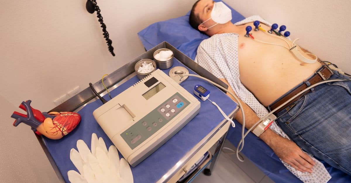
How the Heart Works — Anatomy and Function
How the human heart pumps blood: chambers, valves, electrical conduction, cardiac cycle, and the most important diseases and treatments.
How Skin Works — Your Largest Organ
Skin's three layers, how it protects you, why it ages, and the biology of pigment, sweat, and sensation in your body's largest organ.
How the Liver Works: Detoxification and Beyond
The liver performs over 500 distinct functions and is the only internal organ that can fully regenerate itself from just 25% of its original tissue.

How the Human Heart Works: The Cardiac Cycle Explained
The heart beats more than 100,000 times per day without a single conscious command, regulated by an electrical system so precise that it can be measured from electrodes on the skin. Here is how that system works, from the first spark in the SA node to the pressure wave that reaches your fingertips.