X-ray Discovery: Seeing Inside the Body Explained
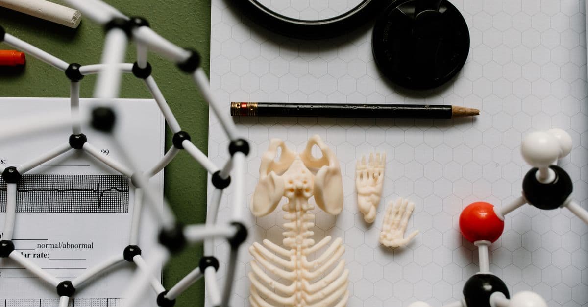
Photo: Tara Winstead / Pexels
On the evening of November 8, 1895, a German physicist named Wilhelm Conrad Röntgen was working alone in a darkened laboratory at the University of Würzburg when he noticed something that should not have happened: a chemically coated screen across the room began to glow, even though the cathode ray tube he was testing was fully wrapped in black cardboard. That faint, unexplained shimmer turned out to be a new form of radiation invisible to the human eye but capable of passing through flesh, paper, and wood while stopping at bone and metal. Within a year, doctors worldwide were using it to look inside living patients without cutting them open. This is the story of how that accident became the backbone of modern diagnosis, and how the physics behind it still shapes the images your doctor reads today.
Educational content, not medical advice. Talk to your doctor before changing any medication, treatment, or routine. Information here reflects current research but every person's situation differs.
What Röntgen actually found in that dark room
Röntgen was not trying to invent a diagnostic tool. He was investigating cathode rays, streams of electrons that vacuum-tube researchers across Europe had been probing for decades without fully understanding what they were or how far they traveled. Physicists knew cathode rays made certain materials fluoresce, but nobody expected any effect to survive a light-tight cardboard shield (History.com).
That is exactly what Röntgen saw. He had covered his Hittorf-Crookes tube in black cardboard specifically to block visible and ultraviolet light so he could study cathode rays in isolation. A barium platinocyanide screen sitting nearly a meter away lit up anyway, every time he switched on the tube's high voltage (Uni-Würzburg exhibition catalog). Cathode rays themselves cannot travel that far through air, let alone through cardboard. Something else was crossing the room.
Over the following seven weeks, Röntgen barely left his lab. He tested what the mysterious rays could and could not pass through: they sailed through paper, wood, and thin sheets of aluminum, but lead and thick bone stopped them cold. He photographed the rays' shadow on treated plates and confirmed they traveled in straight lines, unaffected by magnetic fields (unlike cathode rays, which bend). Because he could not identify what he was dealing with, he called it "X-radiation," borrowing the mathematical convention of X for an unknown quantity. The name stuck in English and German alike, even after physicists fully described the rays' nature as high-energy electromagnetic radiation, similar to visible light but with far shorter wavelengths and correspondingly higher energy.
That photograph of Anna Bertha's hand, bones rendered as pale shadows against darker soft tissue, did more to launch the field of radiology than any scientific paper could have. Röntgen published his findings on December 28, 1895, in a short paper titled "On a New Kind of Rays," and within weeks newspapers across Europe and the United States were reproducing the hand image (American Physical Society). He received the first Nobel Prize in Physics in 1901 for the discovery. Notably, he refused to patent the technology, insisting X-rays belonged to humanity rather than to him (DPMA).
Why did fluorescent screens glow through cardboard?
The screens glowed because X-rays are electromagnetic radiation with enough energy to pass through low-density materials like cardboard and human tissue, then strike phosphor-coated screens and knock their atoms into an excited state. When those atoms relax back down, they release visible light, producing the glow Röntgen observed. Dense materials like lead and bone absorb far more of that radiation, which is why they cast shadows rather than transmitting the rays.
How X-rays actually see through a body
To understand why X-rays reveal bone but blur past muscle, it helps to think of the body as a stack of translucent color filters of different thickness and density, each absorbing a different fraction of light passing through. Bone is dense and rich in calcium, a relatively heavy element, so it absorbs a large share of incoming X-ray photons. Soft tissue and air are much less dense, so more radiation passes straight through them. Wherever more radiation reaches the detector, the image appears darker; wherever tissue blocks radiation, the image appears lighter. That is the entire physical basis of a radiograph.
Producing the beam itself requires an X-ray tube, a descendant of the same cathode ray apparatus Röntgen used, just far more refined. Inside a modern tube, a heated filament (the cathode) boils off electrons, which are then accelerated across a high-voltage gap toward a metal target, usually tungsten, that serves as the anode. When the fast-moving electrons slam into the tungsten, most of their energy becomes heat, but a small fraction converts into X-ray photons through two mechanisms: Bremsstrahlung radiation, produced when electrons decelerate sharply near a tungsten atom's nucleus, and characteristic radiation, produced when incoming electrons knock inner-shell electrons out of tungsten atoms entirely, causing outer electrons to fall inward and release energy at fixed wavelengths (Bushberg et al., The Essential Physics of Medical Imaging).
That beam of photons then passes through the patient and strikes a detector, historically a photographic plate coated in silver halide crystals, and today almost universally a digital flat-panel sensor that converts absorbed X-ray energy directly into an electronic signal. Digital detectors changed the workflow more than the physics: images appear in seconds instead of minutes, can be enhanced digitally, and are stored and shared without a darkroom. But the underlying principle, differential absorption across tissue types, is identical to what Röntgen photographed on Anna Bertha's hand in 1895.
How does a broken bone show up on an X-ray?
A fracture appears as a dark line interrupting the otherwise continuous white shadow of a bone. Where bone has cracked or displaced, there is less calcified material for the beam to pass through at that spot, so more radiation reaches the detector there than at the surrounding, still-intact bone, producing a visible discontinuity that radiologists read as a break.
Two analogies that make the physics click
Think of an X-ray image the way you'd think of a silhouette made by holding a hand up to a bright flashlight against a wall. Thick, opaque objects like your knuckles block more light and cast a darker shadow; thinner, translucent regions between your fingers let more light through and appear brighter on the wall. An X-ray works the same way, except the "flashlight" is a beam of high-energy photons and the "wall" is a detector that records how much radiation made it through your body at every point.
A second useful analogy: imagine pouring water through a stack of sieves with different mesh sizes stacked one on top of another, fine mesh, medium mesh, coarse mesh, and measuring how much water drains out the bottom. A dense material like bone acts like a fine mesh, letting relatively little "water" (X-ray photons) through. Soft tissue acts like a coarse mesh, letting most of it through. The final puddle size, or in imaging terms, the pattern of radiation striking the detector, tells you exactly how the sieves (tissues) were arranged, without you ever touching them directly.
From novelty to the backbone of diagnosis
Physicians recognized the diagnostic value of X-rays almost immediately, a genuinely unusual pace for a nineteenth-century scientific discovery to translate into clinical practice. Within a year of Röntgen's December 1895 publication, hospitals in Europe and the United States were using X-ray equipment to locate fractures, embedded bullets, and swallowed objects without exploratory surgery (Spectroscopy Online).
Tuberculosis, a leading cause of death at the turn of the twentieth century, became one of the earliest and most consequential targets of routine radiography. The disease produces distinctive infiltrates and cavities in lung tissue that show up clearly on a chest X-ray long before physical examination alone could confirm the diagnosis. Mass chest X-ray screening programs in the mid-twentieth century, run in schools, workplaces, and mobile vans parked on city streets, became one of the largest public health surveillance efforts built on a single imaging technology. The same underlying mechanism that reveals antibiotic resistance patterns in how infections spread and adapt (see our explainer on how antibiotic resistance spreads) made radiography essential to tracking TB outbreaks before drug treatments existed at all.
Fluoroscopy, the technique of generating a continuous, real-time X-ray image rather than a single static exposure, arrived almost as quickly as static radiography itself. Early fluoroscopes let surgeons watch a swallowed coin move through a child's esophagus in real time, or watch a barium-coated meal travel through someone's digestive tract to spot obstructions. That live-feed capability eventually became the backbone of interventional radiology: threading catheters through blood vessels during cardiac procedures, guiding needle biopsies with millimeter precision, and placing stents without open surgery. None of that would be possible with a technology that only produced a still photograph.
The dangers of X-rays, however, emerged almost as fast as the benefits. Early radiologists and technicians, working without any concept of a safe exposure limit, developed severe radiation burns on their hands from repeatedly testing tube output against their own skin. Some, including several pioneering radiologists, developed radiation-induced cancers and lost fingers or entire limbs to chronic radiation dermatitis. Marie Curie, whose research into radioactivity ran parallel to and eventually intersected with X-ray technology through her work equipping mobile radiography units for World War I battlefields, died in 1934 of aplastic anemia attributed to decades of unshielded radiation exposure (Discover Magazine). Those early casualties are the reason every modern X-ray room has lead-lined walls, why technologists step behind a shielded barrier before pressing the exposure button, and why patients receive lead aprons during dental and diagnostic scans today.
Where X-rays sit inside modern medicine
Standard radiography remains the first imaging test ordered for suspected fractures, pneumonia, and many chest and abdominal complaints, largely because it is fast, inexpensive, and widely available even in resource-limited settings. A single posteroanterior chest X-ray delivers an average effective dose of roughly 0.1 millisievert (mSv), comparable to about ten days of ordinary background radiation from cosmic rays and the earth itself (RadiologyInfo.org, joint patient-safety resource of the American College of Radiology and Radiological Society of North America).
Computed tomography (CT) takes the same underlying physics, an X-ray source and detector, and rotates them around the patient to collect dozens or hundreds of angled projections, which a computer then reconstructs into cross-sectional slices. CT reveals soft-tissue detail and small lesions invisible on a standard radiograph, but at a real cost: an abdominal CT delivers an average effective dose of about 8 mSv, equivalent to roughly 400 chest X-rays, according to National Council on Radiation Protection and Measurements (NCRP) data compiled by the FDA and reviewed in the medical literature (StatPearls / NCBI Bookshelf). That dose gap is exactly why physicians order plain film first whenever it can answer the clinical question.
Fluoroscopy extends static X-ray imaging into a continuous video feed used to guide catheters, biopsy needles, and orthopedic hardware placement in real time. Because the beam stays on throughout a procedure that can last many minutes, cumulative fluoroscopic dose requires careful monitoring, particularly for complex interventional cardiology cases.
Dental radiography uses very low-dose, narrowly collimated beams to image individual teeth or the full jaw, exposing patients to only a few microsieverts per image, among the lowest doses in medical imaging.
| Imaging type | Typical effective dose | Best for | Real-time capability |
|---|---|---|---|
| Chest X-ray | ~0.1 mSv | Fractures, pneumonia, TB screening | No |
| Dental X-ray | ~0.005 mSv | Cavities, bone loss | No |
| Head CT | ~2 mSv | Stroke, trauma, hemorrhage | No |
| Abdominal CT | ~8 mSv | Internal bleeding, tumors, complex trauma | No |
| Fluoroscopy (per procedure) | Varies widely, often 5–70 mSv | Catheter guidance, GI studies | Yes |
Table: Approximate radiation doses by imaging modality, compiled from FDA and NCRP-referenced data (FDA Radiation Quantities and Units; RadiologyInfo.org). Actual doses vary by patient size, equipment, and protocol.
What people get wrong about X-ray radiation
A common misconception is that any X-ray exposure carries a meaningful, individually noticeable cancer risk. In reality, the excess lifetime cancer risk from a single diagnostic chest X-ray is extremely small, generally estimated in the range of one additional cancer per tens of thousands to hundreds of thousands of exposed people, a figure dwarfed by baseline lifetime cancer risk from all other causes combined (National Cancer Institute). The concern radiologists actually track is cumulative dose across a patient's lifetime, particularly from repeated CT scans, not any single plain film.
Another persistent myth: that X-rays and gamma rays are fundamentally different kinds of radiation. They are not. Both are high-energy electromagnetic radiation occupying overlapping and sometimes identical portions of the electromagnetic spectrum. The practical distinction is about origin, not physics: X-rays are produced artificially, by accelerating electrons into a metal target inside a vacuum tube, while gamma rays are emitted spontaneously from the nucleus of a decaying radioactive atom, the same process explored in depth in the story of Marie Curie's radioactivity research. At the photon level, a gamma ray and an X-ray of the same energy are indistinguishable.
People also frequently assume X-rays can substitute for MRI in evaluating soft-tissue injuries like torn ligaments or muscle strains. They generally cannot. Because muscle, tendon, and ligament tissue have very similar densities, they absorb X-ray photons at nearly the same rate and produce almost no visible contrast on a radiograph. A torn anterior cruciate ligament will typically look like a completely normal knee on plain film even though the joint is seriously injured; magnetic resonance imaging, which relies on the behavior of hydrogen atoms in a magnetic field rather than radiation absorption, is required to see that kind of soft-tissue damage directly. See MRI technology and how magnetic resonance imaging works for how that scan differs.
Finally, many patients believe fever accompanying a chest infection means the infection is automatically visible on an X-ray, or vice versa, that a normal X-ray rules out infection entirely. Neither is reliably true. Early pneumonia can precede visible radiographic changes by a day or more, and the body's own fever response, discussed in our explainer on why fevers help fight infections, is a separate immune mechanism entirely independent of what shows up on imaging. Clinicians combine radiographic findings with symptoms, blood work, and physical exam rather than relying on any single test in isolation.
Where the technology is headed next
Dose reduction continues to be the field's central engineering priority. Advanced digital detectors require far less radiation than the film-based systems of even twenty years ago to produce a diagnostic-quality image, and iterative computer reconstruction algorithms let CT scanners extract usable images from lower-dose acquisitions than earlier reconstruction methods required.
Photon-counting and spectral CT detectors, an emerging generation of scanner hardware, measure the energy of individual X-ray photons rather than simply totaling absorbed radiation, which lets radiologists distinguish between tissue types that would otherwise look identical on a conventional scan, such as differentiating calcium deposits from iodine-based contrast dye. This spectral information promises earlier, more specific diagnosis without necessarily raising patient dose.
AI-assisted interpretation has moved from research demonstration to real clinical deployment at many hospitals, with machine learning models flagging suspicious chest X-ray findings, such as early signs of pneumonia or lung nodules, for radiologist review, functioning as a second set of eyes rather than a replacement for physician judgment. The technology's accuracy varies by task and dataset, and radiologists remain the final decision-makers on diagnosis.
Portable and point-of-care imaging has expanded significantly, with lightweight digital X-ray units now used in ambulances, rural clinics, and disaster response settings where a fixed radiology suite is not practical, extending the reach of a 130-year-old physical principle into settings Röntgen could never have imagined.
Frequently asked questions
Q: How do X-rays penetrate through the body? A: X-rays are high-energy electromagnetic radiation with wavelengths short enough to pass through low-density materials like soft tissue while being largely absorbed by dense materials like bone and metal. That difference in absorption between tissue types is what creates the light-and-dark contrast on an X-ray image.
Q: Are X-rays dangerous? A: X-rays are a form of ionizing radiation, meaning they carry enough energy to damage DNA, and the National Cancer Institute lists ionizing radiation as an established cancer risk factor at sufficiently high cumulative doses. However, the dose from a single diagnostic X-ray, typically around 0.1 mSv for a chest film, is small compared to annual background radiation exposure, and medically necessary imaging is dosed to keep risk as low as reasonably achievable.
Q: Why do doctors still order X-rays when CT and MRI exist? A: X-rays remain first-line imaging for fractures, pneumonia, and many acute complaints because they are faster, cheaper, and deliver a much lower radiation dose than CT, roughly a fortieth to a hundredth the dose of an abdominal CT scan. CT and MRI are typically reserved for cases where plain film findings are unclear or more anatomical detail is needed.
Q: What is the difference between X-rays and gamma rays? A: Both are forms of high-energy electromagnetic radiation and can overlap in energy range, but X-rays are produced artificially by accelerating electrons into a metal target, while gamma rays are emitted spontaneously from the nucleus of a decaying radioactive atom. The distinction is about how the radiation is generated, not a fundamental physical difference.
Q: Can an X-ray show a torn ligament or muscle strain? A: Generally, no. Muscle, tendon, and ligament tissue absorb X-ray photons at nearly the same rate, so they produce little to no contrast on a radiograph, and a seriously torn ligament can look like a normal joint on plain film. MRI, which images soft tissue through a different physical mechanism, is the standard test for that kind of injury.
Q: How was the first X-ray image made, and what did it show? A: Wilhelm Röntgen produced the first X-ray photograph of living human anatomy on December 22, 1895, exposing his wife Anna Bertha's hand to the radiation for about 15 minutes to create a plate showing her bones and wedding ring against her hand's soft outline.
Q: How much radiation does a chest X-ray actually deliver compared to a CT scan? A: A standard chest X-ray delivers an average effective dose of roughly 0.1 mSv, while a CT scan of the abdomen delivers roughly 8 mSv, a difference of about 80-fold, according to FDA-referenced NCRP dose data. That gap is a major reason radiologists reserve CT for cases plain film cannot resolve.
Q: Did Röntgen patent the X-ray, and did he profit from the discovery? A: No. Röntgen deliberately declined to patent X-ray technology, stating that the discovery belonged to humanity rather than to him personally, and he donated his 1901 Nobel Prize money to his university rather than commercializing the invention.
Q: Why did early radiologists get radiation burns before X-ray dangers were understood? A: In the years immediately after 1895, nobody had established a safe exposure threshold, so early radiologists and equipment testers routinely held their own hands directly in front of X-ray tubes to check output, sometimes for extended periods, resulting in chronic radiation burns, tissue damage, and in some documented cases, radiation-induced cancers.
Q: Is fluoroscopy riskier than a standard X-ray? A: Yes, generally. Because fluoroscopy keeps the X-ray beam active continuously to produce a real-time video image, cumulative dose during a lengthy interventional procedure can be substantially higher than a single static radiograph, which is why fluoroscopic dose is monitored closely during long cardiac or vascular procedures.
The takeaway
X-rays were never designed. They were noticed, in a dark room, by a physicist paying close attention to something that shouldn't have happened. What makes the discovery endure 130 years later is not the mystery of what X-rays are, physicists worked that out within a decade, but the elegant simplicity of the mechanism: radiation passes through soft tissue and stops at bone, and that single fact, photographed and read correctly, still tells a doctor within minutes whether your wrist is broken. Every advance since, fluoroscopy, CT, digital detectors, AI-assisted reading, has built on that same shadow-casting principle rather than replacing it, which is why a technology born from an accident in 1895 is still the first imaging test ordered in emergency rooms today.
Read next:
Follow new articles
Add SciHub101 to your feed reader to see new science and technology stories.
About the Author
SciHub101 is an independent science and technology publication founded and edited by Buğra Sözeri, a software engineer and app developer based in Türkiye. AI tools assist with drafting and research. Source documentation in the existing archive is uneven, and articles are being reviewed and corrected. The Editorial Team byline identifies editorial responsibility; it does not certify specialist or independent human review. Where we compare products, anything built by our own team is labelled as such — see the editorial guidelines and About page for how corrections and disclosures work.
Related Articles

Why Vanilla Is So Expensive — The Biology and Economics of the World's Second-Priciest Spice
A flower that stays open for about a day, a pollination step no machine has replaced, months of curing, and a supply base that cyclones can cut by a third: the biology and economics behind vanilla's price.
T-Cell Receptor Structure: How TCRs Read MHC and Peptide
Inside the T-cell receptor: the alpha/beta protein pincer that scans MHC-bound peptides, the thymic gauntlet that filters out self-reactive clones, and why this molecular handshake underlies CAR-T therapy.
MRI Technology: Magnetic Resonance Imaging Explained
How MRI uses magnetic fields to create detailed images of soft tissues and organs

Countries with the Most Doctors Per Capita (Physicians Per 1,000 People Ranked)
Cuba has 9.54 physicians per 1,000 people — the highest doctor-to-population ratio of any large nation on earth. That is nearly three times the OECD average of 3.7, and more than six times the WHO's minimum recommended threshold of 1.5. Behind Cuba sit Monaco, Greece, Belgium, and Lithuania in a top five dominated by an unexpected mix of a Caribbean socialist state and small European nations.

AI in Organ Transplantation: Matching, Allocation, and Saving More Lives
Machine learning is being tested on organ matching, graft-survival prediction, and kidney nonuse. Here is what the evidence shows and where it stops.

Blood Type Classification: Understanding Transfusions Explained
How Karl Landsteiner discovered ABO blood groups enabling safe blood transfusions