AI Cancer Detection: Deep Learning Saves Lives
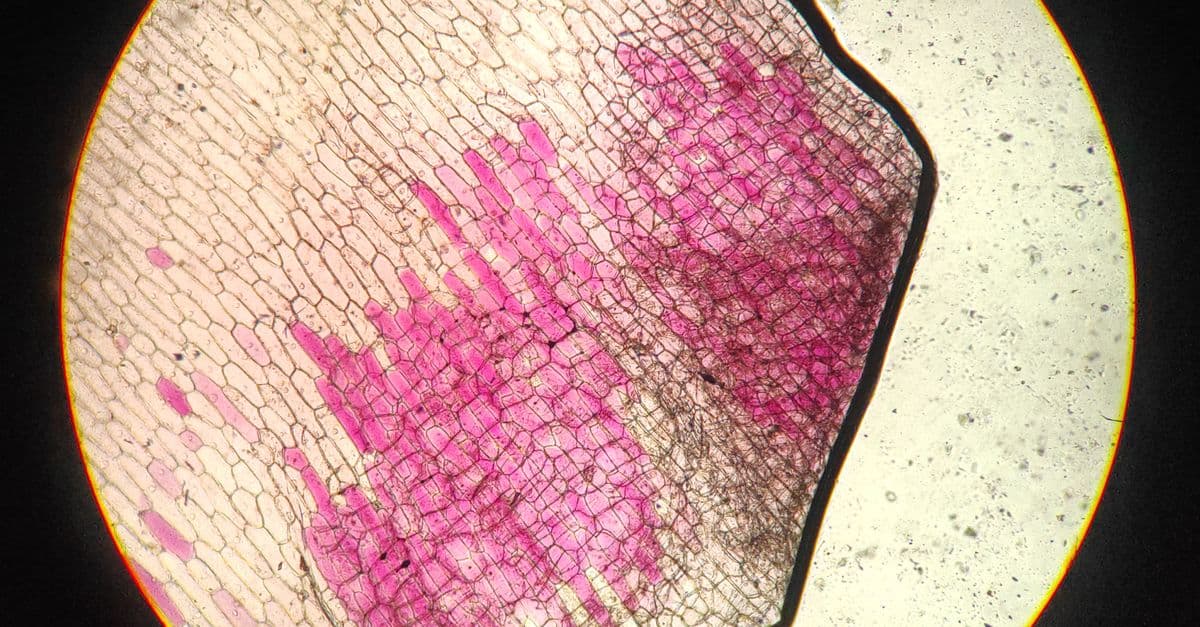
Photo: Leo Freire / Pexels
In 2020, Google Health and DeepMind published a peer-reviewed evaluation in Nature of an AI system for breast cancer screening, tested against real UK and US mammography datasets (McKinney et al., Nature, 2020). The system reduced false positives by 5.7% in the US cohort and false negatives by 9.4%, while matching or beating individual radiologists reading the same scans. That is the shape of the story: not "AI replaces doctors," but "AI changes which cases doctors see first, and how carefully."
Educational content, not medical advice. Talk to your doctor before changing any medication, treatment, or screening routine. Information here reflects published research, but every person's situation differs, and AI tools are used as an aid to clinicians, not a replacement for diagnosis.
This article explains how convolutional neural networks (CNNs, a type of neural network built to recognize visual patterns) learn to spot cancer in mammograms, CT scans, and tissue slides, what the actual published accuracy numbers say, where the technology has been cleared for clinical use, and where it still falls short. Every statistic below is tied to a named peer-reviewed study, regulatory filing, or institutional source, not a vendor's marketing page.
Why early detection is the whole ballgame
Cancer staging is a race against the clock, and the numbers make that concrete. According to the National Cancer Institute's SEER program, the five-year relative survival rate for localized female breast cancer (confined to the breast) is close to 100%, while the rate for distant-stage breast cancer (spread to other organs) drops to roughly 32% (SEER Cancer Stat Facts: Female Breast Cancer). Lung and colorectal cancers show the same pattern: catch it local, and treatment options multiply; catch it distant, and options narrow fast.
Pathologists and radiologists are the frontline of that race, and they are good at it, but they are human. A radiologist reading mammograms for a full clinical day experiences measurable fatigue effects on detection accuracy, and separate studies of interobserver variability, radiologists independently reviewing the same slide or scan, have found meaningful disagreement rates between experienced readers on ambiguous cases. Volume compounds the problem: screening programs in aging populations generate more scans every year, while the pipeline of trained radiologists and pathologists grows far more slowly.
This is the gap deep learning software was built to narrow. Not to replace the reader, but to pre-sort the pile, flag the highest-risk images, and provide a second, tireless set of eyes that never has an off day.
How a CNN actually "sees" a tumor
Talk to an oncologist about pattern recognition and they'll describe cell nuclei that are too large, too irregular, or dividing too fast, plus tissue architecture that looks disorganized compared to healthy structure nearby. A convolutional neural network learns to recognize the same categories of visual evidence, but it gets there through a completely different process: instead of being taught rules, it is shown tens of thousands of labeled examples and gradually discovers which pixel patterns correlate with the "malignant" label.
Think of it like teaching someone to recognize a counterfeit banknote not by handing them a rulebook of security features, but by showing them ten thousand real bills and ten thousand fakes and letting them notice, on their own, which subtle differences keep predicting the difference. The network never explicitly "knows" what a mitotic figure is; it just learns that a certain arrangement of pixel intensities, textures, and shapes tends to appear on the malignant side of its training set.
What happens to the image before the network sees it
A raw digital mammogram can run 4,000 by 3,000 pixels or larger, far too big and too inconsistent to feed a network directly. Preprocessing normalizes contrast and brightness across scanners from different manufacturers, resizes the image to a fixed resolution the network architecture expects (often 512×512 or 1,024×1,024 pixels), and applies data augmentation, small rotations, flips, and crops, so the network sees more effective training variety than the raw dataset provides.
The layer-by-layer climb from pixels to diagnosis
A CNN is organized in stacked layers, and each layer builds on the abstractions of the one before it. Early layers detect simple things: edges, color gradients, blobs. Middle layers combine those into textures and small structures, the graininess of a particular tissue type, the boundary of a nodule. Deep layers assemble those textures into higher-order concepts: cell clusters, tissue architecture, the overall shape of a lesion. The final layer converts all of that into a number, usually a probability that the input belongs to a category like "malignant," "benign," or "normal."
Three architecture families dominate published cancer-detection work. ResNet-50, built around "residual" connections that let information skip layers, made it practical to train much deeper networks without the accuracy degradation that used to plague deep stacks. Inception-V3 processes an image at multiple resolutions simultaneously, useful when a tumor can be either a tiny cluster of cells or a large mass depending on the case. Vision Transformers, a newer approach, break the image into patches and use an attention mechanism (the same core idea behind large language models, explained in our machine learning guide) to weigh which patches matter most for the final decision.
Why an AI's "confidence score" isn't the whole diagnosis
When a trained model processes a new scan, it doesn't hand a radiologist a yes-or-no verdict. It typically outputs a probability ("92% likelihood of malignancy"), plus a heatmap or attention map showing which regions of the image drove that score, so the clinician can verify the AI is actually looking at the lesion and not, say, a scanner artifact in the corner. That transparency layer matters more than the raw accuracy number, because a model that is right for the wrong reasons is a liability the moment the input distribution shifts even slightly.
Where does the training data come from, and why is that a bottleneck?
Training a clinically useful model typically requires tens of thousands to hundreds of thousands of images, each hand-labeled by a pathologist or radiologist marking exactly which regions are cancerous. Patient privacy rules mean this data mostly cannot be pooled into open public datasets the way, say, photos of cats can be. Most hospitals and companies train on internal, de-identified archives and then keep the resulting models proprietary, which is part of why independent, multi-institution validation studies (like McKinney et al.'s UK-to-US test) carry outsized scientific weight: they're one of the few ways to check whether a model generalizes beyond the hospital that built it.
Three deployments with published numbers
Breast cancer: mammography screening
The Google Health/DeepMind system evaluated in Nature in 2020 is the best-documented example in the field. Trained on UK screening data and tested on a separate US population it had never seen, the AI still outperformed the average individual radiologist on true and false positive rates, evidence that the model had learned something closer to "what malignancy looks like" than "what this particular hospital's malignant cases look like" (McKinney et al., Nature, 2020). Commercial tools with FDA clearance in this space include products from Lunit, iCAD, and other vendors on the FDA's published device list; they are typically deployed as a "second reader," flagging scans for radiologist prioritization rather than issuing an independent diagnosis.
Lung cancer: low-dose CT screening
Lung nodules are common on chest CT and the overwhelming majority are benign, which creates a genuine clinical dilemma: order an invasive biopsy on a probably-harmless nodule, or wait months for a follow-up scan while a patient sits with a "you might have cancer" diagnosis hanging over them. Google's model, described in Nature Medicine in 2019, was trained on National Lung Cancer Screening Trial data and achieved a 94.4% area-under-curve (AUC, a standard measure of how well a classifier separates true positives from false positives, where 100% is perfect and 50% is a coin flip) on over 6,700 held-out cases, then validated on an independent clinical set of more than 1,100 cases with consistent performance (Ardila et al., Nature Medicine, 2019).
Skin cancer: dermoscopy image classification
In one of the field's foundational papers, Stanford researchers trained a single CNN on 129,450 clinical images spanning more than 2,000 skin disease categories and showed it matched board-certified dermatologists in classifying skin lesions as malignant or benign, tested against 21 dermatologists on biopsy-confirmed cases (Esteva et al., Nature, 2017). It was an early, striking demonstration that a general-purpose image classifier, not one built with dermatology-specific rules, could reach specialist-level performance purely from labeled examples.
Prostate cancer: the first FDA-cleared AI pathology product
In 2021 the FDA granted De Novo clearance to Paige Prostate, the first AI-based digital pathology product cleared for clinical diagnostic use in the United States. In the clinical validation study submitted to the FDA, the software improved pathologists' cancer-detection accuracy on individual slide images from 89.5% to 96.8%, a 70% reduction in false-negative diagnoses and a 24% reduction in false positives, and it helped generalist pathologists reach the accuracy level of prostate specialists (FierceBiotech, "FDA clears Paige's AI as first program to spot prostate cancer in tissue slides," 2021). That milestone matters beyond prostate cancer specifically: it established the regulatory pathway that later pathology-AI clearances have followed.
| Cancer type | Modality | Study / product | Key published result | Regulatory status |
|---|---|---|---|---|
| Breast | Mammography | McKinney et al., Nature (2020) | US false positives ↓5.7%, false negatives ↓9.4% vs. radiologists | Commercial tools (Lunit, iCAD, others) FDA-cleared as second readers |
| Lung | Low-dose CT | Ardila et al., Nature Medicine (2019) | 94.4% AUC on 6,716 NLST cases; beat 6 radiologists without prior scan | Research model; commercial CT-nodule tools separately FDA-cleared |
| Skin | Dermoscopy images | Esteva et al., Nature (2017) | Matched 21 board-certified dermatologists on biopsy-confirmed cases | Foundational research; several derivative products since cleared |
| Prostate | Tissue biopsy slides | Paige Prostate FDA submission (2021) | Detection accuracy 89.5% → 96.8% with AI assist; false negatives ↓70% | First FDA De Novo clearance for AI digital pathology |
Table: Published accuracy figures for the four most-studied AI cancer-detection applications, drawn from peer-reviewed papers and FDA regulatory filings, not vendor marketing claims.
What deep learning still gets wrong
Dataset bias: a model is only as diverse as its training set
If 80% of a model's training images come from one hospital system, serving one demographic profile, running one brand of imaging equipment, the network can quietly learn shortcuts specific to that environment, contrast settings, scanner artifacts, or population-specific tissue characteristics, that have nothing to do with cancer biology. Deploy that same model at a different hospital and accuracy can drop meaningfully, a well-documented failure mode in medical AI called domain shift. It is exactly why the McKinney et al. UK-to-US cross-validation was scientifically significant: most published cancer-AI studies never test generalization across institutions at all, and the ones that do sometimes reveal the gap.
Adversarial vulnerability: a real but so-far theoretical risk
Researchers at Harvard and MIT demonstrated in Science that imperceptible pixel-level perturbations, invisible to the human eye, could cause a medical image classifier to confidently and incorrectly flip its diagnosis, turning a benign mole into a "malignant" classification or vice versa (Finlayson et al., "Adversarial attacks on medical machine learning," Science, 2019). The paper wasn't reporting an active attack in the wild; it was a warning about a structural vulnerability in how these systems make decisions, and it pushed the field toward more robust training methods and stronger scrutiny of black-box outputs before clinical deployment.
Liability, regulation, and the "who's responsible" problem
If an AI flags a lesion and the radiologist overrides it, then it turns out to be cancer, who is liable? If the AI misses something the radiologist would have caught unassisted, does the hospital bear responsibility for over-relying on the tool? These aren't hypothetical questions; they are actively shaping how the FDA structures its clearance pathways and why the agency's 2024 guidance introduced Predetermined Change Control Plans, a framework for how much a cleared AI model is allowed to keep learning and updating after deployment without triggering a new review. The practical result at most hospitals today: AI functions as a second reader, the radiologist remains the physician of record, and the tool's output is documented as decision support, not a diagnosis.
Where multi-modal AI is headed next
Today's clinically deployed systems mostly work on a single data type at a time: a mammogram, a CT volume, a slide of stained tissue. The next research frontier, already active in academic labs, combines multiple data streams, imaging, pathology slides, blood-based biomarkers, and structured patient history, into a single model that predicts not just "cancer or not" but likely subtype, stage, and even probable response to specific treatments. That shift mirrors what has happened elsewhere in AI: a system that reasons across text, images, and structured data at once tends to outperform narrow single-purpose tools, provided the underlying data can be linked without violating patient privacy protections. It's a genuinely hard engineering problem, not a marketing slide, because merging blood biomarker panels with imaging pixels and free-text physician notes into one coherent model means solving data-alignment and privacy problems that single-modality tools never had to face.
Common misconceptions about AI cancer detection
Doesn't a 95% accuracy number mean the AI is basically infallible?
No, and this is the single most misunderstood number in the field. "Accuracy" on a benchmark test set, curated, cleaned, often from one institution, routinely runs higher than real-world performance across varied equipment, patient populations, and image quality. The Esteva et al. skin cancer study matched dermatologist-level accuracy on a controlled test set of biopsy-confirmed images; that is a meaningfully different claim than "this tool is 95% accurate on any photo taken on any phone in any lighting condition," which no peer-reviewed study has shown.
Will AI replace radiologists and pathologists?
The published clinical evidence points toward augmentation, not replacement. Every major deployment described above, from Paige Prostate to the DeepMind mammography system, is explicitly designed as a second reader or triage tool that surfaces the highest-risk cases for a licensed physician's attention, not a system that issues an unsupervised diagnosis. Radiologists and pathologists using these tools well are becoming faster and more consistent, not obsolete.
Can AI detect cancers that don't show up in imaging or biopsy at all?
No. A model trained on visual pattern recognition can only flag what is visible in the data it receives, a CT slice, a stained slide, a mammogram. It cannot detect a cancer that is too small to appear on the scan, located outside the imaged region, or of a type that presents no visual imaging signature at all. AI narrows the gap on detectable cancers; it doesn't expand what's biologically detectable.
Is AI cancer screening something patients can request today?
Q: Is AI cancer screening available to patients now? A: Yes, in some form, at hundreds of hospital systems across the US, EU, and parts of Asia, most commonly integrated into breast cancer mammography workflows and, increasingly, pathology labs reading biopsy slides. It is not universal, and whether a specific hospital uses an AI-assisted read is rarely something a patient is told unless they ask.
Q: How accurate is AI cancer detection compared to human doctors? A: In peer-reviewed benchmark studies, top AI systems have matched or modestly exceeded average radiologist and dermatologist performance on the specific tasks they were trained for (see the comparison table above for exact figures from named studies). Top human specialists working without time pressure remain highly competitive; the AI's advantage tends to show up most clearly in consistency across large volumes and fatigue resistance, not in beating the single best expert in the room.
Q: What does AI cancer screening cost, and who pays for it? A: Costs are typically absorbed into existing hospital imaging or pathology fees rather than billed separately to patients, though this varies by health system and country. Health-economics analyses generally argue the technology pays for itself through fewer unnecessary biopsies and earlier-stage diagnoses, which are dramatically cheaper to treat than late-stage disease, but large-scale, independent cost-effectiveness studies across diverse health systems are still limited.
Q: Does the FDA "approve" these AI tools the same way it approves a new drug? A: No. The overwhelming majority of AI/ML medical devices, 96.5% as of a 2025 analysis, are cleared through the FDA's 510(k) pathway, which shows a device is "substantially equivalent" to an already-cleared product, rather than the more rigorous premarket approval process used for many drugs and high-risk devices. Only a small share, including the first Paige Prostate clearance, have gone through the stricter De Novo pathway (npj Digital Medicine, 2025).
Q: What happens when an AI model is trained mostly on one hospital's patients and then used somewhere else? A: Accuracy can drop, sometimes substantially, a phenomenon called domain shift caused by differences in scanner hardware, imaging protocols, and patient demographics between the training site and the deployment site. It's the reason multi-institution validation studies, like the UK-to-US test in the McKinney et al. paper, are treated as the gold standard for evidence rather than single-site benchmark results.
Q: Can these systems be fooled or manipulated? A: Researchers have demonstrated in controlled experiments that tiny, human-imperceptible pixel changes can flip a medical image classifier's output, a vulnerability documented in Science in 2019. There is no evidence this has been exploited outside research settings, but it has pushed the field toward more rigorous testing before clinical deployment and more transparent decision-support outputs (like attention heatmaps) that let physicians sanity-check the AI's reasoning.
Q: Are these the same kind of neural networks used in tools like ChatGPT? A: They share the same underlying mathematical foundations, layered artificial neurons trained on labeled examples, but medical imaging models are typically convolutional neural networks or vision transformers built and validated for one narrow task, not general-purpose large language models. A cancer-detection model has no ability to hold a conversation, and a chatbot has no ability to read a mammogram, unless it has been separately trained and validated for that purpose.
Q: How is a pathology AI different from an imaging AI like a mammography reader? A: A mammography or CT model analyzes radiological images captured from outside the body. A pathology model, like Paige Prostate, analyzes digitized microscope slides of actual tissue samples removed during biopsy. Both use CNN-based architectures, but they're trained on completely different image types, at different resolutions, looking for different visual evidence, cell-level detail in pathology versus tissue-mass detail in radiology.
The takeaway
The strongest published evidence for AI cancer detection doesn't claim the technology beats doctors; it shows that AI systems, tested rigorously across institutions and populations, can match or modestly improve on individual specialist performance while working faster and more consistently at scale. The real clinical value shows up in the workflow, not the leaderboard: fewer unnecessary biopsies, earlier flags on the cases that matter most, and physicians who get a second, tireless set of eyes on every image before they sign off. The technology's limits are just as well documented as its strengths, dataset bias, adversarial vulnerability, and unresolved liability questions, which is exactly why every major deployment keeps a licensed physician as the final decision-maker rather than handing the diagnosis to a model.
Read next:
Follow new articles
Add SciHub101 to your feed reader to see new science and technology stories.
About the Author
SciHub101 is an independent science and technology publication founded and edited by Buğra Sözeri, a software engineer and app developer based in Türkiye. AI tools assist with drafting and research. Source documentation in the existing archive is uneven, and articles are being reviewed and corrected. The Editorial Team byline identifies editorial responsibility; it does not certify specialist or independent human review. Where we compare products, anything built by our own team is labelled as such — see the editorial guidelines and About page for how corrections and disclosures work.
Related Articles

Autism Spectrum Disorder: The Neuroscience
Autism spectrum disorder neuroscience explained: DSM-5 criteria, genetics, brain connectivity, sensory processing, and what research says about vaccines.

The Krebs Cycle Explained — Cellular Energy
How the Krebs cycle (citric acid cycle, TCA) powers cells: 8 steps, inputs, outputs (NADH, FADH2, ATP), and regulation explained simply.

Neural Networks and the Microscope: How AI Sees Biology in New Ways
Deep learning now outlines cells, restores noisy images, predicts fluorescent labels, and turns cell photos into numbers. Here is how, and where it breaks.

Neural Networks Decoding the Brain: How AI Reads Neural Activity
Deep learning systems can now turn brain recordings into text, speech, and rough images. Here is how it works, what it needs, and where it stops.
Immunotherapy: How Doctors Train the Immune System to Kill Cancer
How checkpoint inhibitors, CAR-T cells, and cancer vaccines turned the immune system into a cancer treatment — and why it only works for some patients
What Causes Cancer? The Science of Uncontrolled Cells
Cancer isn't one disease — it's hundreds, all sharing one root cause: cells that lose control of how they divide. Here's what triggers it, how it spreads, and what we can actually do about it.