How Does the Sense of Touch Work? The Somatosensory System Explained

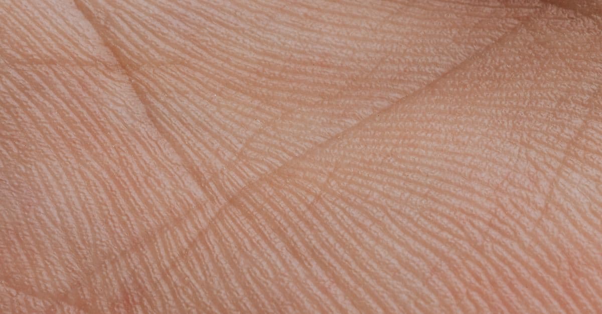
Photo: MART PRODUCTION / Pexels
Touch seems simple. You feel a coffee cup in your hand, the softness of wool, the sting of a pinprick. Yet this apparent simplicity masks one of the most sophisticated sensory systems in the body. Touch is not a single sensation but at least four: light touch, sustained pressure, vibration, and pain. Each is detected by a different type of receptor, each sends signals along different neural pathways, and each occupies distinct territory in the brain. The discovery of how these receptors actually function — how they convert physical pressure into electrical signals — remained unsolved for decades, until 2021 when a Nobel Prize was awarded for the identification of PIEZO channels, the mechanical sensors inside touch neurons. Understanding touch reveals why your fingertips can distinguish textures your back cannot, why you can sense the location of a touch within millimeters, and how your brain maintains a continuous, unconscious map of where every limb is in space.
The Four Touch Modalities and Their Receptors
Touch is not a single sensation but a bundle of related modalities, each mediated by a different sensory receptor optimized for a specific stimulus property. The skin and deeper tissues contain four major types of mechanoreceptors, each sensitive to a distinct frequency or force pattern. Additionally, free nerve endings in the skin detect temperature and pain.
Meissner corpuscles — located in the dermal papillae (the small bumps on the fingertips and palms) and sensitive to light touch and texture discrimination. These receptors are densely concentrated in hairless (glabrous) skin: fingertips, palms, lips, and the soles of feet. They respond best to forces of 0.3 to 3 grams and detect frequencies between 10 and 40 Hz (cycles per second) — a frequency range matching the texture vibrations that occur when a finger slides across a rough surface. Meissner corpuscles are rapidly adapting, meaning they stop firing shortly after stimulation begins. This adaptation is why the sensation of your clothes touching your skin fades within seconds; the receptors have silenced the steady-state signal and fire only when the stimulus changes. Meissner corpuscles are crucial for fine motor control and object recognition through touch alone.
Merkel cells and Merkel disks — located deeper in the epidermis, underlying Meissner corpuscles, and specialized for sustained pressure and edge detection. Unlike Meissner corpuscles, Merkel receptors are slowly adapting, continuing to fire throughout the duration of a sustained stimulus. This allows them to report the presence and location of a constant pressure or the detailed shape of an object edge. Merkel cells have been shown in recent studies to function as mechanoreceptors themselves, not merely as support cells, and they express genes associated with touch transduction. For example, if you hold a comb in your hand, Meissner corpuscles fire during the initial contact and as your finger moves across the texture, while Merkel cells maintain a steady signal encoding the pressure and shape of each tooth edge.
Pacinian corpuscles — large, onion-layered sensory endings located in deep skin layers, fascia, and internal organs, sensitive to vibration and rapid pressure changes. Pacinian corpuscles are exquisitely sensitive to high-frequency vibration, peaking at 200 to 300 Hz. They are rapidly adapting, firing only at the onset and offset of vibration. The anatomy of the Pacinian corpuscle explains this behavior: the receptor nerve terminal is surrounded by concentric layers of connective tissue that dissipate low-frequency stimuli but transmit high-frequency vibrations directly to the nerve ending. This structure acts as a biological high-pass filter. Pacinian corpuscles are responsible for the sensation of buzzing and are used in clinical testing: a vibrating tuning fork held against the skin stimulates Pacinian corpuscles, and loss of this sensation indicates nerve damage.
Ruffini endings — located in the dermis and fascia, sensitive to skin stretch and sustained lateral force. Ruffini endings are slowly adapting and report the direction and magnitude of skin deformation. They are particularly important for detecting hand position during object manipulation and for sensing the orientation of skin folds. Along with Merkel cells, they provide the sustained sensory signal that allows detailed tactile perception and object shape discrimination.
Free nerve endings — unmyelinated or thinly myelinated axons in the skin lacking the specialized receptor structures of mechanoreceptors. Free nerve endings are the primary mediators of temperature sensation and pain. Cold receptors (TRPM8 and TRPA1 channels) respond to temperatures below 25°C. Warm receptors (TRPV3 and TRPV4 channels) respond to temperatures above 30°C. Pain receptors, called nociceptors, respond to noxious mechanical, chemical, or thermal stimuli. C-fiber nociceptors (slow, unmyelinated) conduct at 0.5 to 2 meters per second and mediate the slow, burning pain that follows injury. Aδ-fiber nociceptors (fast, lightly myelinated) conduct at 5 to 30 meters per second and mediate sharp, immediate pain. This dual pain pathway is why you feel a pinprick in two distinct stages: first, a sharp sting, then a delayed, burning sensation.
Source: Abraira, V.E. and Ginty, D.D. (2013), "The Sensory Neurons of Touch," Neuron, Vol. 79 — comprehensive review of mechanoreceptor types, anatomy, and physiological responses.
Source: Eijkelkamp, N. et al. (2013), "Nociceptors: The Sensors of the Pain Pathway," Journal of Clinical Investigation, Vol. 123 — detailed account of temperature and pain receptor physiology.
The PIEZO Channels: How Touch is Transduced into Electrical Signals
For decades, neuroscientists understood the anatomy of mechanoreceptors but not the molecular mechanism by which physical force was converted into electrical signals. In 2021, that mystery was solved with the Nobel Prize in Physiology or Medicine awarded to David Julius and Ardem Patapoutian for the discovery of PIEZO channels.
PIEZO1 and PIEZO2 channels are mechanically gated ion channels — proteins in the nerve cell membrane that open in response to physical deformation. When pressure is applied to a mechanoreceptor, the proteins in the cell membrane are distorted. This mechanical deformation directly opens PIEZO channels, allowing ions (primarily calcium and sodium) to flow into the cell. The influx of ions depolarizes the nerve terminal, triggering an action potential that travels along the axon to the spinal cord. PIEZO channels are so sensitive that they can be opened by membrane tension alone; in some cases, a force as small as a single piconewton (a trillionth of a newton) can open a PIEZO channel.
PIEZO2 is the primary mechanotransducer in touch receptors. Merkel cells, which sense sustained pressure, express high levels of PIEZO2. Meissner corpuscles, which detect light touch, also rely on PIEZO2. The discovery revealed that the transduction process is remarkably direct: mechanical force → PIEZO channel opening → ion influx → action potential. There is no intermediate biochemical cascade; the channel itself is the mechanical sensor.
The structure of PIEZO channels illuminates why they are so sensitive. The channel is a large, three-blade propeller-shaped protein with a dome-shaped central pore. When the membrane is stretched, the blades bend inward, pulling open the pore and allowing ions through. Genetic mutations that prevent PIEZO2 from functioning cause insensitivity to touch, vibration, and proprioception — demonstrating that touch perception is entirely dependent on these channels.
Source: Coste, B. et al. (2010), "Piezo1 and Piezo2 are essential components of distinct mechanically activated cation channels," Science, Vol. 330 — original discovery paper identifying PIEZO1 and PIEZO2 as mechanotransducers.
Source: Patapoutian, A. et al. (2003), "TRP channels and temperature sensation," Nature Reviews Neuroscience, Vol. 4 — foundational work on mechanoreceptor molecular biology, preceding PIEZO discovery.
The Somatosensory Pathway: From Skin to Cortex
The signals generated in mechanoreceptors do not travel directly to the brain. Instead, they follow a stereotyped anatomical pathway with several decision points, each refining the sensory information.
When a mechanoreceptor fires, its axon carries the action potential toward the spinal cord. Sensory axons are organized by body region: the dermatome system divides the body surface into strips, each receiving sensory input from a specific spinal nerve. The dorsal root is the entry point into the spinal cord; sensory neuron cell bodies reside in the dorsal root ganglion just outside the spinal cord.
Once in the spinal cord, sensory axons synapse on second-order neurons in one of two pathways: the dorsal column–medial lemniscus pathway (for touch, vibration, and proprioception) or the spinothalamic tract (for pain and temperature).
The dorsal column–medial lemniscus pathway is the primary route for discriminative touch and vibration. Axons from mechanoreceptors ascend the dorsal columns of the spinal cord, traveling ipsilaterally (on the same side) all the way to the medulla in the brainstem. The pathway is somatotopically organized — axons from the feet are medial (toward the midline), and axons from the arm are lateral. At the medulla, these axons synapse on second-order neurons in the medial lemniscus, which cross the midline (decussate) and ascend to the thalamus. From the thalamus, third-order neurons project to the somatosensory cortex in the postcentral gyrus of the parietal lobe.
The spinothalamic tract handles pain and temperature. Pain axons synapse on second-order neurons in the spinal cord and cross immediately, ascending the opposite side of the spinal cord. This is why a spinal cord lesion causes loss of pain and temperature sensation on the opposite side of the body below the lesion, while touch sensation (which crossed at the medulla) is preserved on the same side — a pattern called Brown-Séquard syndrome.
The somatosensory cortex contains a detailed topographic map of the body's surface: neighboring receptors on the skin project to neighboring neurons in the cortex. This topographic organization preserves the spatial structure of sensory information and allows the brain to localize stimuli. However, the map is not equally magnified across the body. Cortical area is allocated based on sensory importance and receptor density, not body surface area.
Source: Jörntell, H. and Ekerot, C.-F. (2006), "Properties of Somatosensory Synaptic Integration in Cerebellar Granule Cells In Vivo," Journal of Neuroscience, Vol. 26 — anatomical and functional organization of somatosensory pathways.
The Sensory Homunculus: Cortical Magnification of the Fingertips and Lips
In 1937, Canadian neurosurgeon Wilder Penfield discovered something remarkable. While performing brain surgery, he stimulated the somatosensory cortex of awake patients (the brain has no pain receptors, so this is painless) and asked them to report what they felt. By mapping each location and its corresponding sensation, Penfield constructed a map of the human body as represented in the brain. The result was the sensory homunculus — a distorted human figure where body regions are sized according to the cortical area devoted to them.
The homunculus is grotesque: the hands and fingers occupy enormous regions, the lips and mouth even larger, while the trunk and limbs are shrunken. The fingers alone occupy more cortex than the entire back and chest combined. This cortical magnification explains why you can sense two separate touch points on your fingertip when they are only 2 to 3 millimeters apart, while on your back you cannot distinguish two points unless they are 30 to 40 millimeters apart.
Two-point discrimination — the minimum distance at which two simultaneous touches can be felt as two separate stimuli — is a direct consequence of cortical magnification. This threshold depends on the density of mechanoreceptors and, more importantly, on how much cortical area each region occupies. The fingertips have the finest two-point discrimination in the body; the back has the coarsest. This is not because fingertips have more mechanoreceptors per square millimeter (they don't), but because the brain allocates more cortex to processing fingertip signals.
Cortical magnification also explains why finger injuries are perceived as more distressing than injuries to the back — the finger damage disrupts more cortical processing, creating a larger "wound" in the sensory map. Similarly, phantom limb pain (pain felt in a limb that has been amputated) can be alleviated by manipulating the corresponding region on the opposite limb or using a mirror illusion. The brain can reweight its sensory map, reallocating cortical territory.
The homunculus is not fixed from birth. Sensory plasticity — the ability of the cortex to reorganize in response to changing input — allows the homunculus to expand or contract based on experience and use. Musicians who play violin, for instance, have enlarged cortical representations for their left fingers (which are used for precise fingering). Blind individuals have expanded cortical regions for touch, as the somatosensory cortex takes over processing of Braille and tactile information formerly devoted to vision.
| Body Region | Two-Point Discrimination (mm) | Receptor Density (per cm²) | Dominant Receptor Type | Cortical Magnification |
|---|---|---|---|---|
| Fingertip | 2–3 | 240 | Meissner, Merkel | Highest |
| Lips | 5–6 | 200 | Meissner, Merkel | Very high |
| Palm | 8–10 | 120 | Meissner, Merkel | High |
| Forearm | 25–30 | 40 | Meissner, Merkel, Ruffini | Moderate |
| Shoulder | 35–40 | 15 | Merkel, Ruffini | Low |
| Back | 40–50 | 10 | Merkel, Ruffini, Pacinian | Lowest |
Source: Penfield, W. and Boldrey, E. (1937), "Somatic Sensory Representation of the Body in the Sensory and Motor Cortex of Man," Brain, Vol. 60 — classic paper mapping the sensory homunculus through direct brain stimulation in neurosurgical patients.
Source: Duncan, G.H. and Bushnell, M.C. (1994), "Cortical Representation of Pain," Journal of the Neurological Sciences, Vol. 149 — review of cortical plasticity and sensory representation.
Proprioception: The Sixth Sense of Body Position and Movement
While mechanoreceptors in the skin detect touch from the external world, proprioceptors in muscles, tendons, and joints provide the brain with information about limb position and movement in space. Proprioception — literally "sense of self" — allows you to know where your limbs are without looking. Close your eyes and touch your nose with your index finger; proprioception reports your arm's position, and proprioceptors in your hand and neck help estimate the nose's location.
Muscle spindles are sensory organs embedded in skeletal muscle that detect changes in muscle length. A spindle consists of 5 to 15 specialized muscle fibers (intrafusal fibers) surrounded by a connective tissue capsule. When the muscle is stretched, the spindle stretches with it, opening PIEZO and other mechanically gated channels in the sensory nerve ending. The spindle fires in proportion to the stretch rate and magnitude. Spindle signals are used in two ways: they trigger the stretch reflex (a monosynaptic reflex at the spinal cord level that causes muscle contraction), and they provide the brain with proprioceptive information about limb position and movement velocity.
Golgi tendon organs are located at the junction of muscle and tendon and detect the force exerted by the muscle. Unlike muscle spindles, which report length, Golgi organs report tension. When a muscle contracts forcefully, the tendon is pulled and stretched, opening mechanoreceptors in the Golgi organ. This provides the brain with information about how hard a muscle is contracting — the effort required to hold or move an object.
Joint receptors (including Pacinian corpuscles and Ruffini endings in joint capsules) respond to joint angle and stress. These receptors are particularly important at extreme angles of movement and contribute less to proprioception in mid-range positions, where muscle spindle signals dominate.
Proprioceptive signals travel via the same dorsal column pathway as touch information, synapsing on the same second-order neurons. This anatomical convergence means that proprioceptive and touch information are processed together, allowing the brain to integrate limb position with tactile sensory input from the skin.
Proprioceptive deficits are disabling. Patients with loss of proprioceptive feedback (due to spinal cord damage, vitamin B12 deficiency, or certain neuropathies) cannot reliably control limb position or movement without visual feedback. They may be unable to walk smoothly in dim light or to perform fine motor tasks with eyes closed. Rehabilitation of proprioception involves repeated practice and visual feedback, training the remaining sensory modalities to compensate.
Source: Proske, U. and Gandevia, S.C. (2012), "The Proprioceptive Senses: Their Roles in Signaling Body Shape, Body Position and Movement, and Muscle Force," Physiological Reviews, Vol. 92 — comprehensive review of proprioceptive mechanisms and integration with motor control.
Affective Touch and C-Tactile Afferents: Why Touch Feels Good
Not all touch is informational. Some touch is rewarding and emotionally soothing — the experience of being stroked, hugged, or caressed. This affective quality of touch is mediated by a different class of nerve fibers: C-tactile afferents (CTs).
Most mechanoreceptors are innervated by fast-conducting A-beta fibers with diameters of 5 to 12 micrometers. These fibers conduct at 30 to 100 meters per second and mediate discriminative touch — the ability to localize stimuli and perceive fine texture. C-tactile afferents, by contrast, are unmyelinated and only 0.5 to 2 micrometers in diameter, conducting at just 0.5 to 2 meters per second. This slow conduction explains why affective touch sensations are delayed and less localized — the signal takes longer to reach the brain, and the sensory information is less precise.
C-tactile fibers are sensitive to slow, gentle stroking at velocities of 1 to 10 centimeters per second — the speed of a caress. They are not triggered by tapping or rapid movement. Activation of CT fibers triggers activity in the insular cortex and orbitofrontal cortex — areas associated with emotional processing and reward — rather than in the primary somatosensory cortex. This neural distinction explains why a caress feels fundamentally different from a discriminative touch: the brain processes them in entirely different systems.
C-tactile activation also triggers release of oxytocin (the "bonding hormone") and activation of the parasympathetic nervous system, producing feelings of calm and well-being. This may explain the social importance of touch: affective touch provides a direct, neurochemical pathway to emotional reward and social bonding. The loss of affective touch in conditions like autism spectrum disorder or certain forms of neuropathy can impair social connection.
Source: Olausson, H. et al. (2002), "Unmyelinated Tactile Afferents Signal Touch and Payoff," Science, Vol. 297 — discovery that unmyelinated C-tactile fibers mediate affective touch and social bonding.
Sensory Adaptation and Why Touch Fades: Learning to Ignore
When you put on a shirt, you feel the fabric on your skin for a few seconds. Then the sensation fades entirely, even though the shirt remains in contact. This is sensory adaptation — the tendency of sensory systems to reduce their responsiveness to constant stimuli. Adaptation is not a deficit; it is a feature. By suppressing unchanging, uninformative signals, the nervous system can allocate processing resources to novel, behaviorally relevant events.
Adaptation occurs at multiple levels. Peripheral adaptation happens in the receptor itself: rapidly adapting mechanoreceptors (Meissner and Pacinian corpuscles) fire vigorously when a stimulus is first applied, then stop firing even if the stimulus persists. This is because the ion channels in these receptors inactivate — they close spontaneously after opening briefly. Slowly adapting receptors (Merkel and Ruffini) maintain their firing longer but still show some decrease in firing rate over time.
Central adaptation occurs in the brain. Even if a peripheral receptor were to maintain its firing, the spinal cord and cortical neurons reduce their responsiveness to constant, predictable inputs. This is called neural adaptation and reflects a reduction in synaptic transmission and a shift in the frequency response of neurons toward higher-frequency stimuli.
Adaptation is why a sudden poke through clothing is startling (a novel stimulus), but the continuous pressure of the fabric becomes imperceptible. Similarly, light touches are detected readily (they trigger rapidly adapting receptors), while sustained pressure fades into the background (slowly adapting receptors show drift, and central adaptation suppresses the signal further).
The functional benefit is clear: the nervous system can ignore maintained, predictable sensory input and focus on changes. This allows attention to remain available for new information in the environment.
Referred Pain and Cortical Plasticity in Chronic Pain
Pain can originate from internal organs and be felt in remote locations on the skin — a phenomenon called referred pain. For example, a heart attack commonly causes pain in the left arm and shoulder, not in the chest near the heart. This occurs because the pain signals from the heart and the pain signals from the skin of the arm converge on the same second-order neurons in the spinal cord. The brain, which evolved in a world where skin pain was common and visceral pain rare, interprets the signal as coming from the skin.
Chronic pain alters the somatosensory cortex. In patients with chronic pain syndromes, the cortical map becomes distorted: the painful region may expand or shrivel, and the boundaries between body regions may blur. This cortical reorganization can outlast the underlying injury; even after tissue healing, the cortical distortion persists, maintaining the pain experience. Some rehabilitation approaches, such as graded motor imagery and mirror therapy, attempt to reverse this cortical reorganization by gradually retraining the brain's body map. Remarkably, these non-pharmacological interventions can be highly effective at reducing chronic pain.
The takeaway
Touch is a unified sensory system built from multiple specialized receptor types, each tuned to detect a different stimulus modality and frequency. The discovery of PIEZO channels revealed that these receptors use a direct, mechanical transduction mechanism — the pressure itself opens ion channels. Signals ascend the spinal cord in somatotopically organized pathways, reach the thalamus, and project to the somatosensory cortex, where body regions are represented as a distorted map that reflects cortical importance, not body surface area. Proprioceptors in muscles and joints add continuous awareness of limb position, a sense so fundamental that its loss is profoundly disabling. Adaptation ensures that unchanging tactile signals fade, keeping the nervous system responsive to novelty. Understanding touch reveals how the brain constructs the sense of self — the awareness of being a bounded, embodied creature with localized sensations and voluntary movement.
Frequently Asked Questions
Q: Why are fingertips so much more sensitive than other parts of the body?
A: Fingertips have a much higher density of mechanoreceptors than most skin regions, but the major factor is cortical magnification — the somatosensory cortex devotes a disproportionately large area to processing signals from the fingers. This enlarged cortical representation allows finer discrimination of touch (two-point discrimination of 2–3 mm on the fingertip versus 40 mm on the back), higher acuity, and more precise motor control. The fingers are among the most behaviorally important body parts, so evolution allocated them substantial neural processing power.
Q: What does it mean if you lose the ability to feel vibration?
A: Loss of vibration sense indicates damage to large-diameter sensory axons in the dorsal columns of the spinal cord or to the peripheral nerves supplying those axons. This commonly occurs in vitamin B12 deficiency, diabetes (peripheral neuropathy), or spinal cord disorders. Vibration is detected by Pacinian corpuscles deep in the skin and is tested clinically with a vibrating tuning fork. Loss of vibration sense often occurs before loss of light touch and is an early sign of spinal cord or nerve damage.
Q: Can you be born insensitive to touch?
A: Yes, though it is extremely rare. Congenital insensitivity to pain and temperature (CIPA) is a genetic disorder in which TRPV1 channels (heat-sensing nociceptors) are non-functional, rendering the person unable to feel pain or extreme heat. There are also rare cases of PIEZO2 gene mutations that cause loss of mechanoreceptor function, resulting in inability to feel touch or vibration. People with these conditions are in grave danger from undetected injuries and infections, as pain is a crucial warning signal of tissue damage.
Q: Why does a massage feel good, even though it is technically tissue damage?
A: Massage activates C-tactile afferents (unmyelinated fibers that respond to slow, gentle stroking), triggering the insular cortex and reward pathways in the brain. This produces release of oxytocin and activation of the parasympathetic nervous system, creating feelings of calm and well-being. Additionally, massage increases blood flow and reduces muscle tension, providing genuine physiological benefits. The rewarding sensation is distinct from the pain produced by tissue damage; the brain interprets the input as affective touch rather than nociceptive pain.
Q: How do phantom limbs fit into the somatosensory system?
A: After amputation, the somatosensory cortex region representing the missing limb remains intact and active. If the cortex receives input from intact adjacent body parts (the stump, or sensory signals misrouted to neighboring regions), it interprets that input as coming from the phantom limb. This explains why phantom pain can be relieved by stimulating the stump or by mirror therapy (watching the intact limb move in a mirror, creating the visual illusion that the phantom limb is moving). The brain is essentially hallucinating sensory input that matches the missing body part.
Q: What are PIEZO channels and why did they win a Nobel Prize?
A: PIEZO channels are mechanically gated ion channels in the cell membrane that open directly in response to physical force or pressure. Their discovery in 2010 and characterization in the years following solved a century-old mystery: how do nerve cells convert mechanical stimuli into electrical signals? PIEZO2 is the primary mechanotransducer in touch receptors, temperature sensors, and proprioceptors. The 2021 Nobel Prize was awarded to Ardem Patapoutian and David Julius for discovering PIEZO channels and TRP temperature-sensing channels, which are now recognized as the fundamental molecular basis of touch and temperature sensation.
Read next:
Follow new articles
Add SciHub101 to your feed reader to see new science and technology stories.
About the Author
SciHub101 is an independent science and technology publication founded and edited by Buğra Sözeri, a software engineer and app developer based in Türkiye. AI tools assist with drafting and research. Source documentation in the existing archive is uneven, and articles are being reviewed and corrected. The Editorial Team byline identifies editorial responsibility; it does not certify specialist or independent human review. Where we compare products, anything built by our own team is labelled as such — see the editorial guidelines and About page for how corrections and disclosures work.
Related Articles

Why Do We Feel Pain? The Science of Nociception
Pain is not a sensation but a perception — the brain's interpretation of a threat signal from specialized nerve endings called nociceptors. When you touch a hot stove, nociceptors detect the temperature change and fire electrical signals up the spinal cord to the brain. The brain then constructs the feeling of pain, which is why you can feel pain from a phantom limb that no longer exists, or feel no pain during surgery under anesthesia despite injury. Pain serves a critical protective function, preventing injury, but when it persists beyond healing (chronic pain), it becomes a malfunction of the nervous system.

How the Body Processes Alcohol: The Science of Metabolism and Intoxication
Alcohol is absorbed into the bloodstream within minutes of consumption, but the liver metabolizes it at a nearly constant rate of about one standard drink per hour, a rate that cannot be accelerated by food or hydration. The body treats ethanol as a toxin, converting it to acetaldehyde (more toxic than ethanol) and then to acetate through two enzyme-mediated reactions. Blood alcohol concentration (BAC) depends on body weight, sex, food intake, and genetics, making some individuals metabolically unable to process alcohol efficiently, as seen in alcohol flush syndrome. Once intoxicated, alcohol disrupts the balance of GABA and glutamate neurotransmitters in the brain, impairing judgment and coordination. Understanding alcohol metabolism reveals why hangovers occur, why some populations have lower tolerance, and why no trick can accelerate the clearance of alcohol from the body.
Why Do We Have Fingerprints? The Science
Fingerprints exist primarily to improve grip and amplify fine touch. The ridged skin on your fingertips enhances friction against smooth surfaces and channels vibrations toward specialized nerve endings — but the exact patterns those ridges form are shaped by a reaction-diffusion process that plays out in the womb, governed by competing molecular signals that no two people, not even identical twins, experience in quite the same way.

Why Do We Get Thirsty? The Science of Thirst and Fluid Balance
Thirst is not simply a desire for water—it is a complex neural signal triggered by osmoreceptors in the brain that detect changes in blood salt concentration and osmotic pressure. When you lose even 1 percent of your body's water, osmoreceptors in the hypothalamus fire, signaling the anterior pituitary to release vasopressin (ADH), which forces your kidneys to retain water and triggers the conscious sensation of thirst. Remarkably, your brain also drinks before it needs to, through anticipatory thirst, based on learned cues and past dehydration experiences. Understanding thirst reveals how your body maintains the precise electrolyte balance required for every cell to function, and why the myth of '8 glasses a day' is neurobiologically inaccurate.

How Does the Sense of Balance Work? The Vestibular System Explained
Balance is not a single sense but a symphony of three systems working in concert: the vestibular system (inner ear acceleration sensors), proprioception (body position awareness), and vision. The vestibular system contains the semicircular canals (which detect rotation) and the otolith organs (which detect linear acceleration and head tilt). Damage to any component creates the sensation of spinning, nausea, and falls. Understanding how the brain fuses these signals explains vertigo, motion sickness, and why astronauts become disoriented in microgravity where the vestibular system receives no gravitational input.

How We Taste and Smell: The Science of Flavor, 400 Odor Receptors, and the Nose-Brain Connection
Pinch your nose while eating a strawberry and the flavor nearly disappears — what remains is just sweetness with a hint of acid. Flavor, it turns out, is about 80% smell. The tongue detects only five basic tastes; the nose handles everything else through 400 distinct olfactory receptors, making smell the most molecularly diverse sensory system in the human body. Here is how these two ancient senses combine to produce the richness of what we call taste.