Why Do We Get Thirsty? The Science of Thirst and Fluid Balance

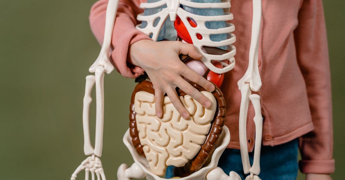
Photo: Gustavo Fring / Pexels
Every human experiences the sensation of thirst, yet few understand that thirst is not a passive request for water but an active, sophisticated neural command issued by osmoreceptors embedded in your brain. The moment your blood becomes just 1 to 2 percent more concentrated in salt, specialized sensory cells called osmoreceptors detect this change and trigger a cascade of neural and hormonal signals that culminate in your conscious desire to drink. This system is so precise that it maintains your blood's salt concentration within a narrow range—between 135 and 145 millimoles per liter—which is essential for muscle contraction, heart rhythm, nerve firing, and every other function that depends on cellular electrolyte balance. Yet your brain does not simply react to dehydration; it also anticipates it, drinking water before your blood osmolarity actually rises, based on learned cues and past experience. Understanding thirst requires understanding osmoregulation, the role of hormones like vasopressin, and the remarkable plasticity of the brain's thirst circuits.
What Is Thirst: A Neural Signal, Not a Desire
Thirst — the conscious desire to drink water, triggered by osmoreceptors that detect changes in blood osmolarity or baroreceptors that detect blood volume loss. Thirst is a homeostatic drive, meaning it is triggered by deviation from a physiological set point and motivates behavior to restore balance.
Thirst is often confused with simple dehydration. Dehydration is an objective decrease in the body's water content; thirst is the subjective sensation that motivates you to restore that water. The two do not always coincide. You can be mildly dehydrated and feel no thirst—athletes in ultramarathons, for instance, sometimes fail to drink enough and develop dangerous dehydration without feeling thirsty, because the intense focus on running overrides the thirst signal. Conversely, you can feel thirsty without being dehydrated—drinking cold soda activates taste receptors and throat sensations that trigger a mild thirst sensation even if your body's water balance is already adequate.
Osmolarity — the total concentration of dissolved particles (solutes, primarily sodium, potassium, chloride, and glucose) in a solution, measured in millimoles per kilogram of solvent (mOsm/kg). Normal blood osmolarity in humans is approximately 290 mOsm/kg, maintained within a range of 285 to 295 mOsm/kg.
The regulation of osmolarity is the primary driver of thirst. Your blood is a solution of water and dissolved particles. As you sweat, urinate, or breathe, you lose water. As you eat salty foods or fail to drink, your blood becomes more concentrated in sodium and other electrolytes. The osmotic pressure of blood—the force with which dissolved particles pull water out of cells—changes. If osmolarity rises above the set point of 290 mOsm/kg, your brain detects this change within minutes and generates thirst. This is why eating salty food immediately triggers thirst; the salt raises blood osmolarity, and osmoreceptors detect the change.
Source: Thrasher, T.N. (1994), "Osmoreception and thirst," Journal of the American Society of Nephrology, Vol. 5 — foundational paper on osmotic regulation of thirst and the role of osmoreceptors.
Osmoreceptors: The Hypothalamus's Salt Sensors
Osmoreceptors are neurons in the hypothalamus—a region at the base of the brain—that respond to changes in osmolarity. Unlike other sensory neurons that respond to external stimuli like light, sound, or touch, osmoreceptors are specialized to sense the internal environment: the concentration of salts in the blood.
The primary osmoreceptors are located in two regions of the hypothalamus: the supraoptic nucleus and the paraventricular nucleus. These regions contain magnocellular neurons (literally "large-celled" neurons) that have dendrites and soma exposed directly to the cerebrospinal fluid that bathes the brain. The cerebrospinal fluid is in osmotic equilibrium with the blood, so its osmolarity changes in parallel with blood osmolarity.
When osmolarity increases, water moves out of the osmoreceptor neurons by osmosis (water molecules move toward higher salt concentration). This loss of water causes the neuron to shrink slightly. The shrinkage activates stretch-sensitive ion channels in the cell membrane, which allow ions to enter the cell. This causes the neuron to depolarize (become electrically excited) and fire action potentials. Increased firing of these osmoreceptor neurons triggers the release of hormones and the conscious sensation of thirst.
The sensitivity of osmoreceptors is remarkable. The threshold for thirst is typically reached when osmolarity exceeds 290 mOsm/kg by just a few units. A person can detect the difference between a 2 mOsm/kg increase and no increase—a sensitivity equivalent to detecting one grain of salt added to approximately 10 kilograms of water.
Source: Bourque, C.W. (2008), "Central mechanisms of osmosensation and systemic osmoregulation," Nature Reviews Neuroscience, Vol. 9 — modern review of osmoreceptor mechanisms and neural osmoregulation.
Vasopressin (ADH) and the Kidney: The Hormonal Response to Thirst
When osmoreceptors in the hypothalamus detect increased osmolarity, they trigger the release of a hormone called vasopressin, also known as antidiuretic hormone or ADH. Vasopressin is synthesized in the same neurons that are osmoreceptors—the magnocellular neurons of the supraoptic and paraventricular nuclei. When these neurons fire in response to osmoreceptor activation, they release vasopressin into the bloodstream from the posterior pituitary gland.
Vasopressin acts on the kidneys, specifically on the collecting duct—the final segment of the renal tubule where the fine-tuning of water reabsorption occurs. In the absence of vasopressin, the collecting duct is impermeable to water; large amounts of dilute urine are produced, and the body loses water. When vasopressin binds to receptors on the collecting duct cells, it triggers the insertion of aquaporin water channels into the cell membrane. Aquaporins are protein pores that allow water to pass through the cell membrane rapidly. With aquaporins in place, water moves back into the blood from the urine via osmosis, and the urine becomes concentrated.
Aquaporins — water channel proteins embedded in cell membranes that allow water molecules to cross the membrane rapidly, down their osmotic gradient. Aquaporins are essential for concentrating urine and reabsorbing water in the kidneys.
This system operates with remarkable precision. A 1 percent increase in blood osmolarity triggers enough vasopressin release to concentrate the urine nearly 10-fold. The concentrated urine reduces water loss, and the body retains water, which dilutes the blood back toward the set point. As blood osmolarity falls back toward 290 mOsm/kg, osmoreceptor firing decreases, vasopressin release drops, aquaporins are removed from the collecting duct, and the body produces dilute urine again to eliminate excess water.
The relationship between osmolarity and vasopressin is so tightly coupled that it serves as one of the most important homeostatic mechanisms in the body. Dysfunction in this system leads to disease. In diabetes insipidus (not related to diabetes mellitus), either the osmoreceptor neurons fail to produce vasopressin (central diabetes insipidus) or the kidneys fail to respond to vasopressin (nephrogenic diabetes insipidus). Either way, the body loses the ability to concentrate urine, and patients can lose 10 to 15 liters of dilute urine per day, leading to severe dehydration unless they drink constantly.
Source: Robertson, G.L. (2006), "Antidiuretic hormone: normal and disordered function," Endocrinology and Metabolism Clinics of North America, Vol. 35 — clinical review of vasopressin physiology and dysfunction.
| Osmolarity Level | Vasopressin Release | Urine Output | Thirst Sensation |
|---|---|---|---|
| 280-285 mOsm/kg | Minimal | High (dilute) | Absent |
| 285-290 mOsm/kg | Low | Moderate | Absent |
| 290-295 mOsm/kg | Increasing | Low | Mild |
| 295-300 mOsm/kg | High | Very low (concentrated) | Strong |
| >300 mOsm/kg | Maximum | Minimal | Intense |
Source: Montani, J.P. et al. (2001), "Water intake and disease," Journal of the American Society of Nephrology, Vol. 12 — systematic review of osmoregulation and disease states.
The Subfornical Organ and Lamina Terminalis: The Conscious Experience of Thirst
The osmoreceptors in the hypothalamus drive the automatic hormonal response—vasopressin release and kidney water reabsorption—but this alone does not explain why you consciously feel thirsty. The conscious sensation of thirst requires signals to reach the cerebral cortex and limbic regions involved in motivation and behavior. Two additional structures play a critical role: the subfornical organ and the lamina terminalis.
The subfornical organ (SFO) is a small, circumventricular organ (a region of the brain outside the blood-brain barrier) located along the midline of the brain, above the fornix. Unlike most of the brain, the SFO is exposed to the general circulation and can sense blood osmolarity directly, without relying on cerebrospinal fluid. The SFO contains osmoreceptive neurons and also receives signals from the hypothalamic osmoreceptors. Neurons in the SFO project to the median preoptic nucleus and other forebrain regions that encode thirst as a conscious drive.
The lamina terminalis is a thin membrane of tissue at the boundary between the third ventricle (cerebrospinal fluid space) and the brain tissue, containing osmoreceptors and neurons that project to thirst-relevant brain regions. The lamina terminalis, like the SFO, can sense osmolarity changes and integrates osmotic information to generate thirst drives.
These structures do not work in isolation. The SFO and lamina terminalis integrate signals from peripheral baroreceptors (which detect blood volume loss) and taste receptors (which detect sodium concentration in food and water), combining these diverse signals into a unified thirst drive. The result is that thirst reflects not just osmolarity, but also blood volume status and the types of fluids available to drink.
This distributed organization means that thirst is not a simple on-off signal but a graded, integrative process. Mild increases in osmolarity trigger mild thirst; large increases trigger intense thirst. A small loss of blood volume (such as from modest sweating) produces thirst, but not as much as the same degree of osmolarity increase, because volume loss is partially compensated by the baroreceptor system, which triggers vasoconstriction to maintain blood pressure.
Source: Johnson, A.K. and Thunhorst, R.L. (1997), "The neuroendocrinology of thirst and salt appetite," Progress in Psychobiology and Physiological Psychology, Vol. 17 — foundational paper on the integration of osmotic and volume-based thirst signals.
Blood Volume and Baroreceptors: The Second Thirst Signal
Osmoreceptors detect changes in water concentration; baroreceptors detect changes in blood volume. Although osmoreceptors are the dominant trigger for thirst in daily life, baroreceptors provide an important secondary signal that becomes critical during severe dehydration, hemorrhage, or excessive sweating.
Baroreceptors — stretch-sensitive mechanoreceptors in the carotid arteries and aorta that detect changes in blood pressure and, indirectly, blood volume. When blood volume decreases, blood pressure drops, and baroreceptors fire less frequently.
Baroreceptors are located in the carotid sinuses (dilated regions of the carotid arteries in the neck) and the aortic arch. These receptors fire continuously when blood pressure is normal; their firing rate encodes the pressure. When blood pressure drops—as occurs during blood loss or severe dehydration—baroreceptor firing rate decreases. This drop in baroreceptor signal is relayed to the brainstem and forebrain, triggering not just vasopressin release, but also thirst.
The threshold for baroreceptor-driven thirst is higher than the threshold for osmoreceptor-driven thirst. A 10 percent loss of blood volume (approximately 500 milliliters in a 70-kilogram adult) is needed to strongly activate baroreceptor-driven thirst, whereas a 1 to 2 percent increase in osmolarity (equivalent to losing only 500 milliliters of pure water) activates osmoreceptor-driven thirst. This asymmetry makes biological sense: osmolarity changes quickly and constantly (from eating salty food, from sweating), so the brain needs a sensitive osmoreceptor system to maintain water balance continuously. Blood volume changes more slowly (from hemorrhage or excessive sweating) and are initially compensated by other mechanisms (vasoconstriction, reduced kidney filtration), so baroreceptor-driven thirst is a backup system that activates when compensatory mechanisms are overwhelmed.
During exercise in hot conditions, both osmoreceptor and baroreceptor signals are engaged. Sweating causes both water loss (which raises osmolarity) and volume loss (which lowers blood pressure and baroreceptor firing). The combined signals produce intense thirst, which motivates the person to drink. This is adaptive: the goal is to restore both osmolarity and blood volume.
Source: Fitzsimons, J.T. (1998), "Angiotensin, thirst, and sodium appetite," Physiological Reviews, Vol. 78 — comprehensive review of baroreceptor-driven thirst and the role of the renin-angiotensin system.
The Renin-Angiotensin-Aldosterone System (RAAS): Thirst and Sodium Regulation
The kidneys have another way to detect blood volume loss besides the nervous system: they sense the drop in blood pressure directly. When renal perfusion pressure (the pressure at which blood enters the kidney) drops, the kidney's juxtaglomerular cells release an enzyme called renin. Renin enters the bloodstream and catalyzes a reaction that produces angiotensin II, a potent hormone.
Angiotensin II acts on multiple targets to restore blood pressure and blood volume. In the kidneys, it increases sodium reabsorption. In blood vessels, it causes vasoconstriction. But critically for thirst, angiotensin II is detected by receptors on neurons in the SFO and other circumventricular organs, directly triggering thirst. Angiotensin II is one of the most powerful thirst stimuli known; a small increase in angiotensin II produces intense thirst, often stronger than the thirst produced by an equivalent degree of osmolarity increase.
The RAAS connects thirst to sodium homeostasis. When blood volume drops, the kidneys release renin, which produces angiotensin II, which triggers thirst and also increases sodium reabsorption in the kidneys. When you drink in response to angiotensin II-driven thirst, you ingest not just water but usually some sodium (from food, saliva, or electrolyte-containing drinks). This restores both blood volume and blood sodium, addressing the underlying deficit that triggered the RAAS in the first place.
In pathological conditions, the RAAS can become overactive. In heart failure, the heart pumps less effectively, and the kidneys interpret the drop in renal perfusion as a signal that blood volume is inadequate. The RAAS activates chronically, releasing renin and angiotensin II, which trigger both intense thirst and sodium and water retention. Patients with heart failure drink excessively and retain salt and water, further overloading the failing heart. This is why heart failure patients are often advised to restrict salt intake and fluid intake, to prevent their activated RAAS from making the heart's burden worse.
Source: Epstein, F.H. (1992), "Cardiovascular disease and kidney disease," New England Journal of Medicine, Vol. 326 — clinical review of RAAS dysregulation in disease.
Anticipatory Thirst: Drinking Before You Need To
One of the most striking aspects of human thirst is that it is not purely reactive—responding to osmolarity or volume changes after they occur—but also anticipatory, occurring before your physiology has actually changed. You drink water before a run, even if your blood osmolarity is normal. You drink when you see others drinking. You drink out of habit at certain times of day. These behaviors are not explained by osmoreceptors detecting a change in blood osmolarity; they are driven by learned cues and expectations.
Anticipatory thirst operates through associative learning. Your brain learns which behaviors lead to dehydration and which environmental cues precede it. When you see running shoes, or when it is hot outside, or when it is time for your regular exercise session, your brain's thirst circuits activate, not because osmolarity has changed yet, but because your brain has learned that these cues precede osmolarity changes. This is why people who exercise regularly and have experienced dehydration develop stronger anticipatory thirst before exercise than people who do not exercise—their brains have learned to predict the osmolarity change and activate drinking behavior in advance.
Recent neuroimaging studies have shown that anticipatory thirst involves different brain regions than osmoreceptor-driven thirst. Osmoreceptor-driven thirst activates the median preoptic nucleus and insular cortex—regions that encode interoceptive (internal bodily) states. Anticipatory thirst additionally activates prefrontal cortex regions involved in learning, prediction, and decision-making. The brain is generating an internal model of the future and initiating drinking behavior based on its prediction of what will happen, not based on what has happened.
This anticipatory system is adaptive but can also go awry. Habit drinkers—people who drink large amounts of water out of learned habit—sometimes drink so much that they develop hyponatremia (dangerously low blood sodium), a condition that osmoreceptors' negative feedback system should prevent. The osmoreceptors do sense the drop in osmolarity and should suppress thirst; in normal conditions, they do. But in habit drinkers, the learned association between cues and drinking is so strong that it overrides osmoreceptor signals, and they continue drinking despite dropping blood osmolarity. This demonstrates that anticipatory thirst is mediated by learned associations and can compete with homeostatic osmoreceptor signals.
Source: Johnson, A.K. (2007), "The sensory psychobiology of thirst and salt appetite," Medicinal Research Reviews, Vol. 27 — review of learned aspects of thirst and osmoregulation.
The "8 Glasses a Day" Myth and Individual Variability
The often-cited recommendation to drink eight 8-ounce glasses of water per day—the "8x8" rule—has no strong physiological basis and does not account for individual variation in water needs. This recommendation likely originated from a 1945 suggestion by the U.S. Food and Nutrition Board, stating that the adequate daily water intake is 2.5 liters, of which about 1.5 liters should come from drinking water and 1 liter from food. This was misinterpreted as a recommendation to drink 8 glasses of plain water daily.
In reality, water needs vary dramatically depending on body size, activity level, climate, diet, and age. A 50-kilogram woman sitting in an air-conditioned office has much lower water needs than an 90-kilogram man running in the heat. A person eating a diet rich in fruits and vegetables, which contain substantial water, needs less water from drinking than a person eating a diet of dry foods. Individual thirst sensation is exquisitely tuned to these differences; the osmoreceptor system adjusts vasopressin release and thirst to match actual water losses from sweat, urine, respiration, and skin evaporation.
Modern physiological evidence suggests that the best approach to hydration is to drink according to thirst. In healthy individuals, thirst is a reliable signal of when water is needed. The osmoreceptor system maintains blood osmolarity within a narrow range even when water intake varies widely; people who drink very little produce concentrated urine, and people who drink large amounts produce dilute urine. The kidneys' ability to adjust urine concentration means that as long as you drink when thirsty and have access to water, your osmolarity will be maintained.
There are exceptions. Athletes exercising in hot conditions can lose fluid faster than thirst signals promote drinking, and they may become dehydrated despite active thirst. This is called the "exercise-induced dehydration paradox"—the same intensity of thirst that protects you during rest inadequately protects you during intense exercise. This is because intense exercise and heat stress activate multiple competing drives (to continue running, to focus on the goal) that can override thirst. Additionally, in elderly people, thirst sensation declines—osmoreceptors remain functional, but connections to thirst-promoting brain regions may weaken, or the osmoreceptor threshold may increase, leaving elderly people at higher risk of dehydration if they rely solely on thirst to guide drinking.
Source: Sawka, M.N. et al. (2007), "American College of Sports Medicine position stand: exercise and fluid replacement," Medicine and Science in Sports and Exercise, Vol. 39 — guideline review of hydration needs in exercise, noting that thirst alone is inadequate in intense exercise.
Dehydration: Osmoreceptor Overload and Cellular Dysfunction
Dehydration occurs when water loss exceeds water intake, leading to an increase in blood osmolarity above the normal range of 290 mOsm/kg. Even mild dehydration—a 1 to 2 percent loss of body water—triggers osmoreceptor-driven thirst and vasopressin release, concentrating the urine to conserve water. But if water loss continues, blood osmolarity rises further, and cells begin to suffer.
When blood osmolarity rises above approximately 300 mOsm/kg, water moves out of cells by osmosis, including neurons in the brain. This cell shrinkage can damage cell membranes and disrupt cellular machinery. Additionally, high osmolarity affects enzyme function; many enzymes evolved to operate at the normal osmolarity of 290 mOsm/kg, and their activity declines when osmolarity deviates too far from this set point.
In moderate to severe dehydration (a loss of 5 to 10 percent of body water), symptoms appear: thirst becomes intense, the mouth and throat become dry, urine becomes dark concentrated, heart rate increases, blood pressure may drop, and cognitive function may decline. The cognitive decline occurs because neurons in the prefrontal cortex and other regions involved in decision-making are particularly sensitive to osmotic stress. People with moderate dehydration often have difficulty concentrating, exhibit slowed reaction times, and make poor decisions—all while osmoreceptors are firing maximally, trying to drive the person to drink.
In severe dehydration (a loss of 15 percent or more of body water), the body enters a crisis state. Blood osmolarity can exceed 310 mOsm/kg, well above the osmoreceptor set point, yet the body has lost so much water that urine output nearly ceases and vasopressin reaches maximum levels. Organ systems begin to fail: the kidneys can no longer filter waste, the heart struggles to pump, and the risk of stroke or seizure rises sharply. Severe dehydration is a medical emergency. In the context of heat stress or illness, dehydration can be fatal.
Notably, the brain regulates temperature alongside fluid balance—excessive heat loss (from sweating) that leads to dehydration also affects the hypothalamus's ability to regulate core body temperature, creating a double threat.
Source: Cheuvront, S.N. et al. (2010), "Dehydration and cognitive performance," Journal of the American College of Nutrition, Vol. 29 — systematic review of dehydration's effects on cognitive function and exercise performance.
Overhydration and Hyponatremia: When Thirst Goes Wrong
While dehydration is a common concern, the opposite problem—overhydration or hyperosmolarity-driven overdrinking—can also occur and is often more dangerous because it is less expected. Hyponatremia is a condition in which blood sodium concentration drops below 135 millimoles per liter (mEq/L), typically caused by drinking too much water or by the kidneys' inability to excrete water.
When a person drinks excessive amounts of water, blood osmolarity drops below the normal set point of 290 mOsm/kg. Osmoreceptors sense this drop and suppress vasopressin release; the kidneys respond by producing large amounts of dilute urine, excreting the excess water and restoring osmolarity. This negative feedback system is so effective that it is rare for a healthy person with access to water and a functioning kidney to develop hyponatremia from drinking alone.
However, hyponatremia can develop in specific conditions. In the syndrome of inappropriate antidiuretic hormone (SIADH), the posterior pituitary releases vasopressin even when osmolarity is low, preventing the kidneys from excreting the water you drink. This can occur in lung cancer, CNS infections, medications, or other conditions affecting the hypothalamic-pituitary system. Patients with SIADH drink normally but cannot excrete water, leading to hyponatremia.
Additionally, in endurance athletes, particularly in marathons, some runners drink large amounts of water and hypotonic (low-salt) electrolyte drinks to prevent dehydration. If they drink more than they lose through sweat, they develop hyponatremia. Symptoms include nausea, confusion, headache, and in severe cases, seizures, coma, and death. This is called exercise-associated hyponatremia.
The danger of hyponatremia is that it affects the brain. Just as cells swell and function declines during hypernatremia (high sodium), cells swell and function declines during hyponatremia. Neurons in the brain swell, intracranial pressure rises, and seizures or brain herniation can result. Unlike mild hyponatremia in some organ systems, cerebral hyponatremia is a medical emergency. Treatment requires carefully raising blood sodium by infusing hypertonic saline, not by restricting water (which would delay correction and worsen brain edema).
The existence of hyponatremia shows that the osmoregulatory system is not foolproof; it assumes that osmoreceptors function properly and that the kidneys respond to vasopressin. When these assumptions are violated, overhydration can occur.
Source: Verbalis, J.G. (2003), "Disorders of body water homeostasis," Best Practice and Research Clinical Endocrinology and Metabolism, Vol. 17 — comprehensive clinical review of hyponatremia and water balance disorders.
The Takeaway
Thirst is not a simple appetite or a mere suggestion to drink; it is a precise homeostatic drive generated by osmoreceptors in the hypothalamus, subfornical organ, and lamina terminalis that sense blood osmolarity with remarkable sensitivity. When your blood becomes just 1 to 2 percent more concentrated in salt, osmoreceptors fire and trigger the release of vasopressin, which forces your kidneys to reabsorb water and concentrate your urine, while simultaneously generating the conscious sensation of thirst. This system is so effective that it maintains your blood osmolarity within a tight range despite dramatic variations in water intake and loss. Yet your thirst is not purely reactive; it is also anticipatory, shaped by learned cues and past experience, allowing your brain to predict and prevent osmolarity changes before they occur. Baroreceptors and the renin-angiotensin system add secondary signals that account for blood volume loss. The "8 glasses a day" myth overlooks the individual variability in water needs that your osmoreceptor system elegantly manages. Both dehydration and overhydration can be dangerous, though osmoreceptors have evolved to prevent both in healthy individuals with access to water. Understanding thirst reveals how your nervous system maintains the precise electrolyte balance on which every cell depends.
Frequently Asked Questions
Q: Can you drink too much water?
A: Yes. Drinking excessive water, especially when combined with conditions that prevent normal water excretion (such as SIADH or certain medications), can lead to hyponatremia—a dangerous drop in blood sodium. Endurance athletes who drink large amounts of hypotonic fluids during prolonged exercise sometimes develop exercise-associated hyponatremia, causing seizures or death. In healthy individuals with normal kidney function, the osmoreceptor system prevents overhydration by suppressing vasopressin when blood osmolarity drops, allowing the kidneys to excrete excess water and restore osmolarity.
Q: Why do you feel thirsty even when drinking water?
A: Drinking water temporarily cools your mouth and throat and activates swallowing, which provides immediate sensory relief. However, osmoreceptors in the hypothalamus do not sense mouth dryness directly; they sense blood osmolarity. The osmolarity change from drinking water takes 10 to 15 minutes to reach osmoreceptors in the brain, so you feel the immediate sensation of relief from mouth/throat stimulation long before osmoreceptors sense that your blood osmolarity has dropped and thirst should cease. This delay is why people often stop drinking before they have fully rehydrated.
Q: Is thirst reduced in older adults?
A: Yes. Thirst sensation declines with age, even in healthy older adults. The osmoreceptor neurons remain functional, but connections to brain regions that generate conscious thirst perception may weaken, or the osmoreceptor threshold may shift higher, requiring a greater degree of osmolarity increase to trigger thirst. This puts older adults at higher risk of chronic dehydration and its complications, including cognitive impairment, kidney dysfunction, and falls. Older adults cannot rely solely on thirst to guide drinking; they need conscious strategies such as drinking water at regular intervals.
Q: Does caffeine or alcohol increase thirst or dehydration?
A: Both can increase urine output and potentially lead to dehydration if not compensated by increased drinking, but through different mechanisms. Caffeine is a mild diuretic that increases kidney filtration rate, and alcohol suppresses vasopressin release, preventing the kidneys from reabsorbing water. Both lead to dilute urine and increased water loss. However, the degree of diuresis is modest; moderate caffeine or alcohol intake does not necessarily cause net dehydration if a person drinks water otherwise normally. Thirst mechanisms generally compensate for these effects, though they do so imperfectly.
Q: Why do you feel thirsty after eating salty food?
A: Eating salty food increases blood sodium concentration, which raises blood osmolarity almost immediately. Osmoreceptors detect this rise in osmolarity within minutes and trigger both vasopressin release (to increase water reabsorption in the kidneys) and the sensation of thirst. The thirst drives you to drink water, which dilutes the blood and lowers osmolarity back to normal. This is an elegant system: eating salt does not dangerously raise blood osmolarity because thirst ensures you drink water to restore balance.
Q: What is "cotton mouth" or dry mouth sensation?
A: Dry mouth sensation is the detection of reduced saliva and moisture in the mouth and throat by sensory receptors in the oral mucosa and pharynx. Dry mouth can be caused by dehydration (osmoreceptors are signaling thirst and the body is reducing saliva production to conserve water), but it can also result from medications (antihistamines, antidepressants, anticholinergics), autoimmune disorders affecting salivary glands, or simply from mouth breathing or low humidity. Dry mouth is not the same as thirst; a person can have a dry mouth without osmoreceptor-driven thirst, or have genuine thirst (high blood osmolarity) without a subjective feeling of dry mouth.
Read next:
Follow new articles
Add SciHub101 to your feed reader to see new science and technology stories.
About the Author
SciHub101 is an independent science and technology publication founded and edited by Buğra Sözeri, a software engineer and app developer based in Türkiye. AI tools assist with drafting and research. Source documentation in the existing archive is uneven, and articles are being reviewed and corrected. The Editorial Team byline identifies editorial responsibility; it does not certify specialist or independent human review. Where we compare products, anything built by our own team is labelled as such — see the editorial guidelines and About page for how corrections and disclosures work.
Related Articles

Why Do We Sneeze? The Science of the Reflex Arc and Explosive Defense
A sneeze expels air from the lungs at speeds approaching 100 kilometers per hour, dispatching up to 40,000 virus-laden droplets up to 8 meters into the air. This explosive defensive response is triggered by irritation of the nasal mucosa and controlled by the trigeminal nerve, which detects irritants and signals the brainstem to initiate a coordinated motor program. Sneezes serve multiple purposes: removing pathogens and allergens, communicating disease state, and protecting the respiratory tract from invasion. The reflex arc involves specialized sensory neurons in the nasal cavity (MrgprC11+ neurons) that respond to capsaicin, histamine, and allergens, transmitting signals through the trigeminal nerve to the medulla oblongata, which then activates motor neurons controlling the diaphragm, intercostal muscles, and facial muscles. Some individuals experience photic sneeze reflex (ACHOO syndrome), triggered by bright light rather than nasal irritation, affecting 18–35 percent of the population. Why eyes involuntarily close during sneezing, why some sneezes are allergic while others respond to pure irritants, and how the brain coordinates the explosive expulsion of air all illustrate the sophistication of this ancient physiological defense mechanism.
Why Do We Get Goosebumps? The Science of Piloerection
Goosebumps are involuntary contractions of arrector pili muscles attached to hair follicles, triggered by cold, fear, or intense emotion. The response is mediated by the sympathetic nervous system and adrenaline, the same system that drives fight-or-flight reactions. In our evolutionary past, when humans were covered in body hair, piloerection served two critical functions: thermal regulation in cold environments and threat display to appear larger and more intimidating to predators. Today, as mostly hairless creatures, goosebumps persist as a vestigial reflex—a relic of our evolutionary history that no longer serves its original purpose but remains embedded in human physiology. Paradoxically, goosebumps can also be triggered by aesthetic and emotional experiences, including music, awe, and profound beauty.


Why Do We Shiver? The Science of Thermoregulation
Shivering is an involuntary muscle contraction that generates heat when your body temperature drops below its set point. Your hypothalamus acts as a thermostat, detecting temperature changes through cold receptors and triggering both shivering and non-shivering heat production. Brown fat provides extra warmth in infants, while shivering during fever occurs because your body's target temperature has been reset upward. Understanding this physiological response reveals how your nervous system maintains the narrow 37°C window needed for survival.

How Does the Sense of Touch Work? The Somatosensory System Explained
Touch is a unified sense that detects four distinct stimulus types — pressure, temperature, pain, and vibration — through specialized nerve endings called mechanoreceptors and nociceptors distributed across the skin and deeper tissues. Each receptor type targets a specific frequency band and depth: Meissner corpuscles detect gentle touch and texture at 10–40 Hz, Merkel cells sense sustained pressure and edges, Pacinian corpuscles detect vibration at 200–300 Hz, and Ruffini endings monitor skin stretch. Signals travel through the spinal cord to the thalamus and then to the somatosensory cortex, where a topographic body map called the sensory homunculus allocates disproportionate cortical real estate to sensitive regions like fingers and lips. Understanding touch explains why two-point discrimination (the ability to feel two separate contacts) is exquisite in fingertips but coarse on the back, how proprioception keeps us aware of limb position, and why the discovery of PIEZO channels in 2021 won a Nobel Prize.

How Does the Sense of Balance Work? The Vestibular System Explained
Balance is not a single sense but a symphony of three systems working in concert: the vestibular system (inner ear acceleration sensors), proprioception (body position awareness), and vision. The vestibular system contains the semicircular canals (which detect rotation) and the otolith organs (which detect linear acceleration and head tilt). Damage to any component creates the sensation of spinning, nausea, and falls. Understanding how the brain fuses these signals explains vertigo, motion sickness, and why astronauts become disoriented in microgravity where the vestibular system receives no gravitational input.