How the Skin Works: The Body's Largest Organ Explained

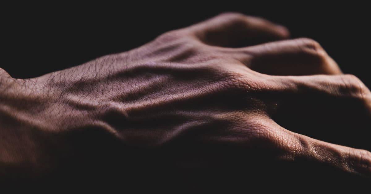
Photo: www.kaboompics.com / Pexels
Your skin is doing at least seven things right now: blocking bacteria and viruses, preventing water from evaporating out of your tissues, detecting the pressure of this page (or screen) against your fingers, regulating your core body temperature, manufacturing vitamin D, serving as a physical barrier against UV radiation, and deploying an active immune system against anything that breaches its surface. It does all of this while continuously replacing itself — the outermost layer turns over completely every 2–4 weeks — and while stretching, flexing, and compressing without tearing. For the organ most people think of as simple packaging, its engineering is remarkably sophisticated.
The Three-Layer Architecture
Skin is not a single sheet. It is three distinct tissue layers, each with different cell types, functions, and depths, stacked into a system about 1.5–4 mm thick depending on body location.
Epidermis — the outermost layer, 0.05–1.5 mm thick (thinnest on the eyelids, thickest on the palms and soles). The epidermis is stratified squamous epithelium: flat, scale-like cells (squamous) stacked in layers (stratified). It has no blood vessels of its own — it is nourished by diffusion from below. The epidermis is primarily composed of keratinocytes, which produce the structural protein keratin.
Dermis — the middle layer, about 1.5–4 mm thick and roughly 10 times thicker than the epidermis. This is the structural core of the skin, made of dense connective tissue (collagen and elastin fibers produced by fibroblasts). The dermis contains blood vessels, lymphatic vessels, nerve fibers, hair follicles, sebaceous (oil) glands, and sweat glands. It is in the dermis that most of the skin's functions — sensation, temperature regulation, immune surveillance — are physically housed.
Hypodermis (subcutaneous tissue) — the deepest layer, not always counted as skin proper but functionally inseparable from it. Composed primarily of adipocytes (fat cells) and loose connective tissue, the hypodermis anchors the dermis to underlying muscles and bones, provides thermal insulation, cushions mechanical impacts, and serves as a caloric reserve.
Source: Cleveland Clinic, Skin: Layers, Structure and Function (reviewed 2022) — Overview of integumentary anatomy and functional organization.
| Layer | Primary cells | Key structures | Thickness |
|---|---|---|---|
| Epidermis | Keratinocytes, melanocytes, Langerhans cells, Merkel cells | Stratum corneum, melanin granules | 0.05–1.5 mm |
| Dermis | Fibroblasts, mast cells, macrophages | Collagen, elastin, blood vessels, nerves, follicles | 1.5–4 mm |
| Hypodermis | Adipocytes, fibroblasts | Fat lobules, major blood vessels | Variable (2–10+ mm) |
The Epidermis: A Continuously Renewing Barrier
The epidermis divides into five distinct sublayers (strata), and understanding them requires following a single keratinocyte from birth to death.
A new keratinocyte is born by cell division in the stratum basale — the deepest epidermal layer, resting on the basement membrane that separates epidermis from dermis. Stem cells here are the source of all keratinocytes; they divide continuously, pushing older cells outward.
As a keratinocyte moves upward into the stratum spinosum, it begins producing keratin filaments internally and forms connections with neighbors via desmosomes (protein "staples" that link adjacent cells). The cell is still alive, still metabolically active.
In the stratum granulosum, the cell begins producing lamellar granules — lipid-filled packages it will eventually secrete into the spaces between cells. This lipid secretion is the foundation of the skin's waterproof barrier. The cell also begins producing loricrin and filaggrin, proteins that crosslink with keratin to create a dense protein envelope inside the cell.
At the stratum lucidum (present only on thick skin, like palms and soles), cells become flattened and begin losing their nuclei.
Finally, the keratinocyte arrives at the stratum corneum — completely dead, filled with keratin, sealed by crosslinked proteins, flattened into a scale-like structure called a corneocyte. Dozens of layers of corneocytes, held together by the lipid matrix from the lamellar granules, form the skin barrier: a brick-and-mortar structure where corneocytes are the bricks and lipids (primarily ceramides, free fatty acids, and cholesterol in roughly equal parts) are the mortar.
This stratum corneum barrier is the body's primary defense against transepidermal water loss (TEWL) — preventing the tissues from drying out — and against the inward passage of microorganisms, allergens, and irritants. Conditions like eczema (atopic dermatitis) involve disrupted filaggrin production and lipid composition, compromising this barrier and allowing irritants and allergens to penetrate.
Eventually, corneocytes shed through a process called desquamation: proteases cleave the connections between cells, and the outermost flakes are lost invisibly. About 30,000–40,000 dead skin cells shed per hour — roughly 3.5 kg per year.
Source: Elias, P.M., Journal of Allergy and Clinical Immunology (2008) — Skin barrier function, lamellar body secretion, and the molecular basis of the epidermal permeability barrier.
Melanocytes: Color, Protection, and UV Defense
Scattered through the stratum basale, roughly one per every 10 keratinocytes, are melanocytes — cells that produce the pigment melanin. Melanocytes extend long dendritic projections that reach dozens of surrounding keratinocytes, transferring melanin-containing packages called melanosomes to them.
Melanin is a polymer that absorbs UV radiation and dissipates its energy as heat, protecting the keratinocyte's DNA from UV-induced damage (primarily pyrimidine dimers, which cause the mutations that lead to skin cancer). After UV exposure, melanocyte activity increases — the biological basis of tanning.
All humans have approximately the same number of melanocytes per unit area of skin. The difference in skin color between individuals and populations comes almost entirely from the type and amount of melanin produced: eumelanin (brown-black) versus pheomelanin (yellow-red), and the size and distribution of melanosomes. This is controlled by several genes including MC1R (melanocortin 1 receptor), which determines which type of melanin is made.
Langerhans cells — dendritic immune cells in the stratum spinosum, derived from bone marrow monocytes. They survey the epidermis, capture antigens (including bacteria, fungi, and allergens), and migrate to lymph nodes to present them to T cells. They are part of the skin's adaptive immune system.
Source: Cui, R. et al., Cell (2007) — Melanocyte regulation of UV tanning response and the role of the MC1R pathway in skin pigmentation.
The Dermis: Infrastructure and Sensation
The dermis is where the skin's operational machinery lives. Its collagen network (primarily type I and type III collagen) gives skin its tensile strength; its elastin fibers (which can stretch to 150% of their resting length before returning) give it flexibility and recoil. The ratio and organization of these fibers change with age: collagen production declines, elastin fibers fragment, and the net result is the thinner, less elastic skin associated with aging.
The dermis contains five mechanoreceptor types, each tuned to different mechanical stimuli:
| Receptor | Location | Responds to | Adapts |
|---|---|---|---|
| Meissner's corpuscle | Papillary dermis (fingertips, lips) | Light touch, texture, slip | Rapidly |
| Merkel's disc | Dermal-epidermal junction | Sustained pressure, fine detail | Slowly |
| Ruffini ending | Deep dermis | Skin stretch, finger position | Slowly |
| Pacinian corpuscle | Deep dermis, hypodermis | Vibration (250 Hz), deep pressure | Very rapidly |
| Free nerve endings | Throughout dermis | Pain, itch, temperature | Variable |
Meissner's corpuscles and Merkel's discs are particularly dense in the fingertips — roughly 241 per cm² in the index finger — giving the hands their exceptional tactile resolution. Reading Braille relies on this density. Pacinian corpuscles, shaped like a small onion bulb, are so sensitive to vibration that they can detect a tool's vibration transmitted through a handle, explaining why skilled surgeons and craftspeople "feel" what their instruments contact.
Source: McGlone, F., Wessberg, J., & Olausson, H., Neuron (2014) — Discriminative and affective touch, mechanoreceptor distribution, and C-tactile afferents in social touch.
Thermoregulation: The Skin as a Radiator
The human body generates heat continuously as a byproduct of metabolism (see how muscles work for the contribution of skeletal muscle). Core temperature must be maintained within roughly 36–38°C for enzyme function; deviations of more than 3–4°C in either direction are life-threatening. The skin is the primary thermoregulatory organ.
Heat dissipation (when hot): The hypothalamus detects rising core temperature and signals two responses via the sympathetic nervous system. First, cutaneous arterioles dilate (vasodilation), directing more blood toward the skin surface, where heat can radiate away into the environment. At peak vasodilation, skin blood flow can increase from a resting 0.2–0.5 L/min to up to 8 L/min. Second, eccrine sweat glands (2–4 million distributed across the body) secrete sweat — a dilute salt solution. As sweat evaporates from the skin surface, it carries heat with it (evaporative cooling). At maximal sweating rates, a human can lose up to 2 liters of sweat per hour, dissipating roughly 580 W of heat.
Heat conservation (when cold): The hypothalamus signals cutaneous arterioles to constrict (vasoconstriction), reducing blood flow to the skin and minimizing heat loss through radiation. Arrector pili muscles in the dermis contract, pulling hair follicles upright — the mechanism of goosebumps (piloerection). In animals with fur, this traps an insulating air layer; in humans, it is vestigial but the mechanism remains.
Source: Charkoudian, N., Mayo Clinic Proceedings (2003) — Skin blood flow in adult thermoregulation: mechanisms and quantitative aspects.
Vitamin D Synthesis: The Sunlight Chemistry
The skin is the only significant endogenous source of vitamin D. Sunlight, specifically UVB radiation (wavelengths 290–315 nm), drives a photochemical reaction in keratinocytes: a cholesterol precursor (7-dehydrocholesterol, stored in cell membranes) absorbs a UV photon and undergoes ring-opening to form previtamin D3, which then isomerizes spontaneously to vitamin D3 (cholecalciferol) with heat. Vitamin D3 enters the circulation and travels to the liver for initial hydroxylation, then to the kidneys for final activation to the biologically active form, calcitriol (1,25-dihydroxyvitamin D3).
Active vitamin D regulates calcium absorption in the gut, bone mineralization, immune function, and several other pathways. About 80–90% of vitamin D in most people comes from this cutaneous synthesis, with food providing the remainder. The synthesis rate depends on the angle of the sun (and thus latitude and season), skin pigmentation (melanin competes with 7-dehydrocholesterol for UVB photons), and the amount of skin exposed.
Sunscreen with SPF 15 blocks about 93% of UVB; SPF 30 blocks about 97%. At moderate SPF use on limited skin area, vitamin D synthesis is not eliminated but is reduced. The tension between UV protection (reducing skin cancer risk) and vitamin D synthesis is real and geographically dependent: in northern latitudes where sun angle is low for much of the year, dietary supplementation becomes more important.
Source: Holick, M.F., New England Journal of Medicine (2007) — Vitamin D deficiency: mechanisms, consequences, and cutaneous synthesis pathway in detail.
Wound Healing: A Four-Phase Cascade
Skin's response to injury is one of biology's most choreographed processes, proceeding through four overlapping phases:
Phase 1 — Hemostasis (minutes to hours): Damaged blood vessels constrict reflexively. Platelets aggregate at the wound site, activated by exposed collagen. They release clotting factors that trigger the coagulation cascade, converting fibrinogen to fibrin and forming a clot. The clot physically seals the wound and provides a provisional matrix for cells to move across.
Phase 2 — Inflammation (hours to days): Immune cells — first neutrophils (arriving within hours), then macrophages (arriving over the next 24–72 hours) — migrate into the wound. Neutrophils phagocytose bacteria and debris. Macrophages clear dead neutrophils and release growth factors (PDGF, TGF-β, VEGF) that recruit fibroblasts and endothelial cells. The redness, heat, and swelling of acute wound inflammation are this phase externally visible.
Phase 3 — Proliferation (days to weeks): Fibroblasts migrate in and lay down new collagen (initially type III — weaker, more flexible). New blood vessels grow into the wound (angiogenesis), fed by VEGF signals. Keratinocytes at the wound edges migrate across the exposed wound surface and proliferate to re-cover it (re-epithelialization). The collagen-and-vessel network fills the wound bed as granulation tissue — the pink, granular tissue visible in healing wounds.
Phase 4 — Remodeling (weeks to months to years): Weaker type III collagen is gradually replaced by type I collagen, and the collagen network reorganizes from a loose, random arrangement to a more ordered structure. The wound contracts as myofibroblasts apply tensile force. Final tensile strength reaches about 80% of unwounded skin — never quite 100%, which is why scar tissue is slightly more fragile.
The balance between these phases determines whether healing produces minimal scarring or forms a hypertrophic scar (excessive collagen deposition) or keloid (scar tissue growing beyond the wound borders).
Source: Gurtner, G.C. et al., Nature (2008) — "Wound repair and regeneration" — review of wound healing phases, cellular players, and the molecular signals that coordinate each transition.
Skin as an Immune Organ
The skin is not merely a passive barrier; it is an active immune tissue. Beyond Langerhans cells, the dermis contains resident T lymphocytes, mast cells, and macrophages — all conducting ongoing surveillance.
Mast cells in the dermis release histamine and other mediators when activated by injury or allergens, driving the redness, itching, and swelling of allergic skin reactions. They are also the cells responsible for urticaria (hives) and the acute phase of allergic contact dermatitis.
Dermal dendritic cells complement Langerhans cells in sampling the skin's antigenic environment.
Sebaceous glands, attached to hair follicles, secrete sebum — an oily mixture of lipids and dead cells. Sebum lubricates the hair shaft and the skin surface, contributes to the slightly acidic acid mantle (skin pH 4.5–5.5), and has antimicrobial properties. When sebaceous glands become blocked and infected by Cutibacterium acnes (formerly Propionibacterium acnes), the result is acne vulgaris.
The skin microbiome — approximately 1,000 bacterial species colonizing the skin surface — also plays an active role in immune education and pathogen exclusion, in ways that researchers are still unraveling.
Source: Gallo, R.L. & Hooper, L.V., Nature Immunology (2012) — Epithelial antimicrobial defence in the skin and gut and the role of the microbiome.
Frequently Asked Questions
Q: How thick is skin in different body locations?
A: Skin thickness varies from about 0.5 mm on the eyelids to over 4 mm on the upper back and soles of the feet. The palms and soles have thicker epidermis due to mechanical stress; the eyelid skin is the thinnest in the body due to the need for flexible movement.
Q: Why does skin wrinkle in water?
A: Aquagenic wrinkling (pruning) occurs because the outermost skin layer absorbs water and swells. For many years it was assumed this was simple osmotic swelling, but current evidence suggests it involves active vasoconstriction of blood vessels in the fingertips, reducing the volume of the tissue beneath the skin and causing the excess surface area to buckle. The response is absent in people with peripheral nerve damage, confirming neural involvement.
Q: Does skin "breathe"?
A: The skin does allow some gas exchange — small amounts of CO₂ escape through the skin surface and some oxygen enters — but this accounts for less than 1–2% of total gas exchange in humans. The lungs handle essentially all respiratory gas exchange (see how the lungs work). The phrase "skin needs to breathe" in cosmetics marketing typically refers to avoiding occlusive products that block sweat evaporation, not to literal gas exchange.
Q: Why does skin tan under the sun?
A: UVB radiation damages keratinocyte DNA. Damaged cells release signals (alpha-MSH, among others) that bind MC1R receptors on melanocytes, stimulating melanin production and its transfer to surrounding keratinocytes. The resulting increase in melanin increases UV absorption in those cells — a delayed protective response. The "immediate" tan visible within minutes of UV exposure is photo-oxidation of existing melanin (darkening without new synthesis), which fades quickly.
Q: Can skin regenerate completely after a severe burn?
A: It depends on depth. Superficial (first-degree) burns damage only the epidermis and heal completely within days. Partial-thickness (second-degree) burns damage into the dermis; if the hair follicle stem cells are preserved, the epidermis can regenerate from follicle remnants. Full-thickness (third-degree) burns destroy all dermal appendages including follicles, eliminating the local stem cell reservoir; these require skin grafting and often heal with scar tissue.
Q: How does sunscreen work?
A: Physical sunscreens (zinc oxide, titanium dioxide) form a mineral layer on the skin surface that scatters and reflects UV radiation. Chemical sunscreens contain organic molecules that absorb UV photons and release the energy as heat rather than allowing it to drive photochemical reactions in skin cells. Modern sunscreens typically combine both classes. SPF (Sun Protection Factor) specifically measures protection against UVB; "broad spectrum" formulations also filter UVA.
Source: Lim, H.W. et al., Journal of the American Academy of Dermatology (2017) — Photocarcinogenesis, photoprotection, and sunscreen mechanisms.
The Takeaway
Skin earns its designation as the body's largest organ not through a single dramatic function but through the sheer breadth of simultaneous operations it performs. It is a physical barrier, an immune organ, a thermostat, a vitamin factory, and a sensory array — all built from a structure that can self-repair a minor cut in days and replace its entire surface every few weeks. Understanding its three-layer architecture shows how elegantly these functions are separated: the epidermis handles barrier and UV defense, the dermis handles sensation and temperature regulation, and the hypodermis handles insulation and structural anchoring. The skin is also, uniquely, the one organ you can directly observe — making it both the most accessible window into health and the site of the most common cancer in humans.
Read next:
Follow new articles
Add SciHub101 to your feed reader to see new science and technology stories.
About the Author
SciHub101 is an independent science and technology publication founded and edited by Buğra Sözeri, a software engineer and app developer based in Türkiye. AI tools assist with drafting and research. Source documentation in the existing archive is uneven, and articles are being reviewed and corrected. The Editorial Team byline identifies editorial responsibility; it does not certify specialist or independent human review. Where we compare products, anything built by our own team is labelled as such — see the editorial guidelines and About page for how corrections and disclosures work.
Related Articles
How the Nervous System Works: Signals, Reflexes, and the Brain-Body Connection
A signal travels from your fingertip to your spinal cord and back in under 20 milliseconds — before your brain has even registered the sensation. The nervous system processes roughly 100 billion neurons, each connected to thousands of others, operating at speeds up to 120 meters per second. Here is how it all works.

How the Kidneys Work: Filtration, the Nephron, and Blood Pressure Control
Your kidneys filter your entire blood supply roughly 40 times every day — about 180 liters of fluid — and return 99% of it to the bloodstream while excreting only 1 to 2 liters as urine. The precision of that selective reclamation process is what keeps you alive.
Why Do We Shiver? The Science of Thermoregulation
Shivering is an involuntary muscle contraction that generates heat when your body temperature drops below its set point. Your hypothalamus acts as a thermostat, detecting temperature changes through cold receptors and triggering both shivering and non-shivering heat production. Brown fat provides extra warmth in infants, while shivering during fever occurs because your body's target temperature has been reset upward. Understanding this physiological response reveals how your nervous system maintains the narrow 37°C window needed for survival.

How Do Wounds Heal? The Science of Tissue Repair
Wound healing is a four-stage biological process triggered immediately after injury and lasting weeks to years. Within seconds, platelets plug the wound; within hours, immune cells flood the area and initiate inflammation; over days to weeks, fibroblasts synthesize collagen and new blood vessels form; over months to years, tissue remodels and scars form. Despite apparent simplicity, the process involves coordinated signaling across dozens of cell types, growth factors, and immune mechanisms. Understanding wound healing is critical for treating chronic wounds (affecting 2 percent of the global population) and improving outcomes after surgery, trauma, and burn injuries.

How the Body Regulates Temperature: The Hypothalamic Thermostat, Sweating, Shivering, and Fever
The human body maintains core temperature within a range of less than 1°C (1.8°F) despite external temperatures swinging 60°C or more across seasons, and despite internal heat generation that — in hard exercise — rises to 20 times the resting rate. This precision requires a thermostat, an active cooling system, an emergency heating system, and dedicated fat that burns itself to generate warmth. None of it is conscious. Here is how thermoregulation actually works.

Why Do Our Ears Pop? The Science of Pressure Equalization
Every airplane climb or diving descent reminds us: the human ear is exquisitely sensitive to pressure change. The familiar pop occurs because the middle ear—a small air-filled chamber behind the eardrum—must equalize its pressure to match the ambient environment. When you yawn or swallow, the Eustachian tube opens to let air flow in or out, restoring balance. Altitude, diving, colds, and even weather shifts can create pressure mismatches that strain the tympanic membrane; when equalization finally succeeds, you feel the sudden relief of the pop.