The Invention of the Pacemaker
Photo: Towfiqu barbhuiya / Pexels
The cardiac pacemaker is a small implanted device that delivers precisely timed electrical pulses to the heart muscle, maintaining a regular heartbeat when the heart's own electrical conduction system fails. The critical breakthrough came in 1958, when Wilson Greatbatch, working in a barn-workshop in upstate New York, accidentally installed the wrong resistor in a heart-monitoring circuit — and recognized that the resulting rhythmic electrical pulse could substitute for the heart's natural pacemaker node. The first human received an implantable pacemaker in Sweden in 1958. His name was Arne Larsson, he was 43 years old, and he lived to 86 — outliving both the surgeon who implanted the device and the engineer who invented it, having worn 26 successive pacemakers over his lifetime.
The Problem: When the Heart Forgets Its Own Rhythm
The human heart beats roughly 100,000 times per day — and it does so without instruction from the brain. The heart has its own internal pacemaker: the sinoatrial (SA) node, a cluster of specialized cells in the upper right chamber that spontaneously generates electrical impulses at 60–100 times per minute. Each impulse spreads across the atria, converges at the atrioventricular (AV) node, and then propagates down specialized conduction pathways — the His bundle and Purkinje fibers — to trigger coordinated contraction of the ventricles.
Complete heart block — a condition in which the electrical impulse from the SA node fails to reach the ventricles at all, because the AV node or the conduction pathways below it are damaged. In complete heart block, the ventricles beat on their own intrinsic rhythm — roughly 30–40 beats per minute — which is insufficient to maintain adequate blood flow to the brain and organs. Symptoms include extreme fatigue, syncope (fainting), and sudden cardiac death.
Before the pacemaker, complete heart block was a death sentence in many cases. It occurred in children who underwent heart surgery to repair congenital defects — the conduction system passes close to the structures that surgeons needed to reach, and accidental injury was common. It also occurred in adults with coronary artery disease, myocarditis, and other conditions. A physician could watch a patient's heart beat too slowly to sustain consciousness and have no way to speed it up.
The only available treatment in the early 1950s was an external electric shock — a large machine that delivered a current through chest-wall electrodes. It was painful, required continuous presence at the bedside, required wall power, and could not be used outside a hospital. Patients with complete heart block were effectively imprisoned in intensive care.
Source: Aquilina, O. (2006). "A brief history of cardiac pacing." Images in Paediatric Cardiology, Vol. 8, No. 2, pp. 17–81. A comprehensive clinical review of pacemaker history.
Earl Bakken and the Power Blackout That Changed Medicine
The immediate predecessor to the implantable pacemaker was a wearable external device built by Earl Bakken, a University of Minnesota electrical engineering graduate student who in 1949 had co-founded a medical device repair company called Medtronic with his brother-in-law Palmer Hermundslie.
C. Walton Lillehei was a cardiac surgeon at the University of Minnesota who in the mid-1950s was pioneering open-heart surgery to repair congenital heart defects in children. He was achieving things that had been thought impossible — closing holes between heart chambers, repairing malformed valves — but losing a significant fraction of his patients postoperatively to complete heart block caused by inadvertent damage to the conduction system during surgery. The only available response was the large, cart-mounted external stimulator that ran on wall power.
On Halloween night, October 31, 1957, a power blackout struck the Twin Cities. One of Lillehei's young patients, dependent on the plug-in pacing machine, died.
Lillehei contacted Bakken and asked whether he could design a small, battery-powered device that could be worn by a patient. Bakken took the circuit diagram for a transistor-based electronic metronome published in Popular Electronics magazine in April 1956, modified it to adjust the pulse rate, and connected it to a wire that a surgeon could thread directly to the heart's surface. Within four weeks, he delivered a small, self-contained pacemaker the size of a large wristwatch that could be taped to the patient's chest.
The Lillehei-Bakken pacemaker was used clinically at the University of Minnesota beginning in January 1958. It was external — the pulse generator was worn outside the body, with a wire passing through the chest wall to the heart. This created infection risk at the entry wound and was impractical for anything but hospital use. But it demonstrated unequivocally that electrical pacing could save lives that would otherwise be lost.
Source: Bakken, E.E. (1990). "One Man's Full Life." Bakken Research Center. Bakken's autobiography documenting the development timeline. Additional documentation in: Bigelow, W.G. (1990). Cold Hearts: The Story of Hypothermia and the Pacemaker in Heart Surgery. McClelland and Stewart.
Earl Bakken later turned Medtronic into the world's largest medical device company. At the time of his death in 2018, Medtronic controlled roughly 40% of the global cardiac rhythm management market. His modified Popular Electronics circuit became, indirectly, a multi-billion-dollar industry.
Wilson Greatbatch: The Wrong Resistor
Working in parallel — and entirely independently — was Wilson Greatbatch, an electrical engineer in Clarence, New York, who had already been interested in cardiac monitoring for years.
In 1951, while working at Cornell Aeronautical Laboratory, Greatbatch had spoken to two surgeons about heart problems, and the conversation planted the idea that electronics could help. In 1956, he took a faculty position at the University of Buffalo and began experimenting with recording circuits for heart sounds. One of his projects was building an oscillator to measure heart sounds — a circuit that would produce a signal at specific intervals.
In 1958, while working on the oscillator in a barn that served as his workshop, Greatbatch reached into his component box to retrieve a 10,000-ohm (10 KΩ) resistor. He pulled out a 1,000,000-ohm (1 MΩ) resistor instead — off by a factor of 100 — and installed it without checking. When he switched on the circuit, instead of the expected continuous tone, the device emitted a rhythmic electrical pulse: one burst, then silence, then another burst. The pulse rate was approximately 70 times per minute.
Greatbatch recognized immediately what he had built. The pulse rate matched the human resting heart rate. The electrical output, while low, was similar to the stimulation that external pacing machines delivered to the heart. And the device was small: the oscillator circuit fit on a board roughly 5 cm × 3 cm.
He spent the next two years refining the device and working with cardiac surgeon William Chardack at the Veterans Administration Hospital in Buffalo. On May 7, 1958, Chardack and Greatbatch implanted a pacemaker in a dog for the first time. The dog survived. They ultimately demonstrated pacing in 10 dogs over 4 months.
Source: Greatbatch, W. and Holmes, C.F. (1991). "History of implantable devices." IEEE Engineering in Medicine and Biology Magazine, Vol. 10, No. 3, pp. 38–49. Greatbatch's own documented account of the accidental discovery and development process.
The challenge of implantation was not just electrical. Any device inside the human body must be biocompatible — it cannot leach toxic materials, cannot provoke an immune response severe enough to cause tissue damage, and cannot fail from corrosion or moisture ingress. Greatbatch encased his early pacemaker circuits in epoxy resin. Later designs would use titanium casing with hermetic sealing — a standard still in use today.
Arne Larsson: The First Human Implant
On October 8, 1958, at the Karolinska University Hospital in Stockholm, Sweden, surgeon Åke Senning implanted the first pacemaker in a living human patient. The patient was Arne Larsson, a 43-year-old civil engineer who had been suffering from Stokes-Adams syndrome — a condition in which complete heart block caused repeated fainting spells, sometimes dozens per day, leaving him largely bedridden and dependent on nursing care. His wife, Else-Marie, had campaigned aggressively to have the experimental procedure done.
The first device failed after three hours. Senning implanted a second one, which lasted about a week before the wire connection degraded. But successive improved devices kept Larsson alive and increasingly active.
By the standards of modern implantable electronics, the first devices were primitive — they paced the heart at a fixed rate regardless of what the heart was doing, and they were large and prone to early failure. But they kept Arne Larsson's heart beating at an adequate rate when his own conduction system could not.
Larsson received 26 pacemakers over the remaining 43 years of his life, as each device was replaced with a new model. He survived to the age of 86, dying in December 2001. At his death, his pacemaker was not one of those originally implanted in 1958 — those had long been replaced — but a modern device of the type that had evolved through decades of engineering refinement.
Åke Senning, the surgeon who performed the implant, died in 2000 at age 79. Wilson Greatbatch died in 2011 at age 92. Arne Larsson outlived them both.
Source: Elmqvist, R. and Senning, Å. (1959). "Implantable pacemakers for the heart." In Proceedings of the 2nd International Conference on Medical Electronics. A review of the first human implants in Stockholm.
The Timeline of Pacemaker Development
| Year | Milestone |
|---|---|
| 1928 | Mark Lidwill (Australia) builds first external pacemaker, used on stillborn infant |
| 1952 | Paul Zoll demonstrates external transthoracic pacing in awake patients at Beth Israel Hospital, Boston |
| 1957 | Earl Bakken builds battery-powered wearable pacemaker for Lillehei's patients |
| May 1958 | Greatbatch and Chardack implant pacemaker in dogs for first time |
| October 1958 | Åke Senning implants first pacemaker in a human — Arne Larsson — in Stockholm |
| 1960 | Greatbatch and Chardack begin human clinical use of their implantable pacemaker in the US |
| 1965 | First demand pacemaker — senses natural heartbeats and only paces when needed |
| 1969 | Medtronic launches first commercial lithium battery pacemaker (previous mercury-zinc batteries lasted 2–3 years) |
| 1980 | First automatic implantable cardioverter-defibrillator (ICD) implanted in human |
| 2013 | First leadless pacemaker human implant (capsule implanted directly in heart, no wires) |
| 2017 | FDA approves Medtronic Micra leadless pacemaker for commercial use |
| 2024 | Medtronic Micra AV2 and VR2 next-generation leadless pacemakers receive FDA approval |
How a Modern Pacemaker Works
A contemporary pacemaker contains a pulse generator — a titanium-encased package about the size of a stack of five quarters — implanted under the skin below the left collarbone. Insulated wire leads thread through the subclavian vein into the right side of the heart, with the tip of each lead lodged against the heart wall. The lead tip contains a small electrode that both senses the heart's natural electrical activity and delivers pacing pulses when needed.
Sensing: The pacemaker continuously monitors the heart's intrinsic electrical activity. When the heart beats on its own at an adequate rate, the pacemaker remains passive — it does not interfere. This is called demand pacing, introduced in 1965. The earliest pacemakers paced at a fixed rate regardless of the heart's own activity; demand pacing eliminated the risk of a pacemaker pulse landing during the vulnerable phase of the heart cycle, which could trigger dangerous arrhythmias.
Pacing: When the pacemaker detects that the heart has not beaten within a programmed interval — say, 800 milliseconds for a rate setting of 75 beats per minute — it delivers a brief electrical pulse through the lead electrode. A typical pacing pulse is 0.4–0.5 milliseconds long and delivers roughly 0.5–1.0 volts. This is enough to depolarize the adjacent heart muscle cells and trigger a propagating wave of contraction.
Rate response: Modern pacemakers contain accelerometers and respiratory sensors. When they detect physical activity — walking, climbing stairs — they increase the pacing rate to match the body's metabolic demand. This is called rate-responsive pacing.
Battery: The modern pacemaker battery is a lithium iodide cell — solid-state, not liquid, which makes it resistant to the forces of body movement and leakage. A pacemaker battery typically lasts 7–15 years before replacement is needed. Battery replacement is a minor surgical procedure: open the pocket under the collarbone, disconnect the leads, swap the generator, reconnect, close.
Source: Mond, H.G. and Freitag, G. (2014). "The Cardiac Implantable Electronic Device Power Source: Evolution and Revolution." Pacing and Clinical Electrophysiology, Vol. 37, No. 12, pp. 1728–1745.
Leadless Pacemakers: Eliminating the Wire
The leads — the wires connecting the pulse generator to the heart — are the most failure-prone component in a pacemaker system. Lead insulation can fracture, metal conductors can break from the stress of 100,000 heart contractions per day, and the entry point into the vein can develop infection. For a device intended to last a decade or more, lead failures requiring reoperation are a significant source of morbidity.
The solution is the leadless pacemaker: a fully self-contained device, about the size of a vitamin capsule, that is delivered through a catheter threaded up through the femoral vein in the leg and screwed directly into the right ventricular wall. There are no leads. There is no surgical pocket under the collarbone. There is no wire threading through the venous system.
Medtronic's Micra device received FDA approval in 2017. The device is 26 mm long, 6.7 mm in diameter, and weighs 2 grams. It paces from inside the heart, senses from inside the heart, and communicates wirelessly with an external programmer through radio frequency. In a 2024 five-year follow-up study published in the European Heart Journal, the Micra system showed significantly lower complication rates than traditional transvenous pacemakers.
The limitation of leadless pacemakers is that current single-chamber devices can only pace one chamber of the heart. The most common pacing indication — sick sinus syndrome — often requires coordination between the atria and ventricles, which single-chamber leadless devices cannot provide without additional hardware. Dual-chamber leadless systems are in clinical trials.
Source: Duray, G.Z. et al. (2024). "Leadless pacemakers at 5-year follow-up: the Micra transcatheter pacing system post-approval registry." European Heart Journal, Vol. 45, No. 14, pp. 1241–1249.
Unintended Consequences and the Shape of Modern Cardiac Care
The pacemaker was the proof of concept for the entire field of cardiac electrophysiology — the study and treatment of electrical disorders of the heart. Once it was established that you could sense the heart's electrical activity with an implanted electrode and deliver corrective energy, the technology expanded in several directions nobody anticipated in 1958.
The implantable cardioverter-defibrillator (ICD) was developed in the 1970s by Michel Mirowski at Sinai Hospital in Baltimore. An ICD monitors heart rhythm continuously and delivers a high-energy shock — 20–35 joules — when it detects ventricular fibrillation, the chaotic electrical activity that causes sudden cardiac death. The first ICD was implanted in a human in 1980. Mirowski had personal motivation: his mentor, Harry Heller, had died suddenly of ventricular fibrillation in 1966. The ICD is now implanted in roughly 200,000 patients per year in the United States and has saved a number of lives too large to count precisely.
Cardiac resynchronization therapy (CRT) addresses heart failure caused by the left and right ventricles contracting out of coordination. A three-lead pacemaker system — with leads in the right atrium, right ventricle, and through the coronary sinus to the left ventricular wall — can synchronize the ventricles, improving pumping efficiency by 15–20% in carefully selected patients. Large clinical trials have shown CRT reduces mortality in patients with heart failure and electrical conduction abnormalities.
Remote monitoring allows a patient's pacemaker to transmit data wirelessly to a secure server nightly, where algorithms flag abnormalities for clinical review. Atrial fibrillation burden, battery status, lead impedance, pacing thresholds — all transmitted automatically. The attending cardiologist reviews a dashboard, not a patient in a waiting room. Multiple studies have shown remote monitoring detects arrhythmia events earlier than clinic-based follow-up, reducing hospitalization.
Source: Moss, A.J. et al. (2002). "Prophylactic Implantation of a Defibrillator in Patients with Myocardial Infarction and Reduced Ejection Fraction." New England Journal of Medicine, Vol. 346, pp. 877–883. The MADIT-II trial establishing ICD benefit in a broad population.
Where the Pacemaker Stands Today
Approximately 1 million pacemakers are implanted worldwide each year; some estimates put the figure at closer to 1.25 million. The global pacemaker market was valued at approximately $4.8 billion in 2022. Medtronic, Abbott, Boston Scientific, and Biotronik are the dominant manufacturers.
The technical challenges that occupied Greatbatch and Bakken in 1958 — power supply, biocompatibility, lead reliability — have been largely solved, though each continues to be refined. Current research focuses on:
Biologically guided pacing — using feedback from the autonomic nervous system to adjust pacing parameters more naturally than current rate-response algorithms. This involves sensing signals from multiple physiological sources simultaneously.
Subcutaneous ICD — a device implanted under the skin of the chest with no leads entering the heart or blood vessels at all, detecting ventricular fibrillation through the skin and delivering a shock through the chest wall. FDA-approved since 2012, indicated for patients who need defibrillator protection but do not need conventional pacing.
Conduction system pacing — threading a lead to the His bundle or left bundle branch, the heart's natural conduction pathways, instead of the right ventricular apex. This produces a more physiologically normal activation sequence, potentially preserving cardiac function better than conventional right ventricular pacing.
The device that began with a misplaced resistor in an upstate New York barn is now one of the most reliable and well-studied pieces of medical technology ever produced. A patient implanted today with a modern pacemaker can expect the device to function reliably for a decade, require minimal follow-up, and restore a near-normal quality of life.
The Takeaway
The pacemaker is a parable about accident, persistence, and collaboration. Bakken responded to a power blackout; Greatbatch grabbed the wrong component from a box. Neither set out to revolutionize cardiology. What made their accidental observations productive was the ability to recognize what they had found — and the presence of clinicians like Lillehei and Chardack who were willing to try it on patients who would otherwise die. Arne Larsson's 43 years of life after his first implant represent, in concentrated form, the impact of the device: roughly 1 million people per year now receive the same gift he received in a Stockholm operating room in October 1958.
Frequently Asked Questions
Q: Who invented the pacemaker? A: Multiple people contributed at different stages. Earl Bakken built the first wearable battery-powered external pacemaker in 1957, working with cardiac surgeon C. Walton Lillehei at the University of Minnesota. Wilson Greatbatch invented the first implantable pacemaker in 1958 after accidentally using the wrong resistor in a circuit. Swedish surgeon Åke Senning performed the first human implant in October 1958. All three are recognized as pioneers; no single person holds exclusive credit.
Q: Who was the first person to receive a pacemaker? A: Arne Larsson, a 43-year-old Swedish civil engineer, received the first implantable pacemaker on October 8, 1958, at Karolinska University Hospital in Stockholm. The surgeon was Åke Senning. Larsson went on to receive 26 successive pacemakers and lived to the age of 86, dying in December 2001. He outlived both his surgeon and the engineer who invented the device.
Q: How does a pacemaker know when to fire? A: A modern pacemaker continuously monitors the heart's natural electrical activity through the lead electrode. It fires only when it detects that the heart has not beaten within a preset interval — typically corresponding to the lower rate limit set by the cardiologist. This is called demand or inhibited pacing. When the heart beats on its own, the pacemaker is inhibited and delivers no pulse.
Q: How long does a pacemaker battery last? A: A modern pacemaker battery — a lithium iodide cell — typically lasts 7–15 years depending on how frequently the device paces and the programmed output settings. Battery replacement requires a minor surgical procedure to open the pocket under the collarbone, swap the generator, and close the wound. The leads are typically left in place and reused.
Q: What is a leadless pacemaker? A: A leadless pacemaker is a self-contained device, about the size of a large vitamin capsule, that is implanted directly inside the right ventricle of the heart via a catheter. It has no leads (wires) threading through the veins. Medtronic's Micra device, FDA-approved in 2017, is the best-known example. Leadless devices eliminate lead-related complications but are currently limited to single-chamber pacing.
Q: Can pacemaker patients have MRI scans? A: Most modern pacemakers are designated "MRI-conditional" — they can be safely scanned under specific conditions, typically at 1.5 T field strength with specific programming settings activated before and during the scan. Older devices (pre-2011, roughly) may be MRI-incompatible. A patient's cardiologist and the MRI facility jointly determine safety and protocol. "MRI-conditional" does not mean "MRI-safe under all circumstances."
Read next:
Follow new articles
Add SciHub101 to your feed reader to see new science and technology stories.
About the Author
SciHub101 is an independent science and technology publication founded and edited by Buğra Sözeri, a software engineer and app developer based in Türkiye. AI tools assist with drafting and research. Source documentation in the existing archive is uneven, and articles are being reviewed and corrected. The Editorial Team byline identifies editorial responsibility; it does not certify specialist or independent human review. Where we compare products, anything built by our own team is labelled as such — see the editorial guidelines and About page for how corrections and disclosures work.
Related Articles
Pasteurization: How Heating Milk Changed the History of Childhood Death
Louis Pasteur invented pasteurization in 1864 to save wine. It took another 50 years to reach milk—and the delay cost hundreds of thousands of children their lives.
The Microwave Oven: How a Melted Candy Bar Rewired the Kitchen
How Percy Spencer's 1945 accident near a Raytheon magnetron produced the Radarange — and eventually the appliance in 90% of US homes.
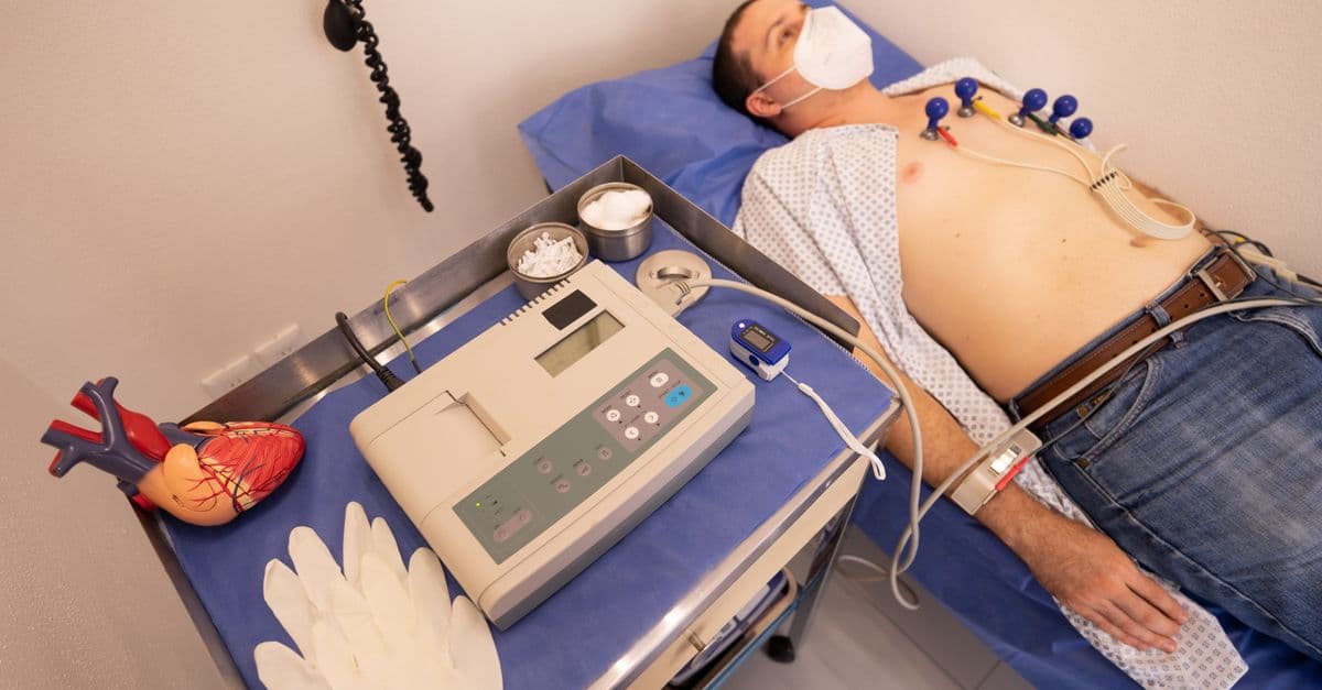
How the Heart Works — Anatomy and Function
How the human heart pumps blood: chambers, valves, electrical conduction, cardiac cycle, and the most important diseases and treatments.