The Invention of MRI
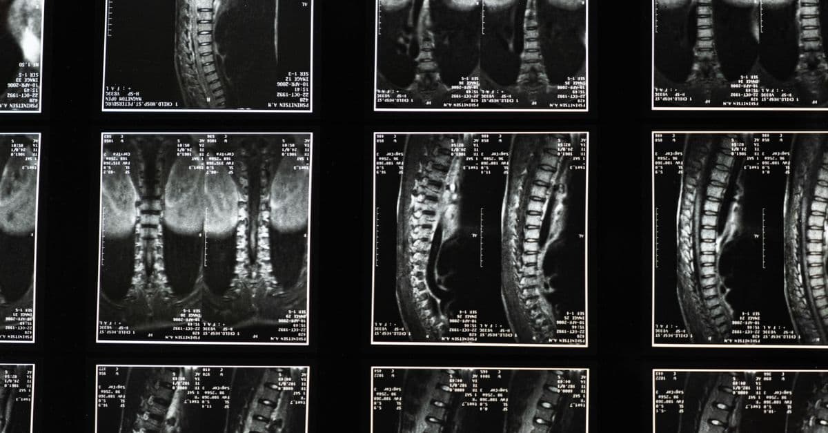
Photo: cottonbro studio / Pexels
Magnetic Resonance Imaging works by placing a patient inside a powerful magnetic field that causes hydrogen nuclei in their body to align like compass needles, then briefly knocking them out of alignment with a radio-frequency pulse and listening to the signal they emit as they return. Paul Lauterbur at the State University of New York Stony Brook published the key insight in Nature in 1973: by superimposing a gradient magnetic field on the uniform field, you can encode spatial position into the signal, turning physics into images. He and Peter Mansfield of the University of Nottingham shared the 2003 Nobel Prize in Physiology or Medicine for this work. Raymond Damadian, who published earlier experimental evidence and holds the first MRI patent, was not included.
The Physics That Makes MRI Possible
Before MRI, there was nuclear magnetic resonance (NMR) — a technique chemists used to identify molecular structures. NMR was discovered independently by Felix Bloch at Stanford and Edward Purcell at Harvard in 1946, work for which they shared the 1952 Nobel Prize in Physics. The underlying physics involves the quantum mechanical property of nuclear spin.
Spin — an intrinsic angular momentum possessed by many atomic nuclei. The hydrogen nucleus (a single proton) has a spin of one-half, meaning it behaves like a tiny bar magnet with a north and south pole. In normal conditions, protons point in random directions and their magnetic moments cancel out. Place them in a strong magnetic field and two things happen: the protons align either with or against the field (a small excess aligns with it, because that orientation is lower energy), and they begin to precess — wobble around the field direction, like a gyroscope wobbling around gravity.
The precession frequency — called the Larmor frequency — is directly proportional to the field strength. For hydrogen in a 1.5 Tesla magnetic field (the most common clinical scanner strength), the Larmor frequency is approximately 63.9 MHz, which sits in the FM radio band. This is not coincidence; MRI scanners are, in physical terms, very sophisticated radio receivers tuned to listen to protons.
If you transmit a radio-frequency pulse at exactly the Larmor frequency, the protons absorb the energy and flip their alignment — they tip away from the main field direction. When the pulse ends, they gradually return to equilibrium, emitting their absorbed energy as a radio signal. This signal is what MRI measures. The time constants that govern the return to equilibrium — called T1 (longitudinal relaxation) and T2 (transverse relaxation) — differ significantly between different tissue types, between healthy and diseased tissue, and between fat and water. This variation in relaxation is what gives MRI its extraordinary soft-tissue contrast. The difference between a brain tumor and surrounding brain tissue, invisible on an X-ray, is legible in T1 and T2 signals.
Source: Bloch, F., Hansen, W.W. and Packard, M. (1946). "Nuclear Induction." Physical Review, Vol. 69, pp. 127. The founding paper of NMR physics, describing the detection of the proton spin resonance signal.
Raymond Damadian: The Man Who Got There First — and Missed the Prize
In 1969, Raymond Damadian, a physician and biophysicist at Downstate Medical Center in New York, had a hypothesis: cancer cells, which have altered membrane transport and higher water content than normal cells, might have distinctly different NMR relaxation times. If so, NMR could serve as a cancer detector.
His 1971 paper in Science — "Tumor Detection by Nuclear Magnetic Resonance" — confirmed the hypothesis experimentally. Damadian measured T1 and T2 relaxation times in several rat tissues, including healthy tissue and two types of tumors, and found that the tumors showed significantly longer relaxation times. He proposed that NMR scanning of the human body could detect cancer.
In March 1972, Damadian filed for a patent on "Apparatus and Method for Detecting Cancer in Tissue." The patent was granted in February 1974, making it the first issued patent for an NMR body scanner — two years before Lauterbur's competing application.
The problem with Damadian's approach — as he described it in 1971 — was that it lacked a method for creating images. His original concept involved mechanically moving the patient through a focused NMR field point by point, like scanning a photograph with a magnifying glass. It would work in principle, but it would take hours per patient. What was missing was the mathematical framework to create images rapidly from the full NMR signal.
Source: Damadian, R. (1971). "Tumor Detection by Nuclear Magnetic Resonance." Science, Vol. 171, No. 3976, pp. 1151–1153. The key experimental paper establishing different NMR relaxation times in healthy and cancerous tissue.
That framework was Lauterbur's contribution.
Paul Lauterbur's Insight: Gradient Fields and Spatial Encoding
Paul Lauterbur was a chemist, not a physician. In September 1971, he was attending a scientific meeting in Pittsburgh when he had an insight that he recorded in his research notebook that evening. If you add a small gradient to the main magnetic field — making it slightly stronger on one side of the sample than the other — then the Larmor frequency of protons will vary with their position. Protons on the stronger-field side will precess faster; protons on the weaker-field side will precess slower. The signal you receive is a superposition of many frequencies, each corresponding to a different spatial position. Unscramble the frequencies mathematically, and you recover the spatial distribution of protons — you get an image.
This is analogous to identifying instruments in an orchestra by listening to a chord: a cello plays at 65 Hz, a violin at 659 Hz, a flute at 1,047 Hz. Even without seeing the orchestra, you can determine which instruments are present by decomposing the sound into its component frequencies. Lauterbur's gradient field turns space into frequency, and Fourier mathematics decodes the spatial information from the frequency spectrum.
In January 1973, Lauterbur produced the first ever NMR image: a two-dimensional map of two small tubes of water. The image was a cross-section, 4 mm across, showing two distinct circles. It was rudimentary. It was also a proof of principle that changed medicine.
His paper, titled "Image Formation by Induced Local Interactions: Examples Employing Nuclear Magnetic Resonance," was submitted to Nature in December 1972. The journal's editors initially were skeptical and asked for revisions. The paper appeared on March 16, 1973.
Source: Lauterbur, P.C. (1973). "Image Formation by Induced Local Interactions: Examples Employing Nuclear Magnetic Resonance." Nature, Vol. 242, pp. 190–191. The paper that introduced gradient field spatial encoding and demonstrated the first NMR image.
Lauterbur called the technique "zeugmatography," from the Greek zeugma meaning "that which is used for joining." The name did not stick. The technique became MRI.
Peter Mansfield's Mathematical Contribution: Making It Fast Enough to Matter
At the same time Lauterbur was working on gradient encoding, Peter Mansfield at the University of Nottingham was developing the mathematical theory that would make MRI clinically practical. Lauterbur's original approach acquired images one data point at a time — useful for proof of concept, inadequate for a hospital.
Mansfield developed echo-planar imaging (EPI) in 1977, a technique that acquires an entire two-dimensional image in a single RF excitation, rather than requiring hundreds of separate pulses. Where Lauterbur's initial approach might need many minutes per image slice, Mansfield's EPI could acquire a full slice in tens of milliseconds. This was the difference between an interesting physics demonstration and a viable medical scanner.
Echo-planar imaging — a rapid MRI acquisition method in which magnetic field gradients are rapidly switched to traverse a two-dimensional grid of k-space (frequency-space) data after a single excitation pulse. The entire image is acquired in one shot. EPI is the technique used in functional MRI (fMRI), which detects brain activity by tracking blood oxygenation changes in real time.
Mansfield also developed the mathematical theory of slice selection — how to excite protons in a specific thin plane within the body without exciting the protons above or below it. This is done by applying the gradient field during the RF pulse, so that only protons precessing at the transmitted frequency (which corresponds to a specific depth) absorb energy. The gradient field and the RF frequency together select a slice as thin as 1 mm.
Source: Mansfield, P. and Maudsley, A.A. (1977). "Medical Imaging by NMR." British Journal of Radiology, Vol. 50, pp. 188–194. Mansfield's key paper on echo-planar imaging and slice selection theory.
The First Human MRI Scan
Raymond Damadian built the first whole-body MRI machine, which he named "Indomitable." On July 3, 1977, after more than four hours of scanning, Damadian and his team produced the first NMR scan of a living human chest. The subject was graduate student Larry Minkoff, who lay inside the apparatus for 4 hours and 45 minutes while data was collected point by point. The image showed a cross-section of his thorax.
Lauterbur's gradient-encoded method — implemented in increasingly sophisticated machines — produced the first cross-sectional images of living animals in 1974 and of the human wrist in 1977. By the early 1980s, commercial scanners using gradient-field imaging were being installed in hospitals.
| Year | Milestone |
|---|---|
| 1946 | Bloch and Purcell discover NMR; Nobel Prize 1952 |
| 1971 | Damadian publishes NMR differences between tumors and healthy tissue |
| 1972 | Damadian files first NMR body scanner patent (granted 1974) |
| 1973 | Lauterbur publishes gradient-field image formation in Nature |
| 1977 | Mansfield develops echo-planar imaging; Damadian scans first human |
| 1980 | First commercial MRI scanners installed in hospitals |
| 2003 | Nobel Prize in Physiology or Medicine awarded to Lauterbur and Mansfield |
| 2023 | Next-generation 7 Tesla scanners achieve 180 times the detail of standard 3T machines |
The Damadian Exclusion: A Bitter Nobel Controversy
When the Nobel Committee announced the 2003 Prize in Physiology or Medicine, the announcement described the prize as recognizing the work of Lauterbur and Mansfield "for their discoveries concerning magnetic resonance imaging." Damadian was not named.
Within days of the announcement, Damadian took out full-page advertisements in The New York Times, The Washington Post, and The Los Angeles Post, with the headline "The Shameful Wrong That Must Be Righted." The ads argued that the Nobel Committee had ignored his foundational 1971 paper, his 1974 patent, and the first whole-body human scan his team performed in 1977.
The Nobel Committee's implicit position — never stated directly, since the Committee does not explain its decisions — appeared to be that Damadian's contribution was the identification of NMR differences in tissue, which is a biological observation, while Lauterbur's gradient encoding and Mansfield's echo-planar mathematics were the inventions that made MRI a usable technology. Damadian's point-by-point scanning method was impractical; the method that became MRI was Lauterbur's.
Supporters of Damadian pointed out that Lauterbur's original 1973 Nature paper made no citation to Damadian's 1971 Science paper, even though Lauterbur's own notebook showed he had read it the day before he developed his gradient-field idea.
The controversy has never been fully resolved. The Nobel Prize rules prohibit posthumous awards and are limited to three recipients per prize. Damadian died in 2022. The Committee's decision stands.
Source: Kevles, B.H. and Kevles, D.J. (2003). "Scenting Trouble." The Lancet, Vol. 362, No. 9394, p. 1494. Commentary on the Nobel Prize controversy and Damadian's response.
How a Modern MRI Scanner Works
A clinical MRI machine has four main systems: the magnet, the gradient coils, the radiofrequency (RF) system, and the computer.
The magnet is the heart of the system. Clinical scanners operate at 1.5 Tesla (T) or 3 T — field strengths 30,000 to 60,000 times stronger than the Earth's magnetic field. These fields require superconducting wire coils, which carry electrical current with zero resistance at cryogenic temperatures. The coils are submerged in liquid helium at 4 Kelvin (−269°C / −452°F). Once energized, a superconducting magnet remains at field indefinitely without requiring external power — the current simply circulates. A 3 T MRI machine contains roughly 1,500 liters (396 gallons) of liquid helium.
The gradient coils sit inside the main magnet bore. Three sets of coils, oriented along the X, Y, and Z axes, can rapidly switch gradient fields on and off to encode spatial position along each dimension. The rapid switching of these coils — currents changing thousands of times per second — is what produces the loud thumping and knocking noise characteristic of MRI scanners. The coils are flexing in the magnetic field, generating sound through the same mechanism as a loudspeaker.
The RF system transmits the excitation pulse at the Larmor frequency and then switches to receive mode to detect the returning signal. Modern scanners use sophisticated phased array coil systems — multiple small receiver coils arranged around the patient — which dramatically improve signal-to-noise ratio compared to a single large coil.
The computer performs the mathematical reconstruction. The raw data acquired by MRI is not an image; it is a grid of numbers in k-space (frequency space). A mathematical operation called the Fourier transform converts k-space data into image space — the anatomical image you see on the monitor. Modern scanners complete this reconstruction in seconds.
Source: McRobbie, D.W. et al. (2017). MRI from Picture to Proton, 3rd ed. Cambridge University Press. The standard clinical reference on MRI physics and technology.
7 Tesla: The Frontier of Whole-Body Imaging
Standard clinical MRI runs at 1.5 T or 3 T. Research institutions have operated 7 T whole-body scanners for two decades, but FDA clearance for clinical 7 T use in the United States came only in 2017. A next-generation 7 T scanner developed by researchers and published in Nature Methods in 2023 achieved functional brain images with 10 times better spatial resolution than previous 7 T systems, and more than 180 times greater detail than conventional 3 T clinical machines.
At 7 T, the Larmor frequency for hydrogen is approximately 297 MHz — nearly five times the frequency of a standard 1.5 T scanner. Higher frequency means shorter wavelength, which means the RF pulses interact with the patient's body differently, producing non-uniform excitation that requires careful management. Specialized RF coil designs address this. The payoff is dramatically improved spatial resolution: 7 T scanners can resolve brain structures at sub-millimeter scale, distinguishing cortical layers and subcortical nuclei that are invisible at 3 T.
Research machines at 10.5 T exist at the University of Minnesota. A 14 T human scanner is under development.
Source: Wald, L.L. et al. (2023). "Next-generation MRI scanner designed for ultra-high-resolution human brain imaging at 7 Tesla." Nature Methods, Vol. 20, pp. 2023–2032.
Unintended Consequences: fMRI and the Imaging of Thought
No one designing MRI in the 1970s anticipated that it would become the primary tool for mapping brain function. Functional MRI (fMRI), developed in 1991 by Seiji Ogawa at Bell Labs, exploits the fact that oxygenated and deoxygenated hemoglobin have different magnetic properties — oxygenated blood is slightly diamagnetic; deoxygenated blood is slightly paramagnetic. When a brain region becomes active, blood flow to that region increases, temporarily raising the local ratio of oxygenated to deoxygenated hemoglobin. This changes the T2* relaxation time, which changes the MRI signal. Map those signal changes over time and you map brain activity.
BOLD signal — Blood Oxygen Level Dependent signal. The basis of fMRI. Not a direct measure of neural firing; rather a proxy that tracks the hemodynamic response to neural activity, which lags by roughly two seconds.
fMRI has been used to study language processing, memory consolidation, pain perception, decision-making, and the neural correlates of consciousness. It has also been misused — by researchers who drew overly confident conclusions from noisy data, and in legal proceedings where "brain scan evidence" was presented with unwarranted certainty. The technique measures blood flow, not thought; the inferential gap between the two is large.
Where MRI Stands Today
Roughly 100 million MRI scans are performed worldwide each year. A standard 1.5 T scanner costs approximately $1 million to $1.5 million, plus installation and siting costs for the magnet shielding and RF shielding. A 3 T scanner costs $2 million to $3 million. Healthcare systems in high-income countries have broadly accessible MRI; in low-income countries, access is severely limited by cost.
Open-bore MRI designs — where the magnet is configured as two disc-shaped poles above and below the patient rather than a cylindrical tunnel — address claustrophobia concerns. Portable MRI systems, using low-field permanent magnets at 0.064 T, have been developed for use at the bedside in intensive care units, though at significantly lower image quality.
Artificial intelligence is accelerating reconstruction: deep learning models can reconstruct diagnostic-quality images from 25% of the normally required k-space data, cutting scan times by three-quarters. FDA-cleared AI reconstruction is now deployed on major manufacturer platforms.
MRI guided focused ultrasound (MRgFUS) combines MRI with high-intensity ultrasound beams that converge on a tissue target with millimeter precision, generating enough heat to ablate tissue without surgery. It is FDA-approved for treating essential tremor and uterine fibroids.
The Takeaway
MRI exists because three people, working mostly independently, each contributed something irreplaceable: Damadian showed that NMR differences in tissue could detect disease, Lauterbur figured out how to turn those differences into images, and Mansfield made the imaging fast enough for clinical use. The Nobel Prize recognized two of the three. The scanner in every major hospital reflects all of them. It is, by any measure, one of the most consequential medical technologies of the twentieth century — a machine that sees inside living tissue without ionizing radiation, with a contrast resolution no other imaging modality approaches.
Frequently Asked Questions
Q: Who invented the MRI scanner? A: Three people made foundational contributions. Raymond Damadian published the first evidence (1971) that NMR could distinguish cancerous from healthy tissue and built the first whole-body NMR scanner in 1977. Paul Lauterbur invented gradient-field spatial encoding in 1973, enabling image formation. Peter Mansfield developed echo-planar imaging in 1977, making MRI fast enough for clinical use. Lauterbur and Mansfield shared the 2003 Nobel Prize in Medicine; Damadian was excluded, which generated significant controversy.
Q: Why is MRI loud? A: The noise comes from the gradient coils inside the main magnet. These coils must switch on and off thousands of times per second to encode spatial position in the MRI signal. Each switching event causes the coil to flex momentarily in the powerful magnetic field — the same mechanism that makes a loudspeaker cone move, but at much higher forces. The resulting vibrations are transmitted as thumping, knocking, and buzzing sounds. Noise-canceling headphones and acoustic insulation reduce but do not eliminate the problem.
Q: Is MRI safe? A: For most patients, yes. MRI uses no ionizing radiation — it uses radio waves and magnetic fields, neither of which damage DNA. The main safety concerns are: (1) ferromagnetic implants or foreign objects, which can be attracted to the magnet or heated by the RF field; (2) certain types of older cardiac devices; and (3) severe claustrophobia. Modern cardiac pacemakers and implantable defibrillators are generally MRI-conditional rather than contraindicated, meaning they can be scanned with specific precautions.
Q: What is the difference between MRI and fMRI? A: Standard MRI produces anatomical images — maps of tissue structure showing where organs, tumors, or injuries are. Functional MRI (fMRI) measures changes in blood oxygenation over time to infer which brain regions are active during specific tasks. fMRI uses the same scanner hardware as standard MRI but acquires data in a rapid time series and measures a different signal property (T2* rather than T1 or T2).
Q: Why was Raymond Damadian excluded from the Nobel Prize? A: The Nobel Committee does not explain its decisions. The implicit distinction appears to be that Damadian identified a biological phenomenon (NMR differences in tissue) while Lauterbur and Mansfield invented the techniques that turned NMR into imaging technology. Damadian's point-by-point scanning method was not the approach adopted by commercial MRI; Lauterbur's gradient encoding was. Damadian protested vigorously, taking out full-page newspaper ads calling the exclusion "The Shameful Wrong That Must Be Righted."
Q: What is the strongest MRI scanner used in humans? A: As of 2026, 10.5 Tesla whole-body scanners are in operation at a small number of research institutions, including the University of Minnesota's Center for Magnetic Resonance Research. The 14 T human MRI project is under development. Clinical scanners are limited to 3 T (FDA-cleared) or 7 T for research use with special clearance.
Read next:
Follow new articles
Add SciHub101 to your feed reader to see new science and technology stories.
About the Author
SciHub101 is an independent science and technology publication founded and edited by Buğra Sözeri, a software engineer and app developer based in Türkiye. AI tools assist with drafting and research. Source documentation in the existing archive is uneven, and articles are being reviewed and corrected. The Editorial Team byline identifies editorial responsibility; it does not certify specialist or independent human review. Where we compare products, anything built by our own team is labelled as such — see the editorial guidelines and About page for how corrections and disclosures work.
Related Articles

mRNA Vaccines: The Technology That Took 30 Years to Arrive
How Katalin Karikó and Drew Weissman solved the immune rejection problem with mRNA in 2005, spent 15 years ignored, then changed medicine during a pandemic.
The Invention of GPS
How Bradford Parkinson, Ivan Getting, and Roger Easton built a satellite navigation system for nuclear bombers that became the infrastructure of everyday life.
How MRI Machines Work: Magnetic Resonance Imaging Explained
MRI sees inside your body without radiation by making hydrogen atoms in your tissues sing at a specific radio frequency.
MRI Technology: Magnetic Resonance Imaging Explained
How MRI uses magnetic fields to create detailed images of soft tissues and organs
Anesthesia: The Invention That Made Modern Surgery Possible
How William Morton's 1846 ether demonstration at Mass General transformed surgery from conscious agony into painless procedure — and why we still don't fully understand it.

The Stethoscope: How a Rolled Tube of Paper Became Medicine's Most Iconic Tool
René Laennec invented the stethoscope in 1816 from rolled paper to examine a patient's heart. Two centuries later, AI is listening through the same basic principle.