Hooke's Cells: How a 1665 Sketch Became Cell Theory
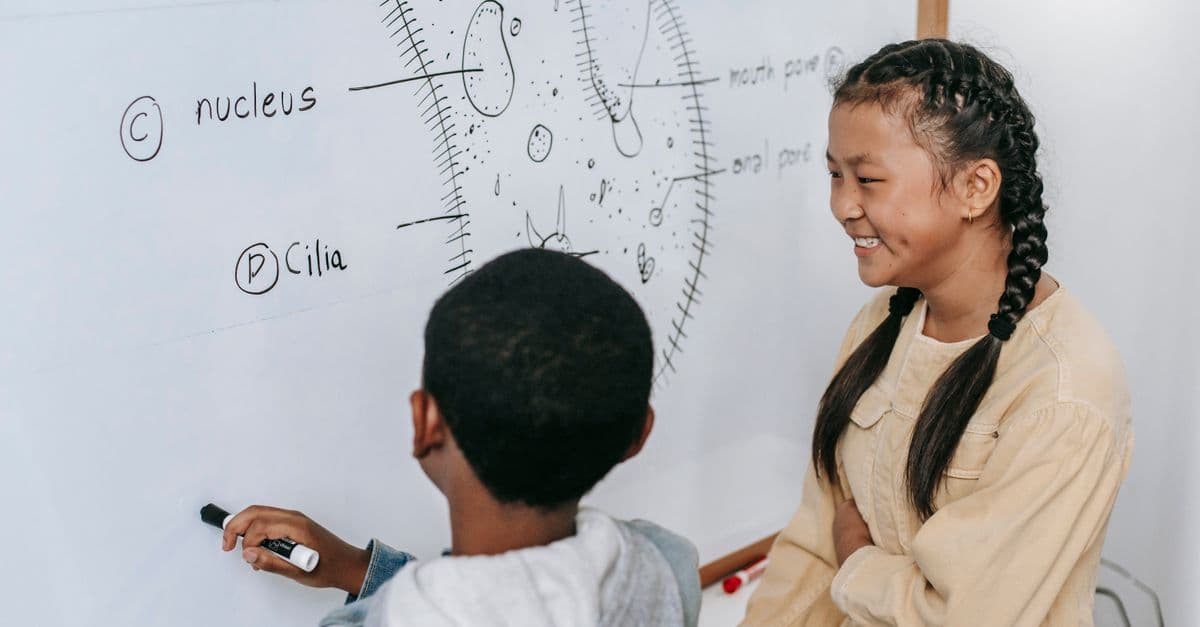
Photo: Katerina Holmes / Pexels
In 1665, Robert Hooke, curator of experiments at the Royal Society of London, pressed a sliver of cork under a homemade compound microscope and saw thousands of tiny, empty compartments arranged in neat rows. He sketched them for his book Micrographia and called them "cells," because they reminded him of the small rooms monks slept in (Royal Society, Micrographia, 1665). That single word survived. The idea behind it took nearly 200 more years, and three more scientists, to become biology's foundational law.
What Hooke actually saw — and what he got wrong
Cell theory — the principle that all living things are made of cells, that the cell is the basic unit of life, and that cells arise only from pre-existing cells — did not spring fully formed from Hooke's cork slide. Hooke saw structure, not life. He needed to explain that distinction before anyone could build a theory on top of it.
Hooke's instrument was a compound microscope of his own design: a leather-and-pasteboard tube holding two lenses, paired with an oil-lamp illumination rig he built himself, magnifying roughly 20 to 50 times depending on the setup (Bodleian Library, Micrographia exhibit notes). Under that lens, a paper-thin slice of cork bark showed a honeycomb of rectangular chambers, walled off from one another, repeating across the entire sample. Hooke drew it in careful cross-hatched detail and gave the chambers a name that had nothing to do with biology and everything to do with architecture: the cellula of a monastery.
What Hooke was actually looking at was dead tissue. Cork is bark — the outer protective layer of a cork oak (Quercus suber) — and by the time it forms cork, the plant cells inside have died and left behind only their rigid cellulose walls. Hooke saw the walls, empty boxes where living cells used to be, not the fluid, organelle-filled interior of a living cell (Royal Society, Notes and Records, "The cells of Robert Hooke," 2024). He had discovered the architecture of life without discovering that life was inside it. Micrographia became the first scientific bestseller of its era — diarist Samuel Pepys reportedly stayed up until 2 a.m. reading it the night it arrived (Hektoen International, "Robert Hooke and Micrographia," 2023) — but the deeper meaning of what Hooke had drawn would sit unclaimed for nearly two centuries.
Hooke himself never generalized his cork observation into a universal law. He used the same book to describe flea anatomy, snowflake geometry, and the surface of a razor's edge — cells were one curiosity among dozens, not a thesis he was arguing for.
Why a name for empty boxes turned into a law of biology
A single observation of dead plant tissue is not a scientific theory. It's a description. What turned "cells exist in cork" into "all life is made of cells" was a 170-year relay of independent discoveries, each one closing a gap the last one left open.
Nine years after Hooke, Dutch draper and lens-grinder Antonie van Leeuwenhoek ground single, tiny spherical lenses capable of roughly 270x magnification — far beyond Hooke's compound instrument — and turned them on pond water. In a September 1674 letter to the Royal Society, he described "little animalcules" swimming in a sample from a nearby lake, the first documented sighting of single-celled living organisms such as protozoa and, later, bacteria (Royal Society, Philosophical Transactions B, "The unseen world," 2015). Where Hooke had seen dead architecture, Leeuwenhoek was watching cells move, feed, and reproduce in real time. It was the missing half of the picture: cells weren't just structural units in cork, they were entire organisms in their own right.
More than a century later, in 1831, Scottish botanist Robert Brown identified a small, dense structure inside plant cells that he named the nucleus — a term that stuck, though Brown himself wasn't sure what it did. That single structure would later turn out to be where a cell keeps its genetic instructions. Then, in 1869, Swiss physiological chemist Friedrich Miescher isolated a phosphorus-rich substance from the nuclei of white blood cells he extracted from pus-soaked surgical bandages. He called it "nuclein" because of where he found it — a name that survives today in the middle of "deoxyribonucleic acid," or DNA (Genome.gov, "1869: DNA First Isolated"). Miescher didn't know he'd found the molecule of heredity. He just knew it wasn't a protein, and that was enough to matter.
What is cell theory, exactly?
Cell theory states three things: all living organisms are composed of one or more cells; the cell is the basic structural and functional unit of life; and all cells arise from pre-existing cells. It was assembled between 1838 and 1855 by three separate scientists working on separate problems, not proposed as a single unified idea by any one of them.
The three scientists who actually wrote cell theory
Hooke gets the name-drop credit, but cell theory as a formal scientific law belongs to three 19th-century Germans who never met Hooke and worked a continent and 170 years away from Micrographia.
German botanist Matthias Schleiden published Beiträge zur Phytogenesis in 1838, arguing from years of plant tissue observation that every part of a plant, from root to leaf, is built from cells or the products of cells (British Society for the History of Medicine, "Matthias Schleiden and the Genesis of Cell Theory"). A year later, physiologist Theodor Schwann — reportedly after a conversation with Schleiden over dinner about the similarity between plant cells and structures Schwann had seen in animal cartilage — extended the claim to animals. In 1839, Schwann declared that both plants and animals are built from the same basic unit: the cell functions as the "elementary particle of organisms," whether the organism is a single cell or trillions of them working together (Hektoen International, "The beginnings of cell theory," 2022).
Schleiden and Schwann's combined claim covered two of the three pillars: all life is cellular, and the cell is life's basic unit. It left one enormous question unanswered — where do new cells come from? Both scientists, working from an older and largely incorrect tradition, initially proposed a kind of free crystallization: that new cells could form spontaneously out of a formless fluid, the way a crystal precipitates out of a solution. That idea was wrong, and it took another physician to correct it.
In 1855, German pathologist Rudolf Virchow, drawing on his own microscope work studying diseased tissue, rejected spontaneous cell formation outright. He proposed that every cell comes from a cell that already existed, division from division, all the way back — a principle he distilled into the Latin phrase omnis cellula e cellula, "all cells from cells" (EBSCO Research Starters, "Schwann and Virchow Develop Cell Theory"). That closed the third pillar and turned cell theory into something falsifiable: if you ever found a cell that hadn't come from cell division, the theory would break. In over 165 years of microscopy since, nobody has.
| Year | Scientist | Contribution | Pillar of cell theory established |
|---|---|---|---|
| 1665 | Robert Hooke | Named "cells" in cork tissue | None — observation only, no generalized theory |
| 1674 | Antonie van Leeuwenhoek | First observation of living single-celled organisms | Cells can be complete, independent living things |
| 1831 | Robert Brown | Identified the nucleus in plant cells | Cells have internal, purposeful structure |
| 1838 | Matthias Schleiden | All plant tissue is built from cells | All living organisms are made of cells (plants) |
| 1839 | Theodor Schwann | Extended the claim to animal tissue | All living organisms are made of cells (animals) |
| 1855 | Rudolf Virchow | Omnis cellula e cellula — no spontaneous generation | All cells arise from pre-existing cells |
It took six scientists and 190 years to turn Hooke's cork sketch into the three-part law taught in every introductory biology course today.
Why "seeing a box" is not the same as "understanding a cell"
Hooke's limitation wasn't a failure of intellect. It was a limitation of the instrument in his hands, and it's worth being precise about the gap, because the same gap — better tools reveal more structure, which in turn demands new theory — repeats throughout the history of cell biology.
A modern light microscope, refined through the 1800s with achromatic lenses and standardized staining techniques, can resolve detail down to roughly 200 nanometers and reach useful magnifications around 1,000x to 1,500x — enough to see a nucleus, a cell wall, and the general outline of larger organelles, but not enough to see individual proteins or the fine architecture of a cell membrane. That ceiling is set by the wavelength of visible light itself, not by lens quality; you cannot resolve detail smaller than roughly half the wavelength of the light you're using, no matter how good the glass is.
Breaking that ceiling required abandoning light altogether. In 1931, German engineers Ernst Ruska and Max Knoll built the first working electron microscope, using magnetic coils to focus a beam of electrons instead of a beam of light (Ernst Ruska Nobel Lecture, 1986). Because electrons have a much shorter effective wavelength than visible light, an electron microscope can resolve detail down to fractions of a nanometer — enough to see individual proteins, viral capsids, and the membrane structures Hooke's compound microscope could never have shown him. Ruska received the Nobel Prize in Physics for the achievement in 1986, 55 years after the fact and only after the technology had already reshaped biology, medicine, and materials science (NobelPrize.org, "The Nobel Prize in Physics 1986").
How much detail can scientists actually see inside a cell today?
Electron microscopes resolve structures down to roughly 0.1–1 nanometer, revealing individual proteins, ribosomes, and viral particles. Cryo-electron microscopy, developed further through the 1980s–2000s, freezes cells in a near-native state before imaging, letting researchers see molecular machines like ATP synthase in something close to their working shape rather than a chemically fixed one.
What a cell actually is, once you can see inside it
Hooke saw a wall. What that wall turned out to contain is, by any honest measure, more intricate than anything he could have imagined from a cork sample.
A living cell is bounded by a lipid membrane that controls what enters and leaves, studded with protein channels and receptors that act like gatekeepers and sensors. Inside, DNA — organized into chromosomes and stored in the nucleus in eukaryotic cells such as those in plants, animals, and fungi — carries the coded instructions for building every protein the cell will ever need. Ribosomes read that code and assemble amino acids into proteins. Mitochondria, believed by most cell biologists to have originated as free-living bacteria absorbed by a larger ancestral cell roughly 1.5 to 2 billion years ago (the endosymbiotic theory), generate the ATP that powers nearly everything the cell does. None of this was visible to Hooke, or even conceivable to him: he had a wooden box with two lenses and a candle. Cell biology as it exists today required three more centuries of instrumentation, chemistry, and genetics layered on top of his one observation.
Scale matters here too. A 2013 systematic estimate published in Annals of Human Biology put the adult human body at approximately 37.2 trillion cells, calculated by tallying published cell counts across every major organ and tissue type rather than relying on the older, loosely sourced estimates that had circulated since the 1960s (Bianconi et al., Annals of Human Biology, 2013). Every one of those 37.2 trillion cells, in Virchow's framing, descends from a single fertilized egg through an unbroken chain of division — the same principle Virchow stated in 1855 based on far cruder microscopy and far fewer data points.
What people still get wrong about Hooke's discovery
The most common misconception is treating Hooke as the founder of cell theory rather than the namer of the cell. Hooke never claimed cells were the universal unit of life; he was cataloging curiosities for the Royal Society, and cork cells were one entry among sketches of fleas, feathers, and frost patterns. The theory came from Schleiden, Schwann, and Virchow, working independently and mostly without direct reference to Hooke's cork sketch at all.
A second, subtler misconception: that Hooke's tools were primitive and therefore unimportant. Hooke's compound microscope, while limited by the lens-grinding technology of the 1660s, was itself a substantial engineering achievement — a two-lens system with custom illumination that he designed and built himself, at a time when even functioning single-lens microscopes were rare specialist instruments. It's more accurate to say Hooke's tools defined the ceiling of what he could observe, not that they were crude. The gap between Hooke's ~30x compound scope and Leeuwenhoek's ~270x single-lens instrument, built within a decade of each other, illustrates how much single-lens design still had left to give even before anyone touched electron optics.
A third confusion worth clearing up: people sometimes assume "cell" was a scientific term before Hooke repurposed it. It wasn't. Hooke borrowed a word from monastic architecture — a monk's small sleeping chamber — purely because the visual resemblance struck him. The word had zero biological meaning before 1665; its entire scientific career started from an architectural metaphor that happened to stick.
Does cell theory need a fourth postulate?
Virchow's 1855 principle closed the third pillar, but it didn't close the conversation. Many modern biology textbooks now teach what's often grouped together as "modern cell theory" — a set of additions layered on top of the original three postulates rather than a replacement of them.
The most commonly cited addition is that energy flow, meaning the capture, conversion, and use of energy through processes such as cellular respiration and photosynthesis, occurs within cells (National Geographic Education, "Cell Theory"). A second addition concerns heredity: DNA is passed between cells during division, making the cell not just the structural unit of life but the unit that carries hereditary information forward. A third addition points to chemical uniformity — cells within a given species tend to be similar in both structure and chemical composition, which is read as evidence of common ancestry (National Geographic Education, "Cell Theory").
None of these additions are formally ratified the way Schleiden, Schwann, and Virchow's three pillars were, and there's no single body that votes cell theory postulates into or out of existence. They function more like a running update: each one closes a gap that the original 1838–1855 framework didn't anticipate, because Virchow had no way to know about metabolism at the molecular level or DNA's role in heredity. Miescher had only just isolated "nuclein" from pus-soaked bandages the same decade Virchow was writing, and it would be another 90 years before Watson and Crick worked out what that molecule actually did.
Does stem cell biology break the "cells only come from cells" rule?
No, but it does stretch what "cell" means functionally within the theory's third pillar. A stem cell is defined by two capacities that most adult cells lack: self-renewal, meaning it can divide and produce more cells with its own properties, and differentiation, meaning it can also produce specialized descendant cells such as heart muscle, blood, or nerve cells (NIH, "Stem Cell Basics"). Stem cells are classified by how much they can differentiate into: totipotent cells can form an entire organism, pluripotent cells can form nearly every cell type in an adult body, and multipotent cells are restricted to one lineage, such as blood cells arising from bone marrow stem cells.
Every one of those divisions still obeys omnis cellula e cellula — a stem cell's daughter cells come from that stem cell, full stop. What stem cell biology adds is a mechanism Virchow couldn't have described in 1855: that a single lineage of cells can hold open the option to become dozens of different specialized types, rather than simply making more of itself. That's less a violation of Virchow's rule than a more detailed picture of what happens inside it, the same way Leeuwenhoek's live pond-water organisms didn't overturn Hooke's cork sketch, just filled in what Hooke's dead sample couldn't show.
The takeaway
Hooke's cork sketch is often taught as the moment biology discovered the cell, but that overstates what one microscope slide of dead bark can tell you. What Hooke actually gave science was a name and a shape — a box, empty and structural. It took Leeuwenhoek to show that cells could be alive on their own, Brown and Miescher to find what was stored inside the nucleus, and Schleiden, Schwann, and Virchow to turn a description into a testable law with three falsifiable parts. Cell theory isn't a single discovery; it's a chain, and every link had to hold before biology could say, with confidence, that a cell is the basic unit of all life.
Read next:
- Microscope Invention: Leeuwenhoek, Hooke, and the Hidden World
- Stem Cells Explained — Biology and Therapies
Frequently Asked Questions
Q: Did Robert Hooke discover cell theory in 1665? A: No. Hooke named the cell after observing dead cork tissue under a microscope, but he never proposed that all living things are made of cells. Cell theory as a formal, three-part scientific law was assembled between 1838 and 1855 by Matthias Schleiden, Theodor Schwann, and Rudolf Virchow — nearly two centuries after Hooke's observation.
Q: Why did Hooke see empty boxes instead of living cells? A: Cork is bark from a cork oak tree, and by the time it forms cork, the living contents of its cells have died off, leaving only rigid cellulose walls behind. Hooke's compound microscope, magnifying roughly 20 to 50 times, showed him the leftover architecture of dead plant tissue, not the fluid, organelle-filled interior of a living cell.
Q: What are the three parts of cell theory? A: All living organisms are composed of one or more cells; the cell is the basic structural and functional unit of life; and all cells arise from pre-existing cells through division. Schleiden and Schwann established the first two parts in 1838–1839, and Rudolf Virchow added the third in 1855 with his principle omnis cellula e cellula.
Q: How many cells are in the human body? A: A 2013 systematic estimate published in Annals of Human Biology calculated approximately 37.2 trillion cells in the average adult human body, based on published cell counts across every major organ and tissue type rather than the rougher estimates used previously.
Q: What let scientists finally see inside a living cell? A: Better light microscopes in the 1800s pushed magnification toward roughly 1,000x, revealing nuclei and larger organelles, but light itself set a resolution ceiling around 200 nanometers. Ernst Ruska and Max Knoll's 1931 electron microscope broke that ceiling by using electron beams instead of light, eventually resolving individual proteins and viral structures.
Q: Who first observed a living cell, rather than a dead one? A: Antonie van Leeuwenhoek, a Dutch draper and self-taught lens-maker, observed single-celled living organisms — which he called "animalcules" — in pond water in 1674, using hand-ground lenses capable of roughly 270x magnification, notably stronger than Hooke's compound microscope.
Follow new articles
Add SciHub101 to your feed reader to see new science and technology stories.
About the Author
SciHub101 is an independent science and technology publication founded and edited by Buğra Sözeri, a software engineer and app developer based in Türkiye. AI tools assist with drafting and research. Source documentation in the existing archive is uneven, and articles are being reviewed and corrected. The Editorial Team byline identifies editorial responsibility; it does not certify specialist or independent human review. Where we compare products, anything built by our own team is labelled as such — see the editorial guidelines and About page for how corrections and disclosures work.
Related Articles
Darwin's Finches: The Evolution Experiment That Changed Biology
Charles Darwin's observations of finch beaks in the Galápagos Islands became the cornerstone of evolutionary theory. A century later, scientists watched evolution happen in real-time on those same islands.
Eratosthenes Measures the Earth: Ancient Genius with Geometry
Around 240 BCE, Greek mathematician Eratosthenes used only sunlight, shadows, and geometry to calculate Earth's circumference with remarkable accuracy. A timeless example of clever scientific thinking.
Leeuwenhoek's Microscope: Discovering a Hidden Universe of Life
In the 1670s, Anton van Leeuwenhoek ground tiny lenses and became the first human to see microorganisms — bacteria, protozoa, and sperm cells. His observations opened the microbial world and laid the groundwork for germ theory.
History of Vaccines — From Jenner to mRNA
From variolation and Jenner's cowpox to Pasteur, polio, smallpox eradication, and mRNA — the full history of vaccines and modern frontiers.
Deep Learning and Microscopy: Seeing Materials at the Atomic Scale
AI is giving electron microscopes superhuman vision—resolving atomic structures, identifying defects, and automating analyses that once took weeks.
Plague and Disease: How Epidemics Shaped History
The Black Death killed a third of Europe. The 1918 flu killed more people than World War I. Epidemics have redirected history more than any army.