Microscope Invention: Leeuwenhoek, Hooke, and the Hidden World
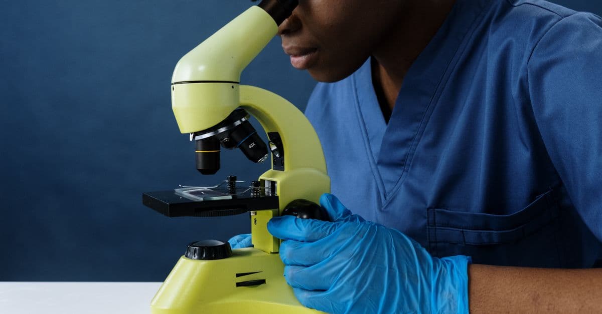
Photo: cottonbro studio / Pexels
In October 1676, a Dutch draper with no university training and no Latin sent the Royal Society of London a letter describing creatures so small that "ten thousand of these living creatures could scarce equal the bulk of a coarse sand grain." He had seen them wriggling in a drop of rainwater through a lens he ground and mounted himself. That draper, Antonie van Leeuwenhoek, and the English polymath Robert Hooke, who a decade earlier had coined the word "cell" while looking at cork, did not invent the microscope. They turned it from a novelty into a scientific instrument, and in doing so revealed that a universe of living things had been invisible, not absent, the entire time.
The problem: a world too small to argue about
Before the 1600s, the smallest unit of biological understanding a person could point to was whatever the naked eye could resolve, roughly a tenth of a millimeter (0.1 mm), about the width of a human hair. Physicians debated the causes of disease, farmers debated why milk soured, and naturalists debated how living things reproduced, all without any tool that could show them the structures actually responsible. Claims about a "hidden" biological world were, by necessity, philosophical guesses rather than observations.
The Roman writer Marcus Terentius Varro had speculated in the first century BCE that swamps bred "certain minute creatures which cannot be seen by the eyes" and caused disease, an idea that anticipated germ theory by more than 1,700 years but that no one could test. The missing piece was not a hypothesis. It was resolving power, the ability of an optical system to distinguish two close points as separate rather than as one blur.
How early lens-makers stumbled into a new instrument
Simple magnifying lenses, single pieces of curved glass, existed for centuries before anyone built a true microscope. Spectacle makers in Europe had been grinding convex lenses for reading glasses since the 13th century. The leap to a compound instrument, one that combines two or more lenses to multiply magnification, happened in the Netherlands around 1590, in the same spectacle-making workshops that a few years later would also produce the first telescopes.
Credit for that leap is genuinely contested. Dutch spectacle makers Hans Janssen and his son Zacharias Janssen, working in Middelburg, are traditionally named as the inventors of the compound microscope, based on a letter written by the diplomat William Boreel in the 1650s, more than 60 years after the fact. Some historians also credit fellow Middelburg lens grinder Hans Lippershey, who is independently credited with the first telescope patent application in 1608, with a competing or parallel claim, according to the American Physical Society's history of the invention.
This is the first place readers run into a common misconception: that a microscope has one clean inventor. What actually happened is closer to a shared trade secret circulating among a handful of spectacle makers in one small Dutch town, none of whom left behind a rigorous account, a working prototype, or a scientific publication describing what they had built. The instrument existed as a curiosity, a toy for the wealthy, for roughly 70 years before anyone used it to produce evidence that changed a scientific argument.
Why didn't the compound microscope matter sooner?
Early compound microscopes suffered from severe optical aberrations, chromatic and spherical distortions that blurred and colored the edges of an image, because 17th-century glass and lens-grinding technique could not correct for them. Paradoxically, this meant single, tiny, carefully ground lenses, like the ones Leeuwenhoek made, often produced sharper images than the multi-lens instruments meant to replace them.
Robert Hooke and the book that named the cell
Robert Hooke, curator of experiments for the Royal Society, published Micrographia in 1665, the first major scientific book built entirely around microscope observations. It contained detailed engraved illustrations of a flea, a louse, and the compound eye of a fly, rendered at a scale no one in England had seen printed before. The diarist Samuel Pepys called it "the most ingenious book that I ever read in my life" and reportedly stayed up until two in the morning finishing it.
Hooke's most lasting contribution came from a mundane material: a thin slice of cork, cut from tree bark for use as bottle stoppers. Looking at it under his compound microscope, he saw a honeycomb of small, empty, box-like compartments. He named them "cells" because they reminded him of the small, bare rooms, cellula in Latin, that monks lived in inside a monastery.
Hooke was actually looking at the cell walls of dead plant tissue, not living cells, and he had no way of knowing that cells were the basic structural unit of all living things. That insight, cell theory, would not be formally established for another 170 years. But the word stuck, and it stuck specifically because Hooke reached for an analogy his readers already understood, monastic architecture, rather than inventing new jargon.
Antonie van Leeuwenhoek and the discovery of a living microscopic world
Antonie van Leeuwenhoek ran a draper's shop in Delft and had almost certainly read Hooke's Micrographia, which likely inspired him to grind his own lenses to inspect the thread count of cloth. He was not trained as a scientist and never learned Latin, the language of European scholarship at the time, which meant he communicated entirely through more than 560 letters written in colloquial Dutch, roughly 200 of which went to the Royal Society over five decades, according to the Royal Society's own archival review of his correspondence.
His technique set him apart from every lens-maker before him. Rather than combining multiple lenses in a tube, Leeuwenhoek fused tiny, nearly spherical single lenses, some just a millimeter across, into small metal plates and held the sample on a mounted pin close to his eye. Surviving examples of his instruments, examined with modern equipment, show single-lens magnifications ranging from roughly 68x to 266x, competitive with, and in some cases better than, commercial compound microscopes built a century later.
In 1674, Leeuwenhoek examined a sample of lake water and described free-swimming organisms he called "animalcules," a term meaning tiny animals. Two years later, in his October 1676 letter, he described animalcules in rainwater, well water, and pepper-infused water in enough anatomical detail, including descriptions matching what modern microbiologists recognize as the bacterium Spirillum volutans, that the Royal Society initially doubted him.
Why was the Royal Society skeptical of Leeuwenhoek at first?
Leeuwenhoek was an untrained tradesman making claims that contradicted centuries of assumption about the limits of the visible world, and he refused to share his exact lens-making methods. The Royal Society only accepted his findings after Hooke and other fellows successfully reproduced similar observations with their own microscopes around 1677, corroborating what Leeuwenhoek had described.
Leeuwenhoek went on to be the first person to observe and describe bacteria, red blood cells, and spermatozoa, among the foundational observations of microbiology. He is widely regarded by historians of science as the father of microbiology, not because he invented the microscope, but because he was the first person to use it systematically enough, and skeptically enough, to build an evidence base rather than a single anecdote.
Why the invention took two separate breakthroughs, not one
Readers often assume a single, tidy moment: someone invents the microscope, and the hidden world opens up. The real sequence required two separate kinds of breakthrough happening a decade apart, and neither alone would have mattered as much.
Hooke's contribution was institutional and communicative. He worked inside the Royal Society, an organization built specifically to formalize observation, illustration, and peer scrutiny, and he produced a widely read, beautifully illustrated book that made microscopy fashionable and credible among the educated public. Micrographia gave the instrument cultural legitimacy.
Leeuwenhoek's contribution was technical and empirical. His superior single-lens grinding technique, which he guarded as a trade secret his entire life, produced images sharper than what contemporary compound instruments could manage, and his patient, repeated, corroborated observation of the same phenomena over years built the kind of evidence base that turns a curiosity into a fact. Neither man could have delivered what the other did. Hooke never matched Leeuwenhoek's resolving power; Leeuwenhoek, an outsider to scholarly institutions, never had Hooke's platform.
A useful way to test this explanation is to ask what happens if you remove either piece. Remove Hooke's book, and microscopy stays a private hobby of a handful of Dutch tradesmen with no scholarly audience. Remove Leeuwenhoek's lenses, and the Royal Society keeps looking at fleas and cork with instruments too blurry to see bacteria, which are roughly 1 to 10 micrometers (0.001 to 0.01 mm) across, well below what 17th-century compound microscopes could resolve.
From cell walls to cell theory: the 170-year gap
Hooke saw cell walls in 1665. It took until 1838 and 1839 for German scientists Matthias Schleiden and Theodor Schwann to independently propose that cells are the basic structural unit of all living tissue, plant and animal alike, a claim now known as cell theory, according to Britannica's history of the concept. The two reportedly compared notes over dinner in October 1838, when Schwann realized that structures he had seen in animal spinal cord tissue matched what Schleiden described in plants.
<ComparisonTable caption="Five turning points in the microscope's 400-year history" headers={["Year", "Person or team", "Contribution", "Why it mattered"]} rows={[ ["c. 1590", "Hans and Zacharias Janssen (disputed, also credited to Hans Lippershey)", "First compound microscope built by combining two lenses in a tube", "Established that stacking lenses could multiply magnification beyond a single lens"], ["1665", "Robert Hooke", "Published Micrographia; coined the word "cell" after observing cork tissue", "Made microscopy scientifically credible and gave biology a founding vocabulary"], ["1674-1683", "Antonie van Leeuwenhoek", "Built superior single-lens microscopes; first to observe bacteria, protozoa, and spermatozoa", "Created the evidence base for microbiology as a discipline"], ["1838-1839", "Matthias Schleiden and Theodor Schwann", "Proposed cell theory: all living things are made of cells", "Turned scattered microscope observations into a unifying biological law"], ["1933", "Ernst Ruska and Max Knoll", "Built the first electron microscope", "Broke past the physical resolution limit of light itself"], ]} />
The physical wall every light microscope eventually hits
For nearly 200 years after Leeuwenhoek, microscope makers assumed that better glass and better grinding could always deliver a sharper image. In 1873, the German physicist Ernst Abbe proved that assumption wrong. He showed mathematically that any microscope using visible light has a hard resolution limit set by the wavelength of light itself and the numerical aperture of the lens, roughly half the wavelength of the light used, according to Abbe's diffraction formula still taught in optics courses today.
This is a genuinely useful analogy: trying to see something smaller than that limit with visible light is like trying to trace the outline of a coin using a paintbrush wider than the coin. No matter how carefully you hold the brush, the bristle width sets the finest detail you can render. Visible light has a wavelength of roughly 400 to 700 nanometers (0.0004 to 0.0007 mm), which puts Abbe's limit at around 200 nanometers, small enough to see a bacterium's outline but far too coarse to see the internal machinery of a cell, let alone a single protein or virus, most of which are 20 to 300 nanometers across.
How did scientists eventually see past the diffraction limit?
Ernst Ruska and Max Knoll solved the problem in 1933 by abandoning light altogether. Because fast-moving electrons behave as waves with wavelengths roughly 100,000 times shorter than visible light, focusing a beam of electrons with magnetic lenses instead of glass ones let Ruska's first working electron microscope reach magnifications around 12,000x, later improved to about 30,000x by 1938, far beyond what any light microscope could achieve, work for which Ruska received the 1986 Nobel Prize in Physics.
Electron microscopes require samples to sit in a vacuum, which kills living cells, so biologists still needed a way to watch life happen in real time at high resolution. That gap was closed decades later by super-resolution fluorescence microscopy, developed independently by Stefan Hell, Eric Betzig, and William Moerner, who shared the 2014 Nobel Prize in Chemistry for techniques that use clever tricks with light-emitting molecules to sidestep Abbe's limit rather than break it outright, effectively turning individual glowing molecules on and off so their positions can be pinpointed one at a time and reassembled into an image far sharper than diffraction alone allows.
What people get wrong about the microscope's origin story
The biggest misconception is that "the microscope" was invented once, by one person, in one moment, the same way popular retellings often flatten the light bulb down to Edison alone. In reality, at least four distinct technical leaps, spanning roughly 350 years, each had to happen before microscopy looked anything like it does in a modern lab: the compound lens tube around 1590, Leeuwenhoek's superior single-lens grinding in the 1670s, the electron microscope in 1933, and super-resolution fluorescence techniques in the 2000s and 2010s.
A second common error is crediting Hooke with discovering cells as living units. He discovered and named the structure, but what he saw under his lens was dead, empty plant cell wall, not a living cell with its internal contents, and he had no theoretical framework connecting his cork observation to the idea that all organisms are built from cells. That framework arrived with Schleiden and Schwann almost two centuries later.
A third misconception, understandable given how often it is retold, is that Leeuwenhoek was a lone genius working in total isolation. He corresponded with the Royal Society for roughly 50 years, had his claims independently verified by other observers including Hooke, and built on lens-grinding knowledge already circulating in the Netherlands' active optical trade. His genius was less the lens itself and more his patience, his skepticism about his own results, and his insistence on repeatable observation, habits that anticipate the scientific method as much as any single instrument does.
Why it changed the world
The microscope's real historical weight is not the instrument, it is the bottleneck it removed: the assumption that anything too small to see could be safely ignored. Once Leeuwenhoek and Hooke demonstrated that an entire tier of living structure existed below the resolution of the human eye, medicine, biology, food safety, and eventually chemistry and materials science all had to reorganize around a simple new fact, that causes of disease, fermentation, decay, and heredity were physical, visible, microscopic entities rather than invisible forces.
That reorganization took time. Full acceptance of germ theory, the idea that specific microorganisms cause specific diseases, did not arrive until Louis Pasteur's and Robert Koch's work in the 1860s through 1880s, nearly 200 years after Leeuwenhoek first described animalcules. The lag illustrates a pattern that shows up across the history of science and technology: observing a phenomenon and building the theoretical, institutional, and technical scaffolding to act on it are two different achievements, often separated by generations.
The takeaway
Neither Robert Hooke nor Antonie van Leeuwenhoek invented the microscope from nothing. Hooke gave microscopy a name, an audience, and scientific legitimacy through Micrographia; Leeuwenhoek gave it the technical precision and patient, corroborated evidence needed to reveal an entire kingdom of living organisms nobody had suspected existed. The instrument itself kept evolving long after both men died, through Abbe's diffraction limit in 1873, Ruska's electron microscope in 1933, and Hell, Betzig, and Moerner's super-resolution fluorescence techniques in the 2000s, each breakthrough pushing back a wall the previous generation had assumed was permanent. The real lesson of the microscope's 400-year history is that "seeing" something for the first time is rarely a single event. It is a slow accumulation of better lenses, better skepticism, and better ways of proving that what you saw was real.
Read next:
- History of Computing: From the Abacus to Artificial Intelligence
- How the Transistor Changed Everything
- The Invention of the Internet
Frequently Asked Questions
Q: Who really invented the microscope? A: No single person did. Dutch spectacle makers, most often credited as Hans and Zacharias Janssen around 1590, built the first compound microscopes, but Robert Hooke and Antonie van Leeuwenhoek transformed the instrument into a genuine scientific tool roughly 75 years later, in the 1660s and 1670s.
Q: What did Robert Hooke actually discover? A: Hooke examined a thin slice of cork under his compound microscope in 1665 and saw small, box-like compartments he named "cells," after the small rooms monks lived in. He published this and dozens of other observations in his 1665 book Micrographia, the first major scientific work built around microscope images.
Q: What did Antonie van Leeuwenhoek discover? A: Leeuwenhoek was the first person to observe and describe bacteria, protozoa, red blood cells, and spermatozoa, using single-lens microscopes he ground and built himself. His 1676 letter to the Royal Society describing "animalcules" in water is considered the founding document of microbiology.
Q: How powerful were Leeuwenhoek's microscopes? A: Measurements of his surviving lenses show magnifications ranging from roughly 68x to 266x, with some estimates reaching 300x, achieved with a single tiny lens rather than the multiple lenses used in later compound microscopes.
Q: Why did it take so long for scientists to accept microorganisms existed? A: Leeuwenhoek's claims contradicted centuries of assumption that nothing smaller than the human eye could resolve was worth taking seriously, and he refused to disclose his exact lens-grinding method. The Royal Society only accepted his findings after other observers, including Robert Hooke, independently replicated them around 1677.
Q: What is cell theory, and who proposed it? A: Cell theory is the principle that all living things are composed of cells, the basic structural and functional unit of life. It was proposed by German scientists Matthias Schleiden and Theodor Schwann in 1838 and 1839, roughly 170 years after Hooke first observed and named cells.
Q: What replaced the light microscope for very small structures? A: The electron microscope, invented by Ernst Ruska and Max Knoll in 1933, uses focused beams of electrons instead of light, taking advantage of their much shorter wavelength to magnify specimens tens of thousands of times beyond what light microscopes can achieve.
Q: What is the diffraction limit, and why does it matter? A: The diffraction limit, described mathematically by Ernst Abbe in 1873, is the physical boundary on how sharp any light-based microscope image can be, set by the wavelength of light itself, roughly 200 nanometers for visible light. It explains why light microscopes cannot resolve individual proteins or viruses without special techniques.
Q: How do modern microscopes see past the diffraction limit? A: Super-resolution fluorescence techniques, developed by Stefan Hell, Eric Betzig, and William Moerner and recognized with the 2014 Nobel Prize in Chemistry, use light-emitting molecules that can be switched on and off to pinpoint their positions individually, reconstructing images far sharper than the diffraction limit would normally allow.
Q: Is Robert Hooke or Antonie van Leeuwenhoek considered the father of microbiology? A: Leeuwenhoek holds that title among historians of science, because he was the first to systematically observe, describe, and get independent verification of microorganisms. Hooke's contribution was foundational but preceded the discovery of microbial life by about a decade.
Sources
- The unseen world: reflections on Leeuwenhoek (1677) "Concerning little animals," Philosophical Transactions of the Royal Society B
- Antonie van Leeuwenhoek and the dawn of microscopic observation, PMC
- Invention of the Microscope, American Physical Society
- Cell theory, Britannica
- Ernst Ruska – Facts, NobelPrize.org
- The Nobel Prize in Chemistry 2014, press release, NobelPrize.org
- What Really Limits Microscopy Resolution? Diffraction, Rayleigh, Aberrations, and Nyquist Explained, Basler AG
Follow new articles
Add SciHub101 to your feed reader to see new science and technology stories.
About the Author
SciHub101 is an independent science and technology publication founded and edited by Buğra Sözeri, a software engineer and app developer based in Türkiye. AI tools assist with drafting and research. Source documentation in the existing archive is uneven, and articles are being reviewed and corrected. The Editorial Team byline identifies editorial responsibility; it does not certify specialist or independent human review. Where we compare products, anything built by our own team is labelled as such — see the editorial guidelines and About page for how corrections and disclosures work.
Related Articles
Telephone Invention: Bell, Gray, Meucci, and the Patent Race
The telephone's invention involved Alexander Graham Bell, Elisha Gray, Antonio Meucci, patents, and the race to transmit speech electrically.
Microchip Invention: Kilby, Noyce, and the Integrated Circuit
The microchip emerged from parallel breakthroughs by Jack Kilby and Robert Noyce. This guide explains the invention that made modern computing possible.
The Invention of the Microscope — SciHub101
From Zacharias Janssen's first compound microscope around 1590 to Nobel-winning super-resolution techniques: how the microscope revealed the hidden universe of cells and microbes.
Camera Invention: From Camera Obscura to Digital Sensors
The camera evolved from the camera obscura to chemical photography and digital sensors. This guide explains the chain of inventions.
Laser Invention: From Masers to Coherent Light
The laser grew from quantum theory, masers, optical engineering, and patent battles. This article explains how coherent light became a world-changing tool.
How Glasses Work — Correcting Vision Explained
How glasses, contacts, and laser surgery correct myopia, hyperopia, astigmatism, and presbyopia — the physics and biology of clear vision.