The Invention of the Microscope
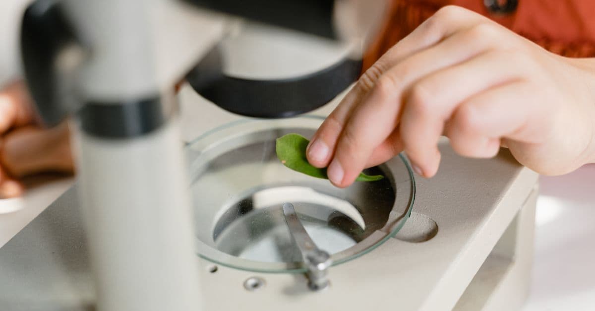
Photo: MART PRODUCTION / Pexels
The microscope is an optical or electronic instrument that produces magnified images of objects too small to resolve with the naked eye. The first compound microscope — using two lenses in sequence to achieve higher magnification than a single lens allows — was constructed around 1590 in Middelburg, in the Dutch province of Zeeland, likely by spectacle maker Zacharias Janssen or his father Hans. Robert Hooke used an improved compound microscope to publish Micrographia in 1665, the first detailed atlas of the microscopic world, and coined the word "cell." Antoni van Leeuwenhoek ground single lenses of extraordinary quality in the 1670s to become the first person to observe living bacteria and protozoa. Today's cryo-electron microscopy resolves individual atoms in frozen biological molecules, and super-resolution fluorescence microscopy — recognized by the 2014 Nobel Prize in Chemistry — bypasses the physical diffraction limit that constrained optical microscopy for 140 years.
The First Compound Microscopes: Middelburg, 1590
The origin of the compound microscope is poorly documented and genuinely disputed. The city of Middelburg in the Netherlands — then a major center of the lens-grinding trade — produced the most credible early candidates.
Compound microscope — a microscope using two or more lenses in series: an objective lens near the specimen that forms a magnified real image, and an eyepiece (ocular) lens that further magnifies that image for the eye; as opposed to a simple microscope, which uses a single lens.
Zacharias Janssen (c. 1585–c. 1632) is the most frequently cited inventor. The evidence rests primarily on a 1655 letter from Willem Boreel, a Dutch diplomat, to the physician and microscopist Pierre Borel, recounting that Janssen had constructed a compound microscope around 1590 when Boreel was a child. The letter was written 65 years after the claimed invention. No microscope clearly attributable to Janssen survives.
Hans Martens, also of Middelburg, has been proposed as an independent or co-inventor. Cornelis Drebbel, a Dutch inventor working in England in the early seventeenth century, constructed compound microscopes of apparent quality by around 1619–1621 and demonstrated them to Galileo in 1624. Galileo himself appears to have built a compound microscope by 1625, calling it the "occhiolino" (little eye).
The attribution problem is typical of inventions from this period: multiple practitioners in the same trade (lens grinding) producing similar devices in approximate contemporaneity, with documentation recorded decades later from fallible memory. What can be said confidently is that functional compound microscopes existed in the Dutch lens-making trade by the 1590s and were being demonstrated in European courts by the 1620s.
Source: Van Helden, A., Dupré, S., van Gent, R., and Zuidervaart, H. (eds.) (2010). The Origins of the Telescope. Royal Netherlands Academy of Arts and Sciences. Chapter on the Middelburg instrument-making tradition, which addresses both telescope and microscope origins.
These early compound microscopes achieved magnifications of perhaps 3 to 9 times — useful for seeing large insects or plant structures, but not for cells. Their optical quality was limited by the grinding technology of the time, which produced spherical aberrations (blurring from imperfectly curved lenses) and chromatic aberrations (color fringing from the different refractive behavior of different wavelengths of light).
Robert Hooke and the Word "Cell"
Robert Hooke (1635–1703) was one of the most versatile experimental scientists of the seventeenth century — mathematician, architect (he participated in the rebuilding of London after the 1666 Great Fire), astronomer, and microscopist. He built or commissioned a compound microscope of improved design and spent years making and drawing detailed observations.
In 1665, Hooke published Micrographia: or Some Physiological Descriptions of Minute Bodies Made by Magnifying Glasses with Observations and Inquiries Thereupon, one of the most influential scientific books in history. It contained detailed copper-plate engravings of microscopic and telescopic observations — a flea (covering a fold-out page), a louse gripping a hair, the compound eye of a fly, textile fibers, and numerous plant and mineral specimens. Samuel Pepys called it "the most ingenious book I ever read in my life."
The observation that gave us the word "cell" was not of a living cell but of a thin slice of cork. Hooke cut cork extremely thin and examined it under his microscope. He saw that the material was divided into small, enclosed compartments, which he described as resembling the cells in a monastery or the chambers of a honeycomb. He published an engraving and wrote: "I could exceeding plainly perceive it to be all perforated and porous, much like a Honey-comb, but that the pores of it were not regular." He called these compartments "cells."
Source: Hooke, R. (1665). Micrographia: or Some Physiological Descriptions of Minute Bodies Made by Magnifying Glasses. J. Martyn and J. Allestry, London. Observation 18, "Of the Schematisme or Texture of Cork." Available via Project Gutenberg and the Royal Society digital archive.
The cells Hooke saw were dead — the living contents of the cork cells had long since dried out, leaving only the empty cell walls. He was looking at biological architecture, not biological life. The concept that cells were the fundamental units of living organisms — cell theory — came nearly two centuries later, articulated by Matthias Schleiden and Theodor Schwann in 1838–1839, and extended to the principle that all cells come from pre-existing cells by Rudolf Virchow in 1855.
Cell theory — the foundational biological principle, established by Schleiden (1838), Schwann (1839), and Virchow (1855), stating that all living organisms are composed of cells, that the cell is the basic unit of life, and that all cells arise from pre-existing cells.
Hooke's microscope achieved magnifications of about 30 to 50 times. His observations were extraordinary for the technology available, but the optics prevented him from seeing anything as small as a bacterium — which would require magnifications above 200 times and better image quality than his lenses could provide.
Antoni van Leeuwenhoek: The First to See Bacteria
Antoni van Leeuwenhoek (1632–1723) was a draper and cloth merchant from Delft who had no formal scientific training and did not read Latin — the language of virtually all seventeenth-century scientific literature. He taught himself lens grinding and produced single-lens microscopes of quality that astonished the Royal Society of London when they first examined his specimens.
Leeuwenhoek did not use compound microscopes. He ground individual small lenses — some barely 1 to 2 millimeters in diameter — to a spherical or near-spherical shape, achieving much smaller radii of curvature and thus much higher magnification than the compound microscopes of his day, which were limited by accumulated aberrations across multiple lens surfaces. His best lenses achieved magnifications of approximately 270 times, with resolution sufficient to observe bacteria and sperm cells — structures no human had ever seen before.
Simple microscope — a magnifying instrument using a single lens; capable of very high magnification if the lens is extremely small and well-made; used by Leeuwenhoek to observe bacteria, a feat compound microscopes of the same era could not achieve.
In 1674, Leeuwenhoek examined water from Berkelse Meer, a lake near Delft, and observed moving organisms he called "little animals" or animalcules — what we would recognize today as protozoa and green algae. In 1676, he examined water from a rain butt, pepper water, and other sources and described organisms so small that they must have been bacteria — the first documented observation of bacteria by any person. He estimated them at about one-thousandth the size of a grain of sand.
Source: Leeuwenhoek, A. van (1677). Letter to the Royal Society of London, October 9, 1677 — describing the "little animals" in various water sources. Published in Philosophical Transactions of the Royal Society, Vol. 12, No. 133. Available via JSTOR digital archive.
Leeuwenhoek communicated with the Royal Society of London from 1673 until his death in 1723, writing 190 letters describing his observations. He was elected a Fellow of the Royal Society in 1680. Despite his lack of formal scientific credentials, his observations were reproducible — Royal Society representatives visited Delft to verify them — and his descriptions of sperm cells (1677), red blood cells (1674), capillary blood flow (1683), and muscle fiber structure fundamentally advanced biology.
He never revealed his lens-grinding technique during his lifetime, and no systematic method for reproducing his highest-quality lenses was known until modern analysis of surviving examples. In 2020, a team at Rijksuniversiteit Groningen showed that Leeuwenhoek likely produced his best lenses by melting glass threads into small spheres rather than grinding — a simpler method than previously assumed.
Source: Cocquyt, T., Zhou, Z., Plomp, J., and van Eijck, L. (2021). "Neutron tomography of Van Leeuwenhoek's microscopes" — Science Advances, Vol. 7, No. 20, eabf2402. Neutron tomography analysis of surviving Leeuwenhoek microscopes, concluding that the lenses were likely formed by melting glass.
The Abbe Diffraction Limit: The Physical Barrier
For two centuries after Leeuwenhoek, compound microscope technology improved steadily — better glass, better grinding, correction of chromatic and spherical aberrations — but a fundamental physical constraint remained unrecognized until 1873.
Ernst Abbe (1840–1905), a German physicist and optics researcher at Carl Zeiss AG in Jena, published a mathematical analysis of optical microscope resolution in 1873 that revealed an absolute physical limit. Light, as a wave phenomenon, cannot be focused to an arbitrarily small spot. When a microscope forms an image, the objective lens must capture the light diffracted by the specimen. Features smaller than roughly half the wavelength of the light used to illuminate them cannot be resolved — their diffracted light overlaps in the image, blending together into indistinguishable blur.
Abbe diffraction limit — the theoretical minimum resolvable distance in an optical microscope, approximately equal to half the wavelength of light divided by the numerical aperture of the objective lens; for visible light, approximately 200 nanometers (0.2 micrometers).
The formula Abbe derived is: d = λ / (2 × NA), where d is the minimum resolvable distance, λ is the wavelength of light, and NA is the numerical aperture (a measure of how widely the objective lens collects light). For visible light (wavelengths of about 400 to 700 nanometers) and the best available objective lenses (NA approaching 1.4 with oil immersion), this gives a resolution limit of roughly 200 nanometers.
This is analogous to trying to read individual letters printed with pixels larger than the eye's resolving power: no matter how sharp the lens, letters smaller than the pixel size cannot be distinguished. The wavelength of light is the "pixel size" of optical microscopy.
Two hundred nanometers sounds small, but it is large relative to the structures of interest in modern biology. A typical bacterium is 1,000 to 3,000 nanometers long — resolvable. A ribosome (the cell's protein-making machinery) is about 25 nanometers — below the Abbe limit. A DNA double helix is about 2 nanometers wide — orders of magnitude below the Abbe limit. To see molecular-scale biology, a different approach was needed.
Source: Abbe, E. (1873). "Beiträge zur Theorie des Mikroskops und der mikroskopischen Wahrnehmung" — Archiv für Mikroskopische Anatomie, Vol. 9, pp. 413–418. The paper establishing the diffraction limit of the optical microscope.
Electron Microscopy: Breaking the Visible Light Barrier
The solution to the Abbe limit came from replacing light with electrons. Electrons, when accelerated to high voltages, behave as waves with wavelengths far shorter than visible light — at 100 kilovolts, an electron has a wavelength of about 4 picometers (0.004 nanometers), roughly 100,000 times shorter than visible light. An electron microscope using electrons instead of light photons could, in principle, resolve features 100,000 times smaller than an optical microscope.
Electron microscopy — microscopy using a focused beam of electrons rather than light; capable of resolving structures down to the sub-nanometer scale; requires the specimen to be in a vacuum.
Ernst Ruska (1906–1988), a German engineer, built the first electron microscope in 1931 with his supervisor Max Knoll at the Technische Hochschule Berlin. The first version achieved a magnification of only about 17 times — inferior to even a basic optical microscope. But Ruska understood that the device's theoretical resolution was far superior to any optical instrument, and development proceeded rapidly. By 1933, Ruska had built an electron microscope exceeding the resolution of the best optical microscopes. In 1939, Carl Zeiss AG produced the first commercial electron microscope, the EM1.
Source: Ruska, E. (1987). "The Development of the Electron Microscope and of Electron Microscopy" — Nobel Lecture, December 8, 1986. Nobel Foundation. Ruska received the Nobel Prize in Physics in 1986 for the development of the electron microscope.
Two types of electron microscopy became dominant:
Transmission electron microscopy (TEM) — electrons pass through an ultra-thin specimen (typically sliced to 70–100 nanometers); the image is formed by the differential absorption and scattering of electrons through the sample. TEM reveals internal structure at nanometer to sub-nanometer resolution. The viral protein coats, ribosomal structures, and membrane systems visible in textbook cell biology diagrams are typically TEM images.
Scanning electron microscopy (SEM) — a focused electron beam scans across the surface of a specimen; secondary electrons emitted from the surface are detected to form a three-dimensional-appearing surface image. SEM produces the dramatic, depth-rich images of pollen grains, insect surfaces, and microchip architecture familiar from popular science illustrations.
Electron microscopy transformed structural biology, virology, and materials science. The double helical structure of DNA, confirmed in 1953 by X-ray crystallography rather than electron microscopy, was later visualized directly by EM. Viruses, which pass through filters that block bacteria and were thus "invisible" to pre-EM science, became directly observable. Cellular ultrastructure — the detailed architecture of organelles, membranes, and molecular complexes — was mapped systematically from the 1950s onward.
The Timeline from Janssen to Cryo-EM
| Year | Event |
|---|---|
| c. 1590 | Zacharias Janssen (Middelburg) constructs first compound microscope |
| c. 1619–1624 | Cornelis Drebbel demonstrates improved compound microscope; Galileo builds "occhiolino" |
| 1665 | Robert Hooke publishes Micrographia; coins "cell" |
| 1674 | Leeuwenhoek observes protozoa in lake water |
| 1676–1677 | Leeuwenhoek first observes bacteria ("little animals") |
| 1830 | Joseph Jackson Lister (father of surgeon Joseph Lister) designs achromatic objective lens, eliminating chromatic aberration |
| 1838–1839 | Schleiden and Schwann formulate cell theory |
| 1855 | Rudolf Virchow: Omnis cellula e cellula (every cell from a cell) |
| 1873 | Ernst Abbe publishes diffraction limit analysis |
| 1931 | Ernst Ruska and Max Knoll build first electron microscope |
| 1939 | First commercial electron microscope (Zeiss EM1) |
| 1944–1945 | Oswald Avery et al. use EM and biochemistry to show DNA carries genetic information |
| 1955 | First high-resolution EM images of virus particles |
| 1967 | Scanning electron microscope (SEM) commercialized by Cambridge Instruments |
| 1975 | Richard Henderson and Nigel Unwin use EM to study bacteriorhodopsin membrane protein |
| 1981 | Scanning tunneling microscope (STM) invented by Binnig and Rohrer at IBM Zurich (Nobel 1986) |
| 1990 | Hell and colleagues publish concept of STED microscopy |
| 2006 | Eric Betzig publishes PALM super-resolution method; Xiaowei Zhuang demonstrates STORM |
| 2013 | Cryo-EM resolution revolution; Henderson's group resolves TRPV1 ion channel at 3.4 Å |
| 2014 | Nobel Prize in Chemistry: Betzig, Hell, Moerner for super-resolution fluorescence microscopy |
| 2017 | Cryo-EM achieves atomic resolution for protein structures below 2 Å |
Four centuries of expanding resolution: from cork cells to individual atoms.
Super-Resolution: The 2014 Nobel and Bypassing Abbe
For 130 years after Abbe's 1873 paper, the diffraction limit was treated as absolute for optical microscopy. The 2014 Nobel Prize in Chemistry was awarded to Eric Betzig, Stefan Hell, and William E. Moerner for demonstrating that it is not — that cleverly designed fluorescence techniques can produce images with resolution far below the diffraction limit.
Fluorescence microscopy — microscopy using fluorescent molecules (fluorophores) that absorb light at one wavelength and emit it at another; the emitted light can be collected against a dark background, providing high contrast; the basis for most modern biological cell imaging.
Stefan Hell (Max Planck Institute, Göttingen) developed the concept of STED microscopy (Stimulated Emission Depletion), published in 1994 and demonstrated experimentally in 1999. The technique uses two laser beams: the first excites fluorophores in a diffraction-limited spot; the second, shaped like a donut, immediately de-excites (depletes) fluorophores in the outer ring of the spot, leaving only a very small central area still emitting. By carefully controlling the depletion beam's power, the emitting area can be reduced to 20–50 nanometers — well below the Abbe limit. The image is built up by scanning this tiny emitting point across the specimen.
Source: Hell, S.W. and Wichmann, J. (1994). "Breaking the Diffraction Resolution Limit by Stimulated Emission" — Optics Letters, Vol. 19, pp. 780–782. The paper proposing STED microscopy, described in the Nobel Committee citation as a foundational theoretical contribution.
Eric Betzig (Janelia Research Campus, Howard Hughes Medical Institute) and independently William E. Moerner (Stanford University) developed the basis of single-molecule super-resolution microscopy. Moerner, at IBM Research in 1989, made the first optical detection of a single molecule — an observation that the Nobel Committee described as the birth of single-molecule spectroscopy. Betzig, building on Moerner's work and on insights from his own research, developed PALM (Photo-Activated Localization Microscopy), published in 2006. PALM uses fluorophores that can be switched between fluorescent and dark states by light. By activating only a sparse, randomly distributed subset of molecules at a time, imaging each activated molecule, localizing each to its center position with nanometer precision, then repeating many times and building a composite image, PALM constructs a super-resolved map of the fluorescent molecule distribution with resolution of 10–20 nanometers.
Single-molecule localization microscopy (SMLM) — a family of super-resolution techniques (including PALM and STORM) based on localizing the centers of individual blinking fluorescent molecules with precision far exceeding the diffraction limit; each molecule's position can be determined to ±10 nanometers.
These techniques collectively broke the barrier that Abbe had identified in 1873. They do not violate the physics of diffraction — the diffraction limit still determines how small a spot can be illuminated. But they use clever photochemistry to extract sub-diffraction-limit spatial information from the fluorescence behavior of individual molecules.
Source: Nobel Committee for Chemistry (2014). Scientific Background: Super-Resolved Fluorescence Microscopy. The Royal Swedish Academy of Sciences. Detailed technical explanation of STED, PALM, and STORM principles. Available at nobelprize.org.
The Cryo-EM Revolution
The most transformative development in structural biology over the past decade has been the "resolution revolution" in cryo-electron microscopy (cryo-EM).
Cryo-EM — electron microscopy of specimens that have been preserved by vitrification: extremely rapid freezing (plunge-freezing in liquid ethane at approximately -180°C) that turns the aqueous environment around biological molecules into amorphous ice, preserving molecular structure in a near-native state.
The technique of vitrifying biological specimens for EM was developed by Jacques Dubochet at the European Molecular Biology Laboratory (EMBL) in Heidelberg in the early 1980s. Dubochet received the Nobel Prize in Chemistry in 2017, jointly with Joachim Frank and Richard Henderson, for developing cryo-EM methodology.
For most of its history, cryo-EM could produce images at 3 to 6 nanometer resolution — good enough to see the overall shape of large protein complexes, but not to determine detailed atomic structure. X-ray crystallography dominated structural determination at atomic resolution, but it required the protein to be crystallized — a process that many important proteins (particularly membrane proteins, which are embedded in biological membranes and are clinically important drug targets) resisted.
The resolution revolution occurred between approximately 2012 and 2015, driven by two technological advances:
Direct electron detectors — new camera technology that detected individual electrons directly rather than converting them to light first, dramatically improving image quality and reducing the electron dose needed per image, which reduced radiation damage to the specimen.
Improved image processing algorithms — particularly maximum-likelihood-based methods developed by Sjors Scheres and colleagues at the MRC Laboratory of Molecular Biology in Cambridge, implemented in software packages such as RELION.
Source: Kühlbrandt, W. (2014). "The Resolution Revolution" — Science, Vol. 343, pp. 1443–1444. The short review article that named and described the cryo-EM resolution revolution, written by a leading practitioner.
In 2013, a team at the University of California San Francisco led by David Julius and Yifan Cheng published a cryo-EM structure of the TRPV1 ion channel — a membrane protein responsible for pain sensation — at 3.4 Ångström resolution (0.34 nanometers). This was the first near-atomic-resolution structure of a membrane protein determined by cryo-EM, demonstrating that cryo-EM could now compete with X-ray crystallography for proteins that could not be crystallized.
By 2020–2021, the best cryo-EM structures were being resolved below 2 Ångström — true atomic resolution, at which individual atoms and their bond lengths are visible. David Julius and Ardem Patapoutian won the 2021 Nobel Prize in Physiology or Medicine for their discoveries of temperature and touch receptors, work that depended critically on cryo-EM structure determination.
Lattice Light-Sheet Microscopy and Live Cell Imaging
The limitation of cryo-EM is its requirement for frozen specimens — living biology is inaccessible. The frontier of live-cell imaging at high resolution belongs to light-sheet microscopy.
Light-sheet fluorescence microscopy (LSFM) — microscopy in which a thin sheet of laser light illuminates a single plane of the specimen, and a camera perpendicular to the light sheet images only that plane; the rest of the specimen is not illuminated, dramatically reducing photobleaching and phototoxicity compared to confocal microscopy.
Eric Betzig, after his Nobel Prize work, developed lattice light-sheet microscopy at Janelia Research Campus, published in 2014. The technique uses a structured, extremely thin light sheet formed by interfering lattices of Bessel beams — mathematical waveforms that reconstruct after obstacles rather than spreading. The result is light-sheet illumination with sub-micron thickness, enabling three-dimensional imaging of living cells at high speed (complete 3D volumes in seconds) with minimal damage.
Lattice light-sheet microscopy has been used to image cell division in three dimensions over multiple hours, the dynamics of immune cells hunting and attacking cancer cells, and the movement of organelles within living neurons — biological processes invisible to any previous technique because earlier methods either required fixation (killing the cell) or caused so much light damage that cells died during extended imaging.
Source: Chen, B.C., Legant, W.R., Wang, K., et al. (2014). "Lattice light-sheet microscopy: Imaging molecules to embryos at high spatiotemporal resolution" — Science, Vol. 346, No. 6208, 1257998. The foundational paper for lattice light-sheet microscopy.
From Cork Cells to Atomic Architecture: What Microscopy Enabled
The microscope is the instrument most responsible for the transformation of biology from a descriptive to a mechanistic science. Every foundational discovery in cell biology depended on the ability to see cells directly.
The germ theory of disease required microscopy. Louis Pasteur's experiments showing that fermentation and putrefaction were caused by microorganisms, and Robert Koch's isolation of the bacteria causing tuberculosis (1882) and cholera (1883), were performed with compound microscopes. Koch received the Nobel Prize in Physiology or Medicine in 1905.
The structure of the nervous system was established by microscopy. Camillo Golgi's silver staining method (1873) and Santiago Ramón y Cajal's neuron doctrine — the claim that the nervous system is composed of discrete cells rather than a continuous network — were based entirely on microscopic observations. They shared the 1906 Nobel Prize in Physiology or Medicine.
The mechanism of cell division — mitosis and meiosis — was observed and described microscopically in the 1870s and 1880s. The chromosomal basis of heredity, connecting Mendel's abstract "factors" to physical structures visible under the microscope, was established by Walter Sutton and Theodor Boveri in 1902–1903.
Virology as a field required electron microscopy. Viruses are too small to be seen with optical microscopes; their existence was inferred from filtration experiments before EM revealed their structure directly. The first EM images of viruses (tobacco mosaic virus, 1939) established structural virology as a field.
The development of CRISPR gene editing — the molecular scissors that allow precise genome modification — relied on decades of structural biology enabled by X-ray crystallography and, later, cryo-EM. The cryo-EM structures of the Cas9 protein in complex with its guide RNA and target DNA, resolved at near-atomic resolution, provided the detailed mechanistic understanding that guided engineering of improved CRISPR tools.
For the relationship between microscopy and the development of vaccines, including mRNA vaccines, see the broader biological context: every step of vaccine development — identifying pathogen targets, validating immune responses, confirming nanoparticle structure — uses microscopy.
Source: Bharat, T.A.M. and Bharat, T.A.M. (2016). "Resolving macromolecular structures from electron cryo-tomography data using subtomogram averaging" — Journal of Visualized Experiments, Vol. 115, e54602. Example of cryo-EM applied to complex biological structures.
Frequently Asked Questions
Q: Who invented the microscope?
A: The compound microscope was most likely constructed first around 1590 in Middelburg, the Netherlands, probably by Zacharias Janssen or his father Hans, based on the lens-grinding expertise concentrated there. The attribution is uncertain because documentation is sparse and written decades after the fact. Galileo independently built compound microscopes by 1625. Antoni van Leeuwenhoek, working in the 1670s, made the most significant early scientific discoveries with microscopes — bacteria, protozoa, sperm — using his own high-quality single-lens instruments.
Q: Who coined the word "cell"?
A: Robert Hooke coined "cell" in his 1665 book Micrographia, describing the box-like compartments he observed in thin slices of cork. He chose the word because the compartments reminded him of monks' cells in a monastery or the chambers of a honeycomb. The cells Hooke saw were dead cell walls; the cell theory explaining that all life is made of cells came from Schleiden, Schwann, and Virchow nearly two centuries later.
Q: What is the resolution limit of optical microscopy, and can it be broken?
A: The Abbe diffraction limit, established in 1873, sets the minimum resolvable distance for optical microscopy at approximately 200 nanometers for visible light. This limit arises from the wave nature of light and cannot be circumvented using classical optics. However, super-resolution fluorescence techniques — STED (Stefan Hell), PALM (Eric Betzig), and STORM (Xiaowei Zhuang) — use photochemistry to extract positional information below the diffraction limit, achieving resolution of 10–50 nanometers. These techniques earned the 2014 Nobel Prize in Chemistry.
Q: How does cryo-electron microscopy work?
A: Cryo-EM involves flash-freezing a thin film of protein solution in liquid ethane at approximately -180°C, forming amorphous (non-crystalline) ice that preserves protein structure. The frozen specimen is imaged with a transmission electron microscope. Thousands to millions of individual particle images are collected, aligned computationally, and averaged to reconstruct a three-dimensional structure. Modern cryo-EM routinely achieves 1.5 to 3 Ångström resolution — sufficient to resolve individual atoms.
Q: What was special about Leeuwenhoek's microscopes compared to compound microscopes of the same period?
A: Leeuwenhoek used single lenses of extremely small diameter — some barely 1 to 2 mm — that he ground or formed by melting glass into spheres. Very small spherical lenses have very short focal lengths and very high magnification (up to 270 times). Compound microscopes of the 1670s suffered from accumulated lens aberrations that degraded image quality, despite their higher nominal magnification. Leeuwenhoek's single-lens instruments were both higher magnification and higher quality for his specific purposes, which is why he achieved observations that no compound microscopist could match until the nineteenth century.
Q: Why did cryo-EM suddenly improve dramatically around 2012–2015?
A: Two advances converged. First, direct electron detectors — cameras that detect electrons directly rather than converting them to light — allowed much better image quality and lower electron doses, reducing radiation damage. Second, improved computational methods for image processing (particularly the RELION software package using maximum-likelihood algorithms) allowed millions of individual particle images to be aligned and averaged more accurately. Together, these advances pushed cryo-EM from ~6-nanometer to sub-2-Ångström resolution within a few years.
Read next:
Follow new articles
Add SciHub101 to your feed reader to see new science and technology stories.
About the Author
SciHub101 is an independent science and technology publication founded and edited by Buğra Sözeri, a software engineer and app developer based in Türkiye. AI tools assist with drafting and research. Source documentation in the existing archive is uneven, and articles are being reviewed and corrected. The Editorial Team byline identifies editorial responsibility; it does not certify specialist or independent human review. Where we compare products, anything built by our own team is labelled as such — see the editorial guidelines and About page for how corrections and disclosures work.
Related Articles
The Microwave Oven: How a Melted Candy Bar Rewired the Kitchen
How Percy Spencer's 1945 accident near a Raytheon magnetron produced the Radarange — and eventually the appliance in 90% of US homes.
The Microchip: How Two Men Independently Shrank a Room
Jack Kilby and Robert Noyce independently invented the integrated circuit in 1958–59. Here's how a germanium sliver and a silicon wafer changed everything.
Microscope Invention: Leeuwenhoek, Hooke, and the Hidden World
The microscope opened the hidden world of cells and microbes. This article explains lenses, early makers, Leeuwenhoek, Hooke, and modern microscopy.
How Apoptosis Works: Why Healthy Cells Choose to Die
Apoptosis is programmed cell death. Learn how caspases, mitochondria, and Bcl-2 proteins carry it out, and what goes wrong in cancer and autoimmunity.
Organoids Explained: What Mini-Organs Can and Cannot Model
Organoids are 3D cell cultures that self-organize into organ-like structures. See how they are grown, what they have taught us, and why they are not tiny organs.
Single-Cell RNA Sequencing Explained: How Scientists Read Gene Activity One Cell at a Time
Single-cell RNA sequencing measures gene activity in individual cells instead of averaging a tissue. See how the workflow runs, what it finds, and where it misleads.