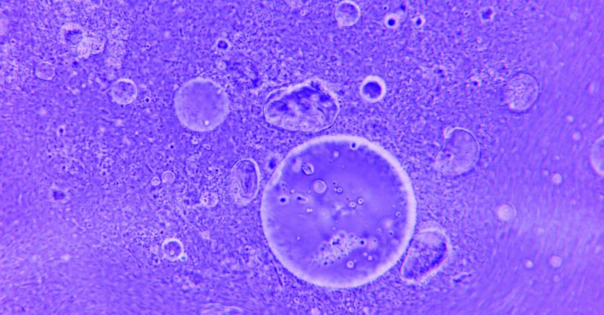
Organoids Explained: What Mini-Organs Can and Cannot Model
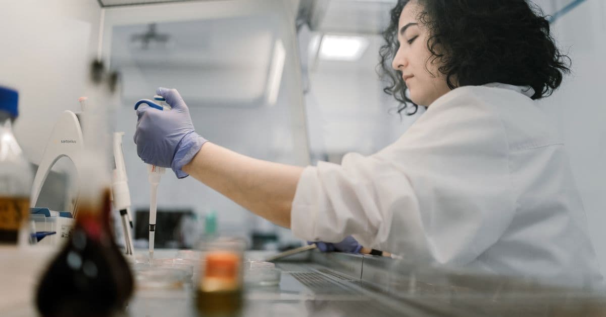
Photo: Cygaretka (CC BY-SA 3.0) / Wikimedia Commons / CC BY-SA 3.0
A layer of cells in a dish can show useful biology, but a flat culture does not reproduce the architecture of a living organ. Organoids are three-dimensional cell cultures that self-organize into structures resembling selected parts of a tissue. A single intestinal stem cell, given the right gel and growth signals, can build a gut-like structure with pockets, a central cavity, and several cell types. This article explains how organoids are grown, what they have taught researchers, and where the "mini-organ" label misleads.
The shorthand is tempting, but it overreaches. An organoid usually lacks a working blood supply, a full immune system, nerves, hormones, and the mechanical forces of a body. It is closer to a scale model in a wind tunnel than to a small aircraft: a deliberately simplified object that answers some questions well and others not at all.
Direct answer: An organoid is a three-dimensional cell model that reproduces selected structural and functional features of an organ through self-organization under controlled culture conditions. Organoids can reveal cell types, tissue architecture, developmental steps and disease mechanisms. They are not complete miniature organs and do not automatically predict how a treatment will work in a particular person.
Medical information disclaimer: This article explains biomedical research and is not medical advice. It cannot diagnose a condition, recommend treatment, select a clinical trial or predict an individual's response. For urgent symptoms or an emergency, contact local emergency services rather than relying on an article or AI system.
How the first organoids were built
The modern organoid field has a traceable starting line. In 2009, a research group reported in Nature that single Lgr5-positive stem cells from the mouse small intestine could build crypt-villus structures in a dish without any surrounding supportive tissue. The cultured structures contained all the differentiated cell types of the intestinal lining, and lineage tracing showed the Lgr5 stem-cell hierarchy was maintained (Sato et al., Nature, 2009). The cells had organized themselves. The researchers had mainly supplied a gel and the right signals.
Two years later, another team showed that aggregates of mouse embryonic stem cells could form an optic cup, the embryonic precursor of the retina. The proximal region became pigment epithelium, while the distal region folded inward and formed layered neural retina, a sequence that resembles normal eye development (Eiraku et al., Nature, 2011). The finding suggested that some of the instructions for tissue shape live inside the cells themselves.
Human brain tissue followed. In 2013, Madeline Lancaster and colleagues grew human cerebral organoids from pluripotent stem cells. These formed several brain regions, reproduced features of human cortical development such as abundant outer radial glia (a type of progenitor cell), and, when made from a patient's cells, showed premature neuron formation in a model of microcephaly (Lancaster et al., Nature, 2013). In 2015, a kidney protocol produced nephrons connected to a collecting-duct network, with proximal tubules that took up dextran and responded to the toxic drug cisplatin (Takasato et al., Nature, 2015).
| Year | Organoid system | What the study showed | Journal |
|---|---|---|---|
| 2009 | Mouse small intestine | Single Lgr5 stem cells built crypt-villus units without a mesenchymal niche | Nature |
| 2011 | Mouse optic cup | Embryonic stem cells formed a folded, layered retina-like structure | Nature |
| 2013 | Human cerebral organoid | Brain regions formed; microcephaly modeled from patient cells | Nature |
| 2015 | Human kidney | Nephrons with tubules and glomeruli; cisplatin response | Nature |
| 2018 | Human breast cancer | More than 100 tumor-derived lines in a living biobank | Cell |
Sources are linked in the list at the end of the article.
What makes an organoid three-dimensional
Cell organization
In a two-dimensional culture, cells grow on a flat plastic surface and receive roughly uniform exposure to nutrients and signals. Think of it as a city drawn on a map: every building is visible and reachable, but nothing is stacked, nothing has an inside, and no street has a back alley. An organoid grows in three dimensions, which creates gradients, layers, cavities and boundaries. Those structures influence gene expression and cell behavior, because a cell at the surface of a sphere senses a different environment from a cell at its core.
Self-organization
Cells can arrange themselves when placed in a suitable environment. Signals in the culture medium and the surrounding matrix guide proliferation, differentiation, and spatial arrangement. The researcher supplies conditions; the cells produce much of the architecture. A useful comparison is a flock of starlings: no bird holds the blueprint of the swirl, but local rules followed by each bird produce the shape.
Partial resemblance
An organoid resembles selected aspects of an organ. A gut organoid may form an epithelial layer and lumen (the hollow interior). A brain organoid may contain regions with neuronal and glial characteristics. A cancer organoid may preserve features of a tumor. A 2016 review in Cell described these systems as ones that can model organ development and disease in the lab, while noting that many applications, such as predicting individual drug response, were still being explored (Clevers, Cell, 2016). The similarity is useful but partial.
Where organoid cells come from
Adult tissue stem cells
Some organoids derive from stem or progenitor cells present in adult tissue, such as the intestinal stem cells in the 2009 work. These cultures can retain aspects of the tissue of origin and are useful for studying maintenance, repair, and disease. Access to tissue, culture success, and patient-to-patient variation all affect the model. Readers curious about how these cells decide their fates can see how stem cells know what to become.
Pluripotent stem cells
Embryonic stem cells and induced pluripotent stem cells (adult cells reprogrammed to an embryo-like state) can differentiate into many cell types. Researchers walk them through a sequence of developmental signals, imitating the order in which an embryo would deliver them. This route can model early development, but it often produces cells that resemble fetal rather than adult tissue. The kidney organoids of 2015, for example, most closely resembled first-trimester human kidney in their gene expression (Takasato et al., 2015).
Patient-derived material
Patient-derived organoids can preserve selected genetic or behavioral features of a tumor or tissue. A 2018 study in Cell built more than 100 organoid lines from primary and metastatic breast cancers. Organoid shape usually matched the original tumor's histology and its hormone-receptor and HER2 status, and DNA changes stayed largely consistent after extended passaging (Sachs et al., Cell, 2018). That is encouraging, but a culture is not a person. Selection during growth can change which cells survive, and the model omits immune, vascular, and whole-body interactions.
How a gut organoid is actually grown
The recipe differs by tissue, but the logic of the intestinal protocol shows the general pattern. It is worth walking through, because each step is also a place where variation can enter.
- Source. Researchers isolate crypts (the small pockets at the base of the intestinal lining that house stem cells) or sorted stem cells from tissue.
- Embedding. The cells are suspended in a gel that imitates the extracellular matrix, the protein scaffold that surrounds cells in a body.
- Feeding. A liquid medium supplies nutrients plus growth factors that stand in for the signals a stem-cell niche normally provides.
- Self-organization. Over days, single cells divide and form a hollow sphere. Bud-like crypt regions appear, and differentiated cells accumulate in the central cavity.
- Passaging. The structures are broken up and replanted so the line can be expanded and stored.
Two features of this recipe deserve attention. First, the lumen of a gut organoid faces inward, so nutrients, microbes, or drugs added to the medium reach the outside of the tissue rather than the side that would face food in a real gut. Second, material shed into the lumen has no obvious route out, because a dish has no flow to carry it away. These are not flaws in the idea, only reminders that the geometry of a dish is not the geometry of a body.
Common organoid models
Gut organoids
Gut organoids can model epithelial organization, barrier function, infection, and interactions among intestinal cell types. They have been used to study how pathogens enter cells and how stem cells replace the lining. Their inward-facing lumen makes some experiments awkward, because anything added to the medium touches the outer surface first.
Brain organoids
Brain organoids can model aspects of neural development, cell differentiation, and selected disease mechanisms, as the microcephaly work showed. They do not reproduce a complete brain, conscious experience, or the network of blood vessels, sensory input, immune surveillance, and body-wide signaling that surrounds a real one. For a broader view of the tissue they imitate, see how the human brain actually works.
Liver and kidney organoids
Liver and kidney models can support research into development, toxicity, transport, and disease. The 2015 kidney organoids responded to cisplatin, which suggests a role in screening for kidney toxicity. A 2026 review of liver organoids for toxicity assessment covers uses in acute injury, chronic toxicity, cholestasis and fibrosis, and notes that functional maturation and standardized derivation remain challenges (Clinical Science, 2026). A compound's behavior in an organoid can inform research without serving as a clinical clearance test. For the organs themselves, see how the kidneys work.
Cancer organoids
Cancer organoids can be derived from tumor material and used to study heterogeneity, growth, drug response, and resistance. Their value depends on how well they preserve the original tumor and which microenvironment components are included. A tumor in a body sits among fibroblasts, immune cells, and blood vessels that most organoid cultures lack. For the underlying disease biology, see what causes cancer.
Newer organ systems
The list keeps growing. A 2026 commentary in Cell Stem Cell introduced two studies of stem cell-derived heart valve models that reproduce aspects of valve development, tissue organization, and disease (Cell Stem Cell, 2026). Each new system arrives with its own questions about maturity and validation, so the headline "organoid of X" tells you the tissue, not the quality.
The extracellular matrix
Cells do not live in empty fluid. The extracellular matrix provides mechanical support and biochemical signals. Many organoid systems use a gel that helps cells organize, and the most commonly used gel, Matrigel, is derived from mouse tumor tissue. A 2026 materials paper describes it as xenogenic (from another species) and variable (Advanced Healthcare Materials, 2026).
Researchers are testing defined replacements. One 2026 study built a light-cured hyaluronic acid and collagen hydrogel that supported intestinal organoid formation about as well as the standard gel, kept its mass over 14 days while the standard gel had fully degraded by day 10, and maintained stable Lgr5 expression across passages (Regenerative Biomaterials, 2026). These are single-laboratory results, but they show where the field is heading: toward matrices whose composition and stiffness can be written down and reproduced.
A model that changes when the matrix lot changes may be biologically sensitive rather than simply unreliable, but the effect must be measured. Standardization and documentation are necessary for comparison across laboratories.
Vascularization and oxygen gradients
A large organ needs blood vessels to deliver oxygen and nutrients and remove waste. Most organoids lack a mature internal vascular network. As they grow, cells near the center may experience low oxygen or nutrient limitation, which limits how large a healthy structure can grow. This is the same physics that limits a colony of bacteria in a clump: diffusion works over short distances and fails over long ones.
Researchers explore endothelial co-culture, perfusion through microfluidic channels, transplantation into animals, and engineered scaffolds. A 2020 study in Nature found that transplanting human cortical organoids into the mouse cortex reduced cellular stress and corrected defects in cell subtype specification, which suggests that the environment, not only the cells, limits how far a dish-grown tissue matures (Bhaduri et al., Nature, 2020). These approaches can improve physiological relevance but add complexity and new variables. A vascularized model still does not reproduce the complete circulatory system.
Immune, nervous, and mechanical context
Organs interact with immune cells, nerves, hormones, blood flow, and mechanical forces. A simple organoid may omit most of these. Co-culture can add selected cell types, but it is difficult to reproduce their proportions, timing, and feedback loops. A gut in a body is also squeezed rhythmically, hosts trillions of microbes, and receives signals from the nervous system, none of which a standard gut organoid experiences. See the gut microbiome explained for what that missing context looks like.
Organ-on-chip systems introduce flow, stretch, or barrier interfaces. They may complement organoids by modeling physical forces. The combined model remains an engineered representation with a particular scope.
What organoids can reveal
Development
Organoids can expose developmental sequences that are difficult to observe directly in human tissue. Researchers can perturb signals, compare genetic backgrounds, and track cell fate. The microcephaly study is an example: patient-derived organoids showed premature neuronal differentiation, a hypothesis for why the disease produces a small brain (Lancaster et al., 2013). Developmental timing in culture may differ from timing in a body.
Infection
Organoids can provide a human-cell context for studying how a pathogen interacts with tissue. The model may reveal entry, replication, barrier disruption, and immune signaling. It does not necessarily predict transmission, symptoms, or treatment response at the organism level.
Disease mechanisms
Gene editing, patient-derived cultures, and controlled perturbations can help connect a mutation or pathway to a phenotype. The model must be validated because culture conditions can create or select phenotypes not present in the original tissue. Gene editing tools are covered in what CRISPR is and how gene editing works.
Drug discovery
Organoids can help identify toxicity, response patterns, and candidate mechanisms. They occupy a middle ground between simple cell culture and animal or clinical evidence. The result is one piece of a chain, not a guarantee of human efficacy. Machine learning is increasingly layered on top of such data; see machine learning in drug discovery.
When a brain organoid looks right but is not
The 2020 Nature study deserves a closer look because it asked a pointed question: how faithfully do cortical organoids copy the real human cortex? The team used single-cell transcriptomics to profile primary human cortical cells across developmental stages and areas, then compared organoids against that reference. Organoids produced the broad classes of cortical cells, but they lacked distinct cell subtypes and normal progenitor maturation. Area-specific molecular signatures appeared in organoid neurons, but the neurons were not spatially segregated. Organoids also showed ectopic activation of cellular stress pathways that impaired cell-type specification (Bhaduri et al., 2020).
Read that as a reminder rather than a verdict. The organoids were not useless; they captured major cell classes. But a reader who sees a labeled map of "cortical neurons" in an organoid paper should ask which subtypes are present, how closely they match a human reference, and whether stress signatures are inflating differences. The same measurement tools that make organoids exciting also make their gaps visible. The technology behind that comparison is explained in our article on single-cell RNA sequencing.
Organoids and personalized research
Patient-derived organoids may be called personalized or precision models because they originate from a particular person. That label should be used carefully. The culture may represent a subset of cells, lose immune context, select for growth advantages, or respond differently under laboratory conditions.
The best-known evidence comes from colorectal cancer. A 2019 study in Science Translational Medicine tested patient-derived tumor organoids from metastatic lesions and reported that the test predicted the response of the biopsied lesion in more than 80% of patients treated with irinotecan-based therapies, without misclassifying patients who would have benefited. The same test did not predict outcomes for 5-fluorouracil plus oxaliplatin (Sci Transl Med, 2019). The result shows why blanket claims fail: a model can work for one drug and not for another, and a single study in a defined cohort is not clinical authorization.
A drug response in an organoid may not translate because absorption, distribution, metabolism, immune effects, toxicity, and dose are absent or incomplete. Conversely, a negative result may reflect an immature or poorly preserved model rather than a patient's likely response. Clinical use requires validated assays, defined turnaround, quality control, regulatory pathways, and evidence that the model changes outcomes. For a related approach that samples tumor DNA from blood instead of growing cells, see liquid biopsy explained.
Why regulators are paying attention
On April 10, 2025, the US Food and Drug Administration announced a plan to reduce, refine, or potentially replace animal testing requirements for monoclonal antibodies and other drugs. The plan names lab-grown human organoids and organ-on-a-chip systems among the tools to be promoted, and says these tests can reveal toxic effects that could easily go undetected in animals. It also describes a pilot program in which select antibody developers could use a mostly non-animal strategy under FDA consultation (FDA, April 10, 2025).
The announcement is a statement of direction, not a finding that organoids have already replaced animal studies. Whether a given organoid assay is accepted will depend on validation for a specific question, which is why the quality-control section below matters more than the enthusiasm around it.
Reproducibility and variability
Organoid cultures can vary between donors, passages, laboratories, matrix lots, media, protocols, and operators. This heterogeneity can reflect real biology, technical variation, or both. Researchers need controls, replicate cultures, blinded analysis where appropriate, quality metrics, and detailed protocols.
A single organoid line or a small sample may not represent the diversity of a disease. Reporting only a successful culture can create publication bias. Public repositories and standardized methods can improve comparison, but biology remains variable. This is why the 100-line breast cancer biobank was notable: a collection spanning many tumors lets researchers see which findings hold across lines and which belong to one.
Organoids versus animal models and 2D cultures
Two-dimensional cultures are simpler, cheaper, and easier to image or manipulate, but they omit 3D architecture. Animal models provide circulation, immune, and whole-body context but may differ from human biology and raise ethical concerns. Organoids offer human-cell architecture in a controlled system but remain incomplete.
| Feature | 2D culture | Organoid | Animal model |
|---|---|---|---|
| Tissue architecture | Flat monolayer | 3D, partial | Complete |
| Human cells | Yes | Yes | No (usually) |
| Blood supply | None | Rarely, limited | Yes |
| Immune system | None unless added | Usually absent | Present |
| Throughput and cost | Highest throughput, lowest cost | Intermediate | Lowest throughput, highest cost |
| Whole-body metabolism | No | No | Yes |
The appropriate model depends on the question. A researcher may use 2D culture for a molecular mechanism, an organoid for tissue organization, an animal model for pharmacokinetics (how a drug moves through the body), and a clinical study for human outcomes. The models complement rather than automatically replace one another.
Ethics and consent
Organoid research can involve human tissues, reprogrammed cells, genomic information, and patient-derived cancer material. Consent should address storage, future use, sharing, genetic analysis, and commercial development where relevant. Governance differs by jurisdiction and institution.
The International Society for Stem Cell Research issued guidelines in 2021 covering recent advances involving embryos, stem cell-based embryo models, chimeras, organoids, and genome editing, and published a targeted update to version 1.2 in August 2025 that refines recommendations on stem cell-based embryo models (ISSCR). Brain organoids raise questions about neural activity, moral status, and the point at which model complexity might require new oversight. There is no single answer independent of the model's properties and evidence. Researchers should follow current institutional and international guidance rather than relying on sensational headlines.
A model-selection framework
| Research question | Useful organoid contribution | Missing context to test separately |
|---|---|---|
| Tissue development | Cell differentiation and spatial organization | Whole-body signals and timing |
| Barrier function | Epithelial structure and permeability | Circulation, nerves and immune response |
| Infection mechanism | Entry, replication and tissue response | Transmission and organism-level symptoms |
| Tumor heterogeneity | Selected tumor-cell growth and resistance | Full immune and vascular microenvironment |
| Drug toxicity | Tissue-specific response signals | Absorption, metabolism and clinical dose |
| Genetic disease | Cell-type phenotype and pathway mechanism | Patient-level variability and environment |
| Personalized research | Donor-derived features | Complete prediction of individual response |
This framework is a way to match a model to a question, not a ranking of organoid types.
Quality control and model qualification
A useful organoid study defines what the model is expected to reproduce before testing it. Researchers can compare morphology, marker expression, cell composition, barrier or contractile function, genetic stability, and response to known perturbations. These checks do not make the model complete, but they show whether it is fit for the stated question.
Quality control also includes contamination testing, passage tracking, matrix and media lots, donor metadata, culture age, and imaging conditions. A line that has changed after many passages may no longer represent the original tissue. Freezing, thawing, and expansion can alter composition. Public reporting of these details helps other laboratories understand why results agree or differ.
Model qualification should be proportional to the claim. A developmental observation may need different controls from a drug-response prediction. If a study claims that an organoid represents a patient tumor, it should report which genomic, cellular, and functional features were preserved and which were absent. Transparency is more informative than calling every successful culture "personalized."
Common misunderstandings
"Organoids are tiny organs." They are not. They lack the integrated circulation, immune system, and nerves that make an organ work in a body, and many resemble fetal rather than adult tissue.
"A bigger organoid is a better organoid." Size is limited by diffusion, and a larger structure often has a starved, stressed core. Complexity is not automatically realism.
"If it works in an organoid, it will work in a person." The colorectal study above shows a model can predict one drug class and fail on another. Prediction has to be validated drug by drug and cohort by cohort.
"Organoids make animal research obsolete." Regulatory interest is growing, but animal and human studies still capture whole-body processes that no current organoid does.
"Brain organoids think." Ordinary brain organoids lack sensory input, a body, and the structure needed for the kinds of activity associated with awareness. More complex neural models raise ongoing scientific and ethical questions that deserve oversight rather than headlines.
Future directions
Researchers are combining organoids with vascular cells, immune cells, neurons, microbiomes, sensors, microfluidics, and spatial measurement. A 2026 report described a device that holds three stretchable microelectrode arrays so that electrical activity can be recorded from three midbrain organoids under standardized perfusion (Lab on a Chip, 2026). Better imaging and single-cell analysis can reveal cell states and interactions, and artificial intelligence is being applied to organoid shape and function; see single-cell AI and cell-state prediction. Biobanks can support larger comparisons.
The more components a model includes, the more biologically rich it may become, but the more difficult it is to standardize and interpret. A model should be judged against a defined question and validated observation.
Limitations
Organoids are partial models. They can be immature, heterogeneous, genetically altered by culture, missing vascular, immune, neural, or mechanical context, and sensitive to matrix and media conditions. Patient-derived organoids may not represent every cell in the original tissue. A drug response in a dish does not establish human efficacy or safety. Ethics, consent, privacy, and regulatory requirements depend on the material and jurisdiction.
This article is biomedical education only. It is not diagnosis, treatment advice, a clinical-trial recommendation, or a prediction of any individual's disease or drug response. For urgent symptoms or an emergency, contact local emergency services.
The takeaway
Organoids earned their place by doing something flat cultures could not: letting human cells build tissue-like structure that researchers can watch, perturb, and compare. The record since 2009 is real, from intestinal crypts to brain regions to patient tumors. The record also contains the corrections, such as stress signatures in cortical organoids and drug-specific prediction in colorectal tumors. The most accurate way to read an organoid result is to ask what the model contains, what it lacks, and what independent evidence supports the link to a living person.
Frequently asked questions
Q: What is an organoid? A: An organoid is a three-dimensional cell culture that reproduces selected structural or functional features of an organ. It is grown from stem cells or tissue-derived cells that self-organize in a supportive gel and a defined medium.
Q: Are organoids miniature organs? A: No. They are organ-like models, not complete organs. They usually lack full blood supply, immune context, nervous-system interactions, and whole-body physiology, and many resemble fetal tissue more than adult tissue.
Q: How are organoids made? A: They are grown from tissue-derived cells or stem cells in media and matrices that guide proliferation and self-organization. In the 2009 intestinal work, single Lgr5 stem cells formed crypt-villus structures containing all differentiated cell types of the intestinal lining.
Q: Can organoids model cancer? A: They can model selected tumor features, growth, and drug-response patterns. A 2018 breast cancer biobank of more than 100 lines largely preserved tumor histology and receptor status, but clinical predictive value must be validated separately for each drug and cancer type.
Q: Can organoids replace animal testing? A: They may reduce or complement some animal experiments. In April 2025 the FDA announced a plan to reduce, refine, or potentially replace animal testing requirements for some drugs and to promote organoids and organ-on-a-chip systems, but organoids do not reproduce every whole-body process.
Q: Can an organoid predict a patient's treatment response? A: Not automatically. A 2019 study predicted response to irinotecan-based therapy in more than 80% of colorectal cancer patients but did not predict outcomes for 5-fluorouracil plus oxaliplatin, which shows that validation is drug-specific.
Q: Do brain organoids have consciousness? A: Ordinary organoid models should not be described as conscious. They lack sensory input, a body, and most of the architecture of a brain. More complex neural models raise ongoing scientific and ethical questions that require careful oversight.
Q: Why do organoids vary between laboratories? A: Cell source, donor genetics, passage number, matrix, media, protocol, operator, and measurement all affect the culture. Common gels are tumor-derived and vary between batches, which is one reason defined replacements are under study.
Sources
- Sato T, et al. Single Lgr5 stem cells build crypt-villus structures in vitro without a mesenchymal niche. Nature, 2009. doi:10.1038/nature07935
- Eiraku M, et al. Self-organizing optic-cup morphogenesis in three-dimensional culture. Nature, 2011. doi:10.1038/nature09941
- Lancaster MA, et al. Cerebral organoids model human brain development and microcephaly. Nature, 2013. doi:10.1038/nature12517
- Takasato M, et al. Kidney organoids from human iPS cells contain multiple lineages and model human nephrogenesis. Nature, 2015. PubMed 26444236
- Clevers H. Modeling development and disease with organoids. Cell, 2016. PubMed 27315476
- Sachs N, et al. A living biobank of breast cancer organoids captures disease heterogeneity. Cell, 2018. PubMed 29224780
- Patient-derived organoids can predict response to chemotherapy in metastatic colorectal cancer patients. Science Translational Medicine, 2019. doi:10.1126/scitranslmed.aay2574
- Bhaduri A, et al. Cell stress in cortical organoids impairs molecular subtype specification. Nature, 2020. doi:10.1038/s41586-020-1962-0
- US Food and Drug Administration. FDA announces plan to phase out animal testing requirement for monoclonal antibodies and other drugs, April 10, 2025. fda.gov
- International Society for Stem Cell Research. Guidelines for stem cell research and clinical translation. isscr.org
This article is educational biomedical information and is not medical advice.
Read next:
Follow new articles
Add SciHub101 to your feed reader to see new science and technology stories.
About the Author
SciHub101 is an independent science and technology publication founded and edited by Buğra Sözeri, a software engineer and app developer based in Türkiye. AI tools assist with drafting and research. Source documentation in the existing archive is uneven, and articles are being reviewed and corrected. The Editorial Team byline identifies editorial responsibility; it does not certify specialist or independent human review. Where we compare products, anything built by our own team is labelled as such — see the editorial guidelines and About page for how corrections and disclosures work.
Related Articles
Personalized Cancer Vaccines Explained: Neoantigens, mRNA and Clinical-Trial Reality
Personalized cancer vaccines are experimental immunotherapies built from a patient's tumor sequence. See how neoantigens are chosen and what trials show.
Liquid Biopsy Explained: What Blood Can Reveal About Cancer
Liquid biopsy reads tumor DNA, cells and vesicles in blood. See what ctDNA can show, where evidence is strong, and why it does not replace tissue biopsy.
Henrietta Lacks: The Woman Behind HeLa, the Cells That Changed Medicine — SciHub101
Henrietta Lacks' cells, taken without consent in 1951, became the first immortal human cell line. HeLa cells helped develop the polio vaccine, cancer treatments, and IVF. Her family learned this 25 years later.
How Apoptosis Works: Why Healthy Cells Choose to Die
Apoptosis is programmed cell death. Learn how caspases, mitochondria, and Bcl-2 proteins carry it out, and what goes wrong in cancer and autoimmunity.
Single-Cell RNA Sequencing Explained: How Scientists Read Gene Activity One Cell at a Time
Single-cell RNA sequencing measures gene activity in individual cells instead of averaging a tissue. See how the workflow runs, what it finds, and where it misleads.