Single-Cell AI: Understanding Cellular Diversity at Scale
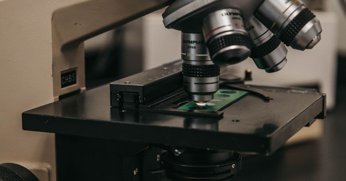
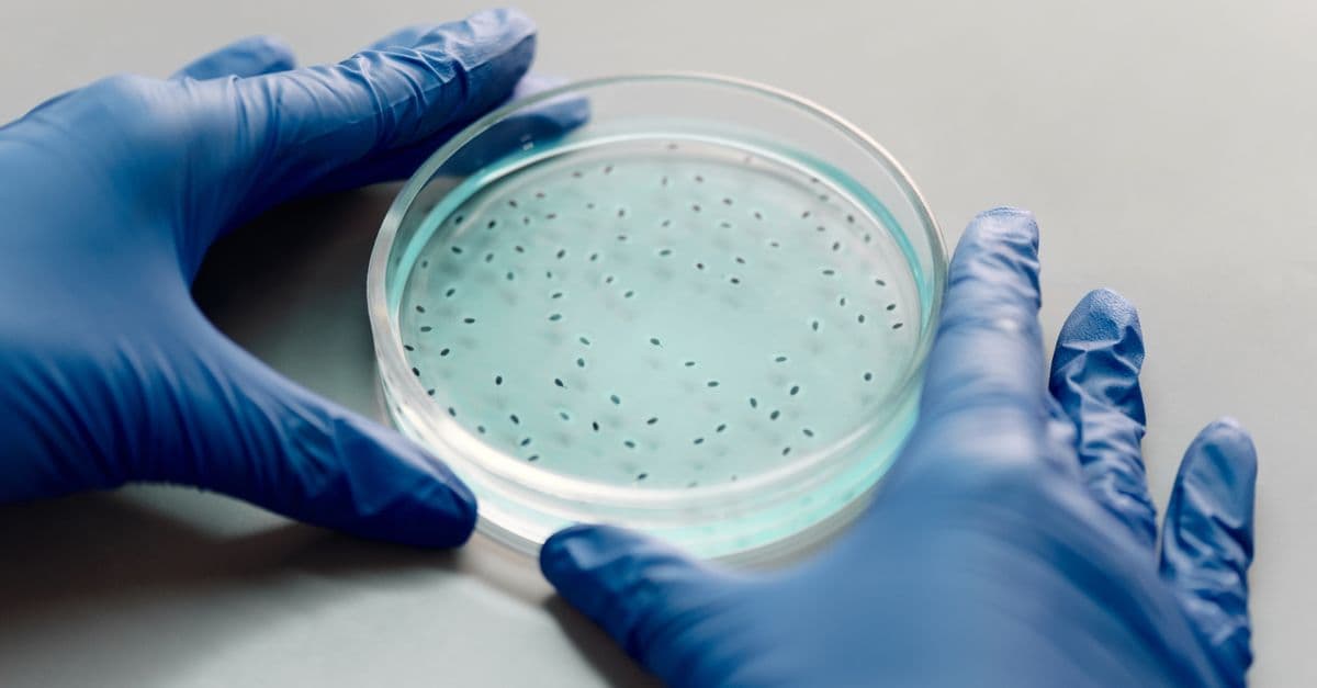
Photo: Jeff Burkholder / Pexels
A human liver is not one thing doing one job. It's a mixture of hepatocytes that process glucose, others that break down drugs, immune cells fresh off the bone marrow assembly line, and immune cells that have spent months fighting infection. Bulk RNA sequencing, the standard method for most of the last 20 years, grinds a tissue sample into a slurry and measures the average gene activity across every cell in it. That average erases the differences that actually explain how a tissue works, and how it fails. Single-cell RNA sequencing (scRNA-seq) reads each cell separately instead, but a single 100,000-cell experiment generates roughly two billion data points. No human can eyeball that. Machine learning is what turns the flood of numbers into a map of cell types, developmental paths, and disease states.
Educational content, not medical advice. Talk to your doctor before changing any medication, treatment, or routine. Information here reflects current research but every person's situation differs.
Key Takeaways
- Single-cell RNA-seq resolves dozens to hundreds of distinct cell types per tissue that bulk sequencing collapses into a single average signal.
- Neural network-based clustering and classification methods now assign cell types with accuracy competitive with expert manual annotation on well-characterized tissues, though performance drops sharply on rare or novel cell states.
- Trajectory inference algorithms reconstruct differentiation pathways, showing how a stem cell becomes a mature neuron or immune cell, from static snapshots rather than time-lapse footage.
- Large single-cell "foundation models" trained on tens of millions of cells, such as Geneformer and scGPT, are starting to predict how cells will respond to gene edits and drugs they have never been directly tested against.
- Global reference atlases, including Tabula Sapiens and the Human Cell Atlas, have already logged tens of millions of annotated human cells and are the training ground for most of these models.
The problem: cells are individuals, not averages
Traditional bulk RNA sequencing works like this: take a chunk of tissue, homogenize it, and sequence the combined RNA from every cell inside. The output is a single number per gene, an average across possibly a million cells. That number is useful for coarse comparisons, but it hides which specific cells are doing what.
Consider a tumor biopsy that contains cancer cells tangled up with immune cells. Bulk sequencing might report that the gene for the immune checkpoint protein PD-1 is expressed at a middling level across the sample. That single number is compatible with wildly different biological realities: T cells expressing PD-1 heavily while B cells express none, or a more even spread across all immune cells. Which reality is true changes how a doctor might think about an immunotherapy response. Bulk sequencing cannot tell the two apart.
Single-cell RNA-seq solves this by dissociating the tissue into individual cells before sequencing, so each cell produces its own expression profile. A typical experiment on the 10x Genomics Chromium platform, the most widely used commercial single-cell system, profiles a few thousand to tens of thousands of cells per sample, with each cell's data spanning roughly 20,000 genes. That is where the scale problem starts. A 50,000-cell run generates a matrix with a billion entries, and every one of those entries carries noise from sequencing depth, cell size, and technical dropout, where a gene is genuinely on in a cell but the sequencer simply misses the transcript.
How AI decodes single-cell data
Dimensionality reduction and automatic clustering
The first challenge is visual and computational: a 20,000-gene expression profile per cell cannot be plotted, compared, or reasoned about directly. Researchers have used t-SNE (introduced in 2008) and UMAP (2018) for years to compress that 20,000-dimensional space down to two dimensions for a scatter plot, then eyeball the resulting islands of dots and guess, "that cluster in the corner looks like T cells based on its marker genes."
Machine learning changes the compression step itself. Instead of a fixed mathematical projection, an autoencoder, a neural network trained to reconstruct its own input after squeezing it through a narrow bottleneck layer, learns which combinations of genes actually distinguish cell types in the data at hand. The bottleneck typically holds 10 to 50 dimensions instead of 20,000. Because the network is trained on the data itself rather than a generic formula, it tends to preserve biologically meaningful structure, like the gradual transition between a progenitor cell and its mature descendant, that a one-size-fits-all projection can blur together.
scVI (single-cell Variational Inference), published by Romain Lopez and colleagues at UC Berkeley in Nature Methods in 2018, is the most cited example of this approach. It models the raw count data directly, accounting for the fact that scRNA-seq counts are sparse and noisy by construction, and it scales to datasets with more than a million cells. Downstream of that compressed representation, standard clustering algorithms, k-means or Gaussian mixture models, group similar cells together, and a classifier assigns each cluster a cell type based on the genes it expresses.
Cell type assignment: how accurate is automated annotation, really
Once clusters exist, the next question is what to call them. The standard approach trains a classifier on reference data, meaning cells that have already been labeled by experts using known marker genes (CD3 for T cells, CD19 for B cells, CD68 for macrophages), and then applies that classifier to new, unlabeled data.
A 2019 Nature Methods benchmark by Abdelaal and colleagues compared 22 automated annotation methods against manual expert labeling across multiple datasets and found that the best-performing tools matched expert annotation closely on common, well-represented cell types, but accuracy dropped substantially for rare populations and for cell types absent from the training reference. That gap between "common cell type in a well-studied tissue" and "rare or novel cell type" is the single biggest caveat to keep in mind whenever a paper reports a headline accuracy number for automated cell typing.
Trajectory inference: reconstructing development from a single snapshot
Cells are not static. A hematopoietic stem cell in bone marrow differentiates step by step into, say, a mature T cell, passing through intermediate progenitor states along the way. A single-cell RNA-seq experiment captures a snapshot of a tissue at one moment, not a movie, so it cannot show that process directly. But if a large enough population of cells is sampled at once, that snapshot will contain cells caught at every stage of the transition simultaneously.
Trajectory inference algorithms exploit that fact. The general logic, formalized across dozens of published methods and reviewed comprehensively by Saelens and colleagues in Nature Biotechnology in 2019 (technically Nature Biotechnology, not Nature Reviews Molecular Cell Biology as sometimes cited), works roughly like this: build a graph connecting each cell to its most transcriptionally similar neighbors, then find the longest coherent path through that graph. Cells along that path are ordered into "pseudotime," a relative ordering that approximates developmental progression even though no clock was ever running during the experiment.
It's a bit like reconstructing a movie from a single photograph of a crowded street where people are frozen mid-stride at every possible position between two points. If enough pedestrians are captured, you can infer the walking path even without ever filming anyone moving.
How AI decodes disease states in single cells
In diseased tissue, entire cell populations can shift their behavior in ways bulk sequencing would never separate from the healthy background. In Alzheimer's disease research, healthy neurons typically express genes tied to synaptic signaling and normal energy metabolism, while neurons under disease stress upregulate stress-response and apoptosis-related genes and downregulate synaptic genes. A classifier trained to distinguish these two profiles, then applied to a new patient's brain tissue, can estimate what fraction of that patient's neurons have already crossed into the dysfunctional state, which researchers are exploring as a potential marker of disease progression speed. That link between cell-state composition and clinical trajectory is still an active area of research rather than a settled clinical tool.
Real-world applications
Human Cell Atlas and Tabula Sapiens: building the reference maps
The Human Cell Atlas is an international consortium effort to catalog every cell type in the human body. As of recent public reporting, the project has profiled more than 70 million cells from over 11,000 donors across more than 100 countries, spanning more than 500 individual research projects (Human Cell Atlas). A related but distinct effort, Tabula Sapiens, published in Science in 2022 by the Chan Zuckerberg Biohub-led consortium, sequenced roughly 500,000 cells from 24 tissues and organs taken from the same set of donors, identifying more than 400 distinct cell types in a single coordinated dataset (Tabula Sapiens, Science, 2022).
Machine learning is what makes these atlases usable as references rather than just enormous spreadsheets. Batch correction algorithms, including Harmony and scVI, remove technical differences between samples processed on different days or by different labs so that a T cell sequenced in a Boston lab in 2021 and a T cell sequenced in a Shanghai lab in 2023 land in the same computational neighborhood rather than being misread as biologically distinct.
Single-cell foundation models: Geneformer and scGPT
The newest development in this field borrows directly from large language models. Geneformer, introduced by Theodoris and colleagues, is a BERT-style transformer originally trained on around 30 million single cells and later scaled to roughly 95 million, learning to represent genes based on the regulatory context they typically appear in, much like a language model learns word meaning from surrounding text. scGPT, published in Nature Methods in 2024 by Cui and colleagues, takes a GPT-style generative approach trained across more than 33 million cells and can be fine-tuned for cell type annotation, batch integration across studies, and predicting how a cell's gene expression will shift in response to a genetic perturbation it has never actually been exposed to in the lab (scGPT, Nature Methods, 2024).
The practical promise of these models is prediction without an experiment: instead of physically knocking out a gene in a dish and waiting weeks for results, a researcher can ask the model to simulate the likely transcriptional consequence first, then prioritize which of dozens of candidate experiments are actually worth running.
Single-cell cancer genomics
Tumor biopsies analyzed with scRNA-seq routinely reveal that a single tumor is not one cancer but a mixture of genetically distinct cancer cell subpopulations, some of which may already carry resistance mutations to a drug before treatment even begins. Layered on top of that, single-cell profiling of the immune cells infiltrating a tumor, particularly the density of CD8+ cytotoxic T cells, has been studied as a candidate signal for predicting which patients are more likely to respond to checkpoint immunotherapy, though this remains an active research area rather than a routine diagnostic test in most clinical settings.
COVID-19 immune response mapping
A widely cited single-cell immune atlas study by Lee and colleagues, published in Science Immunology in 2020, compared immune cell states between patients with severe and mild COVID-19 and found that severe cases were associated with dysregulated, hyperinflammatory monocyte populations and altered T cell states, distinguishing the immune signature of severe disease from a more controlled, coordinated response in mild cases (Lee et al., Science Immunology, 2020). That kind of cell-state fingerprinting became one of the clearest public demonstrations of what single-cell resolution adds over a bulk blood test during the pandemic.
What single-cell AI gets wrong, and why it matters
Batch effects are a moving target, not a solved problem
Cells processed on different days, by different technicians, or on different sequencing runs pick up systematic technical differences in gene expression that have nothing to do with biology. Algorithms like Harmony and scVI correct for a large share of this, and Tian and colleagues benchmarked correction methods head to head using controlled cell-line mixtures in Nature Methods in 2019, but no method removes batch effects perfectly, and over-correction carries its own risk: erasing a real biological difference between, say, healthy and diseased tissue because the algorithm mistook it for a technical artifact.
Rare cell types are systematically undercounted
Clustering algorithms are tuned to find the dominant populations in a dataset, the cell types that make up 5 percent or more of the sample. A cell type present at 0.1 percent of a 20,000-cell sample corresponds to roughly 20 cells, a number too small for most clustering methods to confidently separate from noise. Finding genuinely rare cell populations, which are frequently the most clinically interesting ones (a handful of drug-resistant cancer cells, for instance), usually requires deliberately oversampling a tissue or running targeted follow-up sequencing on a suspected rare population.
Transitional cells resist clean labels
Not every cell fits neatly into "T cell" or "B cell." Cells caught mid-differentiation, or mid-apoptosis, occupy a genuine biological gray zone, and forcing them into a single discrete label misrepresents the underlying biology. The more honest computational approaches assign probability distributions across multiple possible cell states rather than a single confident answer, but many downstream analyses still simplify that distribution down to "most likely label" for the sake of a clean plot, quietly discarding uncertainty that was real.
Sample size limits what conclusions are statistically defensible
Most published single-cell studies profile somewhere between 10,000 and 50,000 cells per condition. That is more than enough to characterize dominant cell types confidently, but it is thin for anything occurring below roughly 1 percent frequency. Detecting a population present at 0.1 percent with reasonable statistical confidence generally requires sampling on the order of a million cells, which is why the largest reference atlases, not individual disease studies, are usually where rare cell discovery happens.
Comparison: single-cell RNA-seq vs. bulk RNA-seq vs. spatial transcriptomics
| Method | What it measures | Resolution | Typical cost driver | Best use case |
|---|---|---|---|---|
| Bulk RNA-seq | Average gene expression across the whole sample | Whole tissue, no cell-level detail | Lower per-sample cost, simple prep | Comparing overall tissue states across many samples cheaply |
| Single-cell RNA-seq (scRNA-seq) | Gene expression per individual cell | Individual cell, expression only | Cell isolation + per-cell library prep, scales with cell count | Discovering and classifying cell types and states within a tissue |
| Spatial transcriptomics | Gene expression per cell or small region, with physical tissue location preserved | Individual cell or small spot, plus X/Y position | Highest, specialized imaging + sequencing hardware | Mapping which cell types sit next to each other in intact tissue |
Table: How the three main transcriptomic profiling methods trade off resolution, cost, and what question they can actually answer.
What people get wrong about single-cell AI
A common misreading of headline accuracy numbers, like "95 percent accurate cell classification," is treating them as a single fixed property of the algorithm rather than a number that depends heavily on which tissue, which cell types, and which reference dataset were used. A classifier that hits high agreement with expert annotation on well-studied blood cell types will typically perform far worse on a poorly characterized tissue with few labeled reference cells, simply because it has almost nothing to learn from.
Another frequent confusion is assuming single-cell RNA-seq shows where cells are physically located in a tissue. It does not. Standard scRNA-seq requires dissociating the tissue into a cell suspension first, which destroys the spatial arrangement entirely. Spatial transcriptomics, a separate and more recently matured technique, is what preserves physical position alongside gene expression, at a higher cost and lower per-cell sequencing depth than standard scRNA-seq.
Finally, "AI discovered a new cell type" headlines deserve a skeptical read. Clustering algorithms will always produce clusters, including some that reflect nothing more than technical noise, a stressed-cell artifact from tissue dissociation, or two genuinely different cell types that happen to look similar in the reduced-dimension space the algorithm used. Distinguishing a real, reproducible novel cell type from an artifact still requires independent validation, typically with a different technique like imaging or protein staining, before the finding is considered established.
FAQ
Q: How many cells do I need to sequence for a useful single-cell RNA-seq experiment?
A: For basic cell type discovery in a moderately diverse tissue, 10,000 to 100,000 cells is a common working range on platforms like 10x Genomics Chromium. Detecting genuinely rare cell populations, below roughly 1 percent frequency, generally requires sampling into the hundreds of thousands or millions of cells.
Q: Can single-cell RNA-seq replace tissue imaging?
A: No. Standard scRNA-seq requires dissociating tissue into a cell suspension, which destroys physical location information entirely. Imaging shows where cells sit relative to each other; scRNA-seq shows what genes they express. Spatial transcriptomics is the technique built specifically to combine both.
Q: How much does a single-cell RNA-seq experiment cost?
A: Per-sample library preparation costs on 10x Genomics platforms run roughly in the hundreds of dollars per sample before sequencing, with total project costs varying widely by cell target count, sequencing depth, and whether the work is done through a university core facility or a commercial vendor. Institutional core facility price lists, such as Harvard's Single Cell Core, are the most reliable current pricing reference for a specific project.
Q: Can machine learning actually discover new cell types, or does it just relabel known ones?
A: Clustering algorithms can surface groups of cells with expression patterns that don't match any known reference, which is how some genuinely novel cell states have been identified in large atlas projects. But distinguishing a real new cell type from a technical artifact of tissue dissociation or sequencing noise requires independent validation, so any single clustering result should be treated as a hypothesis rather than a confirmed discovery.
Q: What is pseudotime, and is it the same as real time?
A: Pseudotime is a computationally inferred ordering of cells along an inferred differentiation path, based on how similar their gene expression profiles are to each other. It approximates a developmental sequence but is not measured in actual minutes or hours, and it assumes the sampled cells genuinely span a single coherent developmental trajectory, an assumption that does not always hold.
Q: What's the difference between scVI and a simple PCA-based clustering pipeline?
A: PCA (principal component analysis) is a fixed linear projection that reduces dimensionality using the same mathematical rule regardless of the dataset. scVI is a neural network trained specifically on the dataset at hand, explicitly modeling sequencing noise and dropout, which generally preserves more biologically meaningful structure, particularly for large, noisy, or multi-batch datasets, at the cost of being more computationally intensive to train.
Q: Are single-cell foundation models like Geneformer and scGPT actually used in real research yet?
A: Yes, though mostly in research and early translational settings rather than routine clinical use. They are being applied to tasks like predicting gene perturbation responses and improving cell type annotation across studies, but as of 2026 they function primarily as tools that help researchers prioritize which wet-lab experiments to run, not as replacements for those experiments.
Q: Why do cell type classifiers perform worse on rare cell populations?
A: Classifiers learn from labeled reference data, and rare cell types are, by definition, underrepresented in almost any reference dataset. A 2019 Nature Methods benchmark by Abdelaal and colleagues found this gap directly: strong agreement with expert labeling on common cell types, and a measurable drop in accuracy for rare or reference-absent populations.
Q: Does single-cell analysis work on any tissue, or are some tissues harder than others?
A: Some tissues dissociate into single-cell suspensions cleanly; others, particularly tissues with tough extracellular matrix like bone or certain fibrotic tissue, are harder to break apart without damaging or losing cells, which can bias which cell types actually make it into the final dataset. Single-nucleus RNA-seq, which profiles nuclei instead of whole cells, is one common workaround for tissues that resist standard dissociation.
The takeaway
Single-cell RNA sequencing turned tissue biology from an averaged signal into a cell-by-cell census, and machine learning is the only practical way to read that census at the scale modern experiments produce. The tools work well where they have abundant, well-labeled training data, common cell types in well-studied tissues, and they work considerably worse at the edges, rare populations, novel cell states, poorly characterized tissues. The newest generation of single-cell foundation models is starting to close that gap by learning from tens of millions of cells at once, but the fundamental trade-off has not disappeared: a model's confidence is a reflection of how much labeled data it has seen, not a proof of biological truth. Machine learning is applied to other areas of biology too, such as AI-driven antibiotic discovery against resistant bacteria.
Read next:
- The CRISPR Gene Editing Breakthrough
- The AlphaFold Protein Folding Breakthrough
- The Human Genome Project Explained
- How Scientists Use AI to Discover New Drugs
Sources
- Human Cell Atlas Consortium. https://www.humancellatlas.org/
- Saelens, W., et al. (2019). "A comparison of single-cell trajectory inference methods." Nature Biotechnology. https://www.nature.com/articles/s41587-019-0071-9
- Lopez, R., et al. (2018). "Deep generative modeling for single-cell transcriptomics." Nature Methods. https://www.nature.com/articles/s41592-018-0229-2
- Lee, J. S., et al. (2020). "Immunophenotyping of COVID-19 and influenza highlights the role of type I interferons in development of severe COVID-19." Science Immunology. https://www.science.org/doi/10.1126/sciimmunol.abd1554
- 10x Genomics Chromium Platform. https://www.10xgenomics.com/
- Tian, L., et al. (2019). "Benchmarking single cell RNA-sequencing analysis pipelines using mixture control experiments." Nature Methods. https://www.nature.com/articles/s41592-019-0425-8
- Abdelaal, T., et al. (2019). "A comparison of automatic cell identification methods for single-cell RNA sequencing data." Genome Biology. https://genomebiology.biomedcentral.com/articles/10.1186/s13059-019-1795-z
- The Tabula Sapiens Consortium (2022). "The Tabula Sapiens: A multiple-organ, single-cell transcriptomic atlas of humans." Science. https://www.science.org/doi/10.1126/science.abl4896
- Cui, H., et al. (2024). "scGPT: toward building a foundation model for single-cell multi-omics using generative AI." Nature Methods. https://www.nature.com/articles/s41592-024-02201-0
- Theodoris, C. V., et al. (2023). "Transfer learning enables predictions in network biology" (Geneformer). Nature. https://www.nature.com/articles/s41586-023-06139-9
Follow new articles
Add SciHub101 to your feed reader to see new science and technology stories.
About the Author
SciHub101 is an independent science and technology publication founded and edited by Buğra Sözeri, a software engineer and app developer based in Türkiye. AI tools assist with drafting and research. Source documentation in the existing archive is uneven, and articles are being reviewed and corrected. The Editorial Team byline identifies editorial responsibility; it does not certify specialist or independent human review. Where we compare products, anything built by our own team is labelled as such — see the editorial guidelines and About page for how corrections and disclosures work.
Related Articles
AI & Viral Evolution: Predicting Pandemics Before They Spread
How machine learning models forecast viral mutations, flag vaccine-escape variants, and speed up pandemic preparedness before outbreaks peak.

H5N1 Bird Flu Explained — How the Virus Works in 2026
H5N1 bird flu explained: how avian influenza infects birds, cattle, and people, what the US dairy outbreak changed, and what the evidence says on risk.
Personalized Medicine & AI-Driven Genomics
How genetic data and machine learning guide drug choice, and what trials of cancer, warfarin, and antidepressant testing actually show.
Machine Learning for Biomarker Discovery: Finding Disease Signals in Biological Noise
AI is combing through genomic, proteomic, and metabolomic data to identify novel biomarkers—measurable biological signals that predict disease, drug response, and patient outcomes.

AI Meets CRISPR: How Machine Learning Is Supercharging Genetic Engineering
Machine learning is making CRISPR more precise and predictable, designing better guide RNAs and anticipating off-target edits before they happen.
Machine Learning in Drug Discovery: AI Finds Medicines
How neural networks accelerate drug discovery from years to months.