How Do Teeth Work? The Science of Dental Anatomy
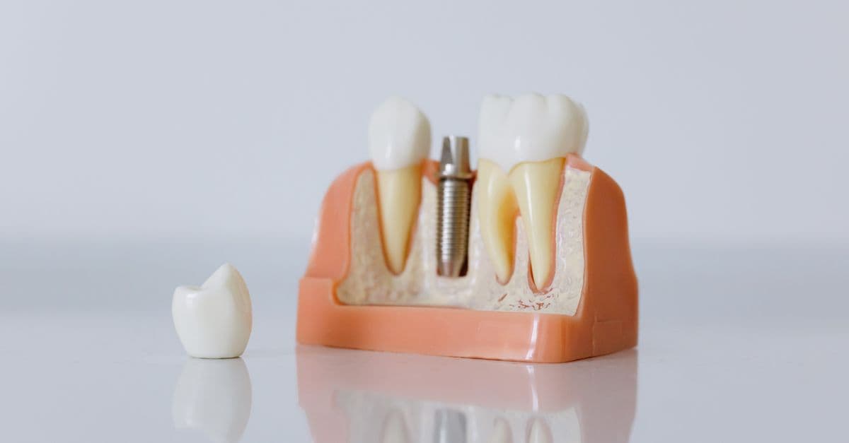
Photo: cottonbro studio / Pexels
Human teeth must withstand 400 metric tons of cumulative bite force over a lifetime while resisting acid, bacteria, and abrasion. Enamel—the hardest tissue in the human body—covers the crown, but once it erodes, it cannot regenerate because the cells that built it (ameloblasts) self-destruct after completing their work. A single cavity that penetrates enamel and reaches the dentin below triggers pain as fluid inside microscopic tubules moves, signaling danger to the nerve. Understanding how teeth work requires learning why they are not bones, what makes enamel irreplaceable, and how the alignment of hard and soft tissues creates a system of remarkable durability that is, paradoxically, vulnerable to a single bacterium's waste product: acid.
The Four-Layer Structure: From Hardest to Soft
A tooth is not a monolithic structure but a composite of four distinct tissues, each serving a separate function. Beginning at the surface and moving inward, the organization from hardest to most vulnerable reveals the evolutionary priorities baked into dental anatomy.
Enamel — the translucent, crystalline outer layer of the tooth crown that is harder than bone and the hardest substance naturally produced by the human body. Enamel consists of approximately 96% hydroxyapatite (a crystalline calcium-phosphate mineral) and only 4% water and organic material. This extremely high mineral content gives enamel its hardness (ranked 5 on the Mohs hardness scale, comparable to steel), but it also makes enamel brittle and inflexible. Unlike bone, enamel contains no living cells; it is an acellular tissue. Once enamel is damaged, the body cannot repair it because there are no cells available to secrete new material. Enamel wears at approximately 10 micrometers per year under normal function. Over 80 years, this adds up to nearly 1 millimeter of wear, which is why elderly people who kept their teeth often have shorter crowns and more worn-down biting surfaces.
The second layer, dentin, lies directly beneath enamel. Dentin is softer than enamel (measuring around 3 on the Mohs scale, roughly the hardness of bone) and comprises about 70% hydroxyapatite and 30% water and collagen. Unlike enamel, dentin is cellular and contains microscopic tubules—fluid-filled channels that radiate from the pulp chamber at the center of the tooth outward toward the enamel-dentin junction. These dentinal tubules contain fluid and processes (extensions) of odontoblasts, specialized sensory cells that live at the inner edge of the dentin. When dentin is exposed (through enamel erosion, gum recession, or cavity formation), changes in the fluid pressure inside these tubules trigger the odontoblasts and pain receptors, causing the sharp, shooting sensation of tooth sensitivity.
Source: Marshall, G.W. (1993), "Dentin: microstructure and characterization," Quintessence International, Vol. 24, No. 9 — comprehensive description of dentinal tubule anatomy and mechanosensitivity.
The third layer, cementum, covers the root of the tooth below the gum line. Cementum is similar to bone in composition (approximately 45% mineral and 55% water and collagen) and, like bone, is a living tissue containing cells called cementocytes. Cementum is softer than enamel or dentin and serves as an attachment point for the periodontal ligament, the elastic fibers that anchor the tooth to the jawbone. Because cementum is softer and exposed in gum recession (where gum tissue pulls away from the tooth), cementum erodes more easily than enamel, and root cavities in older adults with receding gums are common.
At the core of the tooth lies the pulp, a soft tissue chamber containing blood vessels, nerves, and connective tissue. The pulp extends from the pulp chamber in the crown down through a root canal to the tip of the root, where it opens to the bone. During development, the pulp is critical for tooth formation because it contains odontoblasts (which produce dentin) and ameloblasts (which produce enamel). In an adult, the pulp's primary function is sensory—it detects temperature, pressure, and pain. Blood vessels in the pulp deliver nutrients to the tooth, though mature teeth with fully developed roots can survive for years even if the pulp is removed (root canal therapy). Nerve fibers in the pulp transmit pain signals, which is why decay that reaches the pulp causes severe pain. The pulp gradually shrinks with age as secondary dentin is deposited, which is one reason teeth become less sensitive to temperature as people age.
| Tissue | Hardness | Mineral Content | Living Cells | Regenerates | Location |
|---|---|---|---|---|---|
| Enamel | Hardest (5 Mohs) | 96% | No | No | Crown surface |
| Dentin | Medium (3 Mohs) | 70% | Yes (odontoblasts) | Limited | Crown and root interior |
| Cementum | Softer than enamel | 45% | Yes (cementocytes) | Limited | Root surface |
| Pulp | Soft (not mineralized) | 0% | Yes (nerves, blood) | Yes | Core chamber and canal |
Source: Nanci, A. (2017), Ten Cate's Oral Histology: Development, Structure, and Function, 9th ed., Mosby — detailed histological descriptions of all four tooth tissues.
Tooth Types and Their Specialized Functions
The human mouth contains 32 permanent teeth (in adults with all wisdom teeth) organized into four types, each adapted for a different mechanical role in eating. Baby teeth follow the same pattern but with only 20 total (no second molars or wisdom teeth).
Incisors (8 total: 4 upper, 4 lower) are the front teeth with a sharp, blade-like crown and a single root. The flat, straight edge of the incisor crown is optimized for cutting and slicing food. Incisors are used to bite into apples, bread, and leafy vegetables. Because they are exposed during speech and smiling, incisors receive the most cosmetic scrutiny, and damage to incisors causes significant social concern in ways that damage to molars does not.
Canines (4 total: 2 upper, 2 lower) are the pointed teeth located at the corners of the mouth. Canines have the longest, strongest single root of any tooth type, allowing them to bear high bite forces. The pointed cusp (tip) is designed to tear fibrous foods like meat and to grip and rip. In many mammal species, canines are greatly enlarged as weapons and dominance signals; in humans, canines are reduced in size but are still specialized for piercing and tearing.
Premolars (8 total: 4 upper, 4 lower) are intermediate teeth with two cusps (peaks) on the crown and usually one or two roots. Premolars transition from the cutting function of incisors to the grinding function of molars. Premolars have wider, flatter crowns than canines but narrower surfaces than molars. They are adapted for crushing and grinding food with moderate force.
Molars (12 total: 8 permanent molars + 4 wisdom teeth/third molars, or 8 without wisdom teeth) have four cusps (in upper molars) or five cusps (in lower molars) and two to three roots. Molars have the largest surface area and lowest cusps relative to crown size, optimized for grinding. The broad, flat occlusal (chewing) surface of molars is designed to comminute (break down) food into small particles suitable for swallowing. Molars are where the highest bite forces are generated—the average bite force on a molar is approximately 1000 newtons (about 100 kilograms of force), compared to 300 newtons on incisors.
This specialization by tooth type is the reason that missing molars impair chewing ability disproportionately to missing incisors. A person missing one incisor can still eat relatively normally by using other incisors and shifting food to the side. A person missing both lower molars must compensate using premolars, which generate less force and wear out more quickly.
Source: Kaidonis, J.A. (2001), "Tooth wear: a review of the literature," Australian Dental Journal, Vol. 46, No. 4 — functional and evolutionary perspective on tooth type specialization.
Why Enamel Cannot Regenerate: The Ameloblast Paradox
The irreversibility of enamel damage is rooted in the bizarre biology of enamel formation. During early childhood, cells called ameloblasts secrete enamel matrix proteins and then mineralize that matrix into crystals. But ameloblasts do not simply rest quietly after their work is done; they die. The entire cell layer—the ameloblasts—undergoes apoptosis (programmed cell death) once the enamel surface reaches its final form, typically by age 12 for permanent teeth. Once ameloblasts are gone, the body has no way to produce new enamel. Unlike skin, which continuously regenerates from stem cells, or bone, which remodels throughout life using osteoblasts, enamel is a static tissue.
This death of ameloblasts is an evolutionary trade-off. Enamel's hardness and insolubility come from its extremely high mineral content and low water content—properties that require ameloblasts to work extremely hard during the mineralization phase, at great metabolic cost. The removal of ameloblasts after enamel is complete reduces the ongoing metabolic burden of maintaining a tooth. But it also means that enamel damage—whether from cavity-causing acid, physical trauma, or erosion from stomach acid in gastric reflux—cannot be naturally repaired.
A cavity that penetrates into enamel will not close on its own, even after the bacterial infection is removed. The only option is a filling, which is an artificial patch. This is why prevention is far more cost-effective than treatment in dentistry: every cavity avoided saves years of complications and repair. A single untreated cavity can progress to the dentin, then to the pulp, leading to infection, abscess, and extraction—a cascade that could have been prevented by a single application of fluoride or a dental sealant.
The timing of enamel formation also explains why childhood fluoride exposure is critical for lifelong dental health. Fluoride incorporated into enamel during the years of active ameloblast secretion (ages 0–12, primarily 0–7 for the outer enamel layers) permanently strengthens the enamel. Fluoride after enamel is complete cannot penetrate to the interior of the enamel crystal and offers minimal benefit. This is why public water fluoridation (beginning in the 1940s) reduced childhood cavities so dramatically in countries where it was implemented.
Source: Lacruz, R.S. et al. (2017), "Dental enamel formation and implications for oral health and disease," Physiological Reviews, Vol. 97, No. 3 — comprehensive review of ameloblast function and enamel biomineralization.
Tooth Decay: How Bacteria Engineer a Cavity
Tooth decay is the result of a metabolic process: bacteria in the mouth use dietary sugars as fuel and produce acid as waste. This acid bath demineralizes enamel and dentin over time, creating a cavity.
The culprit is primarily Streptococcus mutans, a cariogenic (cavity-causing) bacterium that thrives in the acidic biofilm (plaque) on tooth surfaces. When dietary carbohydrates—sugars and refined starches—reach the tooth surface and are trapped in plaque, S. mutans metabolizes them through fermentation, producing lactic acid and other organic acids. These acids lower the pH in the plaque to below 5.5, the critical pH at which enamel begins to demineralize (lose mineral content).
Demineralization is a chemical process in which the acidic environment dissolves the hydroxyapatite crystals that compose enamel. Hydrogen ions (H+) from the acid penetrate the spaces between enamel crystals and leach out calcium and phosphate. If the pH remains low for extended periods (as it does after consuming sugary food and not cleaning the teeth), the demineralization becomes irreversible, and a microscopic pit forms. Repeated acid attacks over days, weeks, and months enlarge this pit into a cavity.
The process is reversible in its early stages—a phenomenon called remineralization. If the pH in the plaque is raised above 5.5 (by saliva buffering the acid or by rinsing), and if dietary calcium and phosphate are available, enamel crystals can regain the lost minerals and the lesion can heal. This is why saliva is one of the body's most underrated defenses: it buffers acid, provides calcium and phosphate, and contains antimicrobial proteins (lysozyme, lactoferrin) that limit bacterial growth.
However, once a cavity has penetrated beyond the early "white spot lesion" stage and into the dentin, the process is no longer reversible. Dentin is softer and more porous than enamel, and bacterial acids dissolve it more quickly. Additionally, once bacteria reach the dentin, they can invade the dentinal tubules and march toward the pulp. If the bacterial infection reaches the pulp, the pulp becomes infected and inflamed—a condition called pulpitis—which causes severe pain.
| Decay Stage | pH Required | Tissue Affected | Reversible? |
|---|---|---|---|
| White spot lesion | <5.5 (24–48 hours) | Outer enamel only | Yes, with remineralization |
| Enamel cavitation | <5.5 (weeks) | Through enamel layer | No, requires filling |
| Dentin involvement | <5.5 (weeks to months) | Dentin tubules | No, requires treatment |
| Pulpal infection (abscess) | <5.5 + bacteria | Pulp and root | No, requires root canal or extraction |
Source: Fejerskov, O. et al. (2015), Dental Caries: The Disease and its Clinical Management, 3rd ed., Blackwell Publishing — comprehensive pathophysiology of tooth decay.
Source: Takahashi, N. and Nyvad, B. (2011), "Ecological hypothesis of dental caries," Journal of Dental Research, Vol. 90, No. 3 — ecological model of cariogenic biofilm dynamics.
Why Teeth Are Not Bones: A Fundamental Difference
A common misconception is that teeth are bones, or that teeth regenerate like broken bones do. This is incorrect, and the differences are fundamental.
Bone is a living tissue composed of a mineralized matrix (calcium and phosphate crystals) and living cells—osteocytes, osteoblasts, and osteoclasts—that constantly remodel bone throughout life. When you break a bone, osteoblasts secrete new bone matrix, and osteoclasts resorb old bone, gradually restoring the structure. Bone adapts to the stresses placed on it; the more heavily loaded a bone, the stronger it becomes. This is why astronauts lose bone mass in microgravity (no loading stress) and why weight-bearing exercise strengthens bones.
Teeth, by contrast, are not uniformly alive. The enamel is completely acellular (no living cells at all). The dentin is mostly dead, with only a thin layer of living odontoblasts at its inner edge. The cementum and pulp are living, but once a tooth is fully developed, the tooth as a whole does not remodel. The biting surface wears down over decades, but the body does not replace this lost tissue. The tooth simply becomes shorter, like a pencil worn down by writing.
Because teeth lack the osteoclast and osteoblast cells that remodel bone, teeth cannot heal fractures the way bones do. A tooth cracked deep into the dentin will not grow back together. The crack must be sealed with a restoration (crown or filling) to prevent bacterial infection of the pulp. A severely fractured tooth often requires extraction. This is why traumatic injuries to teeth (bike accidents, sports impacts) result in permanent damage in ways that the same impact to bone often does not.
Additionally, bone is internally vascularized (has blood vessels running through it) and can tolerate pressure changes and loading stress. Teeth are only vascularized in the pulp at their core; the bulk of the tooth structure (enamel and dentin) relies on diffusion and tubular fluid movement for any transport of minerals. This makes teeth mechanically unique—they are very hard and brittle, excellent at distributing bite force, but vulnerable to permanent damage.
Baby Teeth vs. Adult Teeth: A Developmental Sequence
Human dental development spans nearly two decades, from the first tooth eruption around 6 months of age to the eruption of the third molars (wisdom teeth) between ages 17 and 25.
Infants are born with tooth buds (the earliest stages of tooth formation) buried in the jawbones. The first teeth to erupt are the lower central incisors, typically around 6 months of age. By age 3, most children have the full set of 20 primary (baby) teeth: 8 incisors, 4 canines, and 8 molars. Primary molars are slightly larger and more spread out than permanent molars, occupying the space needed for the developing permanent teeth beneath them.
Primary teeth serve two critical functions: they allow the child to eat and speak while the jaw is still growing, and they act as placeholders for permanent teeth. The roots of primary teeth are shallower and more easily resorbed (broken down) than permanent teeth roots. As the jaw grows and the permanent tooth bud beneath each primary tooth enlarges, pressure on the primary tooth root triggers osteoclasts to resorb the root. The primary tooth becomes loose and eventually falls out or is extracted. The permanent tooth then erupts into the space left behind.
This shedding process is called exfoliation. The timing is generally predictable: lower incisors are lost around age 6, upper incisors around age 7, canines and first premolars around ages 9–11, and second premolars around age 11–12. The first permanent molars erupt around age 6 (before any primary teeth are lost, so they erupt behind the primary molars—the "first permanent molars" are sometimes called "six-year molars"). The second molars erupt around age 12. The third molars (wisdom teeth) erupt between ages 17 and 25, if at all; approximately 35% of people never develop wisdom teeth.
The reason wisdom teeth often cause problems is their late eruption and variability in size and position. By the time wisdom teeth erupt, the jaw has finished growing and the space available for teeth is fixed. If the wisdom tooth is large or the jaw is small, the wisdom tooth will be impacted (unable to fully erupt) or will erupt at an angle, pushing neighboring teeth. This is why wisdom tooth extraction is one of the most common oral surgical procedures.
Source: Proffit, W.R. et al. (2018), Contemporary Orthodontics, 6th ed., Mosby — timing and mechanisms of tooth eruption and exfoliation.
Tooth Sensitivity: The Exposed Dentinal Tubule Problem
Tooth sensitivity—the sharp, short-lived pain triggered by cold, sweet, or acidic stimuli—is caused by exposed dentin. When enamel is lost or gums recede, the dentin beneath becomes exposed to the oral environment. The microscopic tubules in dentin connect the outer surface to the inner nerve endings in the odontoblasts, creating a direct pathway for stimulus transmission.
The mechanism of sensitivity was long unclear until Henry Brannström proposed the hydrodynamic theory in 1964. The theory states that when a stimulus (cold liquid, acidic food, even air) reaches exposed dentin, it causes fluid inside the dentinal tubules to move. This fluid movement deforms the odontoblast at the inner end of the tubule, triggering the sensory nerve fiber. The sensation is rapid and sharp because the stimulus is transmitted directly through fluid movement rather than requiring chemical signaling. This explains why cold and tactile stimuli cause sensitivity while warm stimuli (which move fluid in the opposite direction) often relieve it.
Dentinal sensitivity is one of the most prevalent oral health problems, affecting approximately 12% of the adult population worldwide and up to 57% in some populations. The condition is more common in adults over 40 and increases with age, even though people tend to become less sensitive to cold as they age (due to pulp shrinkage). The increase in sensitivity prevalence is due to increased gum recession with age.
Sensitivity can be temporarily relieved by desensitizing toothpastes, which contain compounds (potassium nitrate, arginine, strontium salts) that either block the opening of dentinal tubules or desensitize the nerve. Permanent relief requires covering the exposed dentin with a restoration (filling or graft) or treating gum recession surgically. However, many cases of sensitivity are simply managed with awareness and prevention of further enamel loss and gum recession.
Source: Brännström, M. (1964), "The elicitation of pain in human teeth and its application to the investigation of dentinal pain-sensitive elements," Archives of Oral Biology, Vol. 9, No. 4 — original hydrodynamic theory of dentin sensitivity.
Source: West, N.X. et al. (2013), "Dentin hypersensitivity: prevalence and etiology," Journal of Oral Rehabilitation, Vol. 40, No. 4 — epidemiology and mechanisms of tooth sensitivity.
The Periodontal Ligament: How Teeth Are Anchored
While the tooth itself—enamel, dentin, pulp—gets most of the attention, the periodontal ligament is equally critical to long-term tooth survival. The periodontal ligament (PDL) is a specialized connective tissue that attaches the cementum on the root surface to the alveolar bone of the jaw. Rather than being a simple glue, the PDL is a complex network of elastic fibers, blood vessels, and nerve endings that serves multiple functions.
The PDL accomplishes three critical tasks. First, it anchors the tooth to bone while allowing microscopic movement (about 25 to 100 micrometers) that distributes and absorbs bite force. This slight give prevents the tooth from being driven into the bone like a nail hammered into wood, which would cause bone damage. Second, the PDL contains mechanoreceptors (sensory nerve endings) that detect the force and direction of bite, allowing the nervous system to modulate bite force automatically. This is why dentures (artificial teeth not connected to the PDL) feel odd initially—users must consciously modulate bite force without the proprioceptive feedback from the PDL. Third, the PDL contains immune cells and is rich in blood vessels, making it critical for healing after tooth injury and for maintaining the tooth's attachment.
Periodontal disease—also called gum disease or gingivitis and periodontitis—targets the PDL. Bacterial biofilms in plaque trigger an inflammatory response in the gum tissue and PDL. If the inflammation persists unchecked, osteoclasts activate and begin resorbing the alveolar bone. The PDL fibers detach, and the tooth loses attachment to bone. The tooth becomes loose and mobile. Eventually, the tooth will fall out or require extraction because it no longer has a functional attachment. This is the primary cause of tooth loss in adults over age 30 worldwide—not cavities, but bone loss due to periodontitis.
Interestingly, the PDL can regenerate to some degree if the inflammatory stimulus (bacterial biofilm) is removed. This is why scaling and root planing (professional cleaning below the gum line) can stop the progression of periodontitis and allow some reattachment of the PDL. However, bone that has been resorbed does not fully regenerate, which is why early treatment of gum disease is critical.
Source: Bartold, P.M. and Narayanan, A.S. (2006), "Biology of the periodontal ligament," Periodontology 2000, Vol. 24 — comprehensive review of PDL structure and function.
Fluoride: How It Works and Why It Matters
Fluoride is one of the most extensively studied and most effective preventive agents in dentistry, yet it remains one of the most misunderstood and debated. The evidence for fluoride's benefit is overwhelming, but understanding how it works requires understanding its chemistry.
Fluoride ions (F−) are incorporated into the hydroxyapatite crystal structure of enamel during mineralization (especially during childhood development, as discussed above). When fluoride replaces some of the hydroxyl groups in hydroxyapatite, the resulting crystal is called fluorapatite, which is more resistant to acid dissolution than hydroxyapatite. In experimental conditions, fluorapatite enamel requires a lower pH to demineralize than hydroxyapatite enamel. Studies have consistently shown that children with lifelong fluoride exposure (from water fluoridation, fluoridated toothpaste, or dental treatments) have 50–70% fewer cavities than those without fluoride exposure.
Fluoride is also effective after enamel has formed, though less dramatically. When applied topically (in high-concentration fluoride gel or varnish), fluoride can deposit on enamel surface and even remineralize early-stage white spot lesions. Additionally, fluoride inhibits bacterial metabolism—it interferes with the glycolytic pathways that oral bacteria use to produce acid. Low-concentration fluoride in toothpaste (1000–1500 ppm in standard formulations) provides both topical remineralization and mild antimicrobial effects.
The optimal dose of fluoride is narrow. Too little offers minimal benefit. Too much, especially during enamel formation (ages 0–8), can cause dental fluorosis—a cosmetic condition where the enamel develops white spots or brown staining due to irregular mineralization. Severe fluorosis can cause pitting of enamel. However, severe fluorosis is rare in countries where water fluoridation is controlled (typically 0.7–1.0 parts per million), and it is easily managed with cosmetic treatments if it occurs. The absolute risk of fluorosis from optimal water fluoridation is minimal compared to the benefit of cavity prevention.
The safety of water fluoridation has been studied for over 80 years. No credible scientific evidence has established that water fluoridation at recommended levels (0.7 ppm in the United States) causes bone disease, kidney disease, or cancer. The claim that fluoride is toxic is based on the toxicology principle that "the dose makes the poison"—any chemical at extremely high doses is toxic, but the recommended fluoride exposure is orders of magnitude below doses that cause systemic toxicity.
Source: CDC (2019), "Water Fluoridation Basics," Centers for Disease Control and Prevention — public health review of fluoride safety and efficacy.
Source: Lipari, G. and Davenport, E.S. (2021), "The effectiveness and safety of water fluoridation: a systematic review," Community Dentistry and Oral Epidemiology, Vol. 50, No. 4 — evidence-based review of fluoride benefit and risk.
What This Means
Teeth are a masterpiece of evolutionary design and a paradox of durability and vulnerability. Enamel is harder than any metal alloy, yet it cannot regenerate. The tooth is anchored to bone by a ligament that distributes force but is vulnerable to bacterial attack. Baby teeth make way for permanent teeth through a coordinated process of root resorption and eruption. And tooth sensitivity reveals the hidden mechanical world inside the tooth—where fluid movement in microscopic tubules transmits signals at the speed of stimulus, not the slower speed of chemistry.
What emerges from this anatomy is clear: dental health is a prevention-first system. Once a cavity penetrates enamel, it cannot heal on its own. Once bone is lost to periodontitis, it cannot fully regenerate. Once enamel erodes, it is gone forever. This is why fluoride, dental sealants, plaque removal, and dietary limitation of sugar are not luxuries but necessary defenses against the permanent loss of hard tissues. A person with excellent preventive dental habits will keep their natural teeth for life, while a person with poor prevention will spend decades and considerable money on restorations and eventual tooth replacement.
Frequently Asked Questions
Q: Why can't the body just regrow a tooth like it regrows skin or bone?
A: Enamel is produced by ameloblasts that die once their work is complete, leaving no living cells to regenerate enamel. Dentin can be produced throughout life (secondary dentin forms slowly), but the body prioritizes keeping the tooth's internal space protected rather than thickening or rebuilding enamel. Unlike bone, which continually remodels using osteoblasts and osteoclasts, teeth are static once fully developed. Scientists are investigating stem cell therapies and regenerative approaches, but natural regeneration is extremely limited.
Q: Is it true that every time you eat something sugary, your teeth are under acid attack?
A: Yes, but the timeframe is important. After consuming sugar, it takes several minutes to about 30 minutes for bacteria to metabolize the sugar and produce acid. The acid attack lasts until saliva neutralizes the pH. If you consume many sugary snacks throughout the day, the teeth are continuously bathed in acid and demineralization can outpace remineralization. This is why continuous sipping of sugary drinks is worse than eating one sugary meal—the pH never returns to safe levels.
Q: Does whitening damage enamel permanently?
A: Whitening (bleaching) does not damage enamel structure the way acids or cavities do, but it can cause temporary sensitivity because it opens pores in the enamel and may temporarily dehydrate teeth. Multiple whitening treatments can cause incremental enamel loss if the whitening acid is left in contact too long. Professional whitening, done properly, is considered safe. At-home whitening strips at over-the-counter concentrations are also generally safe when used as directed, though some people experience sensitivity.
Q: Why do wisdom teeth have to be extracted so often?
A: Wisdom teeth erupt after the jaw has finished growing, so there is often insufficient space in the jaw for four additional molars. Impacted wisdom teeth (unable to fully erupt) can pressure neighboring teeth and cause orthodontic relapse (movement). Additionally, impacted or partially erupted wisdom teeth are extremely difficult to clean, making them cavity and gum disease hotspots. However, if a wisdom tooth erupts fully, has sufficient space, and the person can clean it, extraction is not necessary—approximately 35% of people never develop wisdom teeth, and some people keep all four teeth throughout life.
Q: Is tooth loss inevitable with age?
A: No. Modern evidence shows that tooth loss is preventable with consistent oral hygiene, regular professional care, dietary limitation of sugar, and avoidance of tobacco. Many people live into their 80s and 90s with their natural teeth intact. Tooth loss is not an automatic consequence of aging but rather a consequence of accumulated dental disease—decay and periodontitis—both of which are preventable. The high rate of tooth loss in elderly populations is due to decades of suboptimal prevention, not aging itself.
Q: Can fluoride cause skeletal fluorosis or other systemic diseases?
A: At the doses used for water fluoridation (0.7 mg/L in the US), fluoride does not cause skeletal fluorosis, kidney disease, or cancer. Skeletal fluorosis occurs only with prolonged exposure to fluoride at levels 10 times higher than water fluoridation levels, typically in areas with natural water fluoride contamination. Decades of research have found no credible evidence of systemic toxicity from community water fluoridation at recommended concentrations. However, in infants, excess fluoride during enamel formation can cause mild dental fluorosis (cosmetic white spots), which is why fluoride-free infant formula is recommended for babies whose water is heavily fluoridated.
Read next:
Follow new articles
Add SciHub101 to your feed reader to see new science and technology stories.
About the Author
SciHub101 is an independent science and technology publication founded and edited by Buğra Sözeri, a software engineer and app developer based in Türkiye. AI tools assist with drafting and research. Source documentation in the existing archive is uneven, and articles are being reviewed and corrected. The Editorial Team byline identifies editorial responsibility; it does not certify specialist or independent human review. Where we compare products, anything built by our own team is labelled as such — see the editorial guidelines and About page for how corrections and disclosures work.
Related Articles

How Do Wounds Heal? The Science of Tissue Repair
Wound healing is a four-stage biological process triggered immediately after injury and lasting weeks to years. Within seconds, platelets plug the wound; within hours, immune cells flood the area and initiate inflammation; over days to weeks, fibroblasts synthesize collagen and new blood vessels form; over months to years, tissue remodels and scars form. Despite apparent simplicity, the process involves coordinated signaling across dozens of cell types, growth factors, and immune mechanisms. Understanding wound healing is critical for treating chronic wounds (affecting 2 percent of the global population) and improving outcomes after surgery, trauma, and burn injuries.
Why Do We Have Fingerprints? The Science
Fingerprints exist primarily to improve grip and amplify fine touch. The ridged skin on your fingertips enhances friction against smooth surfaces and channels vibrations toward specialized nerve endings — but the exact patterns those ridges form are shaped by a reaction-diffusion process that plays out in the womb, governed by competing molecular signals that no two people, not even identical twins, experience in quite the same way.

Why Do Our Ears Pop? The Science of Pressure Equalization
Every airplane climb or diving descent reminds us: the human ear is exquisitely sensitive to pressure change. The familiar pop occurs because the middle ear—a small air-filled chamber behind the eardrum—must equalize its pressure to match the ambient environment. When you yawn or swallow, the Eustachian tube opens to let air flow in or out, restoring balance. Altitude, diving, colds, and even weather shifts can create pressure mismatches that strain the tympanic membrane; when equalization finally succeeds, you feel the sudden relief of the pop.
Why Do Our Limbs Fall Asleep? The Science of Paresthesia
Sustained pressure squeezes a nerve and its blood supply, so the signals that carry sensation stop. When the pressure lifts, the recovering fibers fire on their own, and the brain reads that noise as pins and needles. The episode is common and usually harmless, but the same mechanisms underlie nerve compression syndromes, and the pattern of symptoms helps separate the two.

Why Do We Get Motion Sickness? The Science of Sensory Conflict
Motion sickness strikes when your inner-ear vestibular system, eyes, and body position sensors send the brain conflicting motion signals. The leading explanation is the sensory-conflict theory, supported by the evolutionary toxin-detector hypothesis: your brain interprets the contradiction as a sign of poisoning and triggers nausea, sweating, and dizziness as a purge reflex. Reading in a moving car, standing below deck on a boat in rough seas, or piloting a flight simulator worsens the conflict. This response is ancient, rooted in survival.
Why Do We Shiver? The Science of Thermoregulation
Shivering is an involuntary muscle contraction that generates heat when your body temperature drops below its set point. Your hypothalamus acts as a thermostat, detecting temperature changes through cold receptors and triggering both shivering and non-shivering heat production. Brown fat provides extra warmth in infants, while shivering during fever occurs because your body's target temperature has been reset upward. Understanding this physiological response reveals how your nervous system maintains the narrow 37°C window needed for survival.