How Pregnancy Works — The Biology of New Life
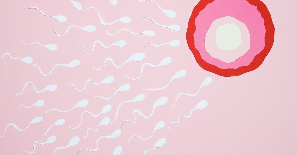
Photo: Nadezhda Moryak / Pexels
Building a human takes about 40 weeks. In that time, a single fertilized cell becomes roughly 26 billion cells organized into 200 different cell types, a beating heart, a brain with 80+ billion neurons, and a complete immune and digestive system. It is one of the most extraordinary biological processes we know of — and the more closely you look, the stranger and more elegant it becomes.
Educational content, not medical advice. Talk to your doctor before changing any medication, treatment, or routine. Information here reflects current research but every person's situation differs.
This article walks through what actually happens, week by week, organ by organ, and why pregnancy is biologically risky in ways that modern medicine has dramatically — but not completely — tamed.
Fertilization: One Sperm, One Egg, One Cell
Each menstrual cycle releases a single egg (sometimes two) from one of the ovaries. The egg is swept into the fallopian tube, where it is fertilizable for roughly 12–24 hours. Sperm, after ejaculation, can survive in the female reproductive tract for up to about 5 days. The window of possible fertilization per cycle is therefore about 6 days.
A typical ejaculate contains 200–500 million sperm; perhaps 200 reach the fallopian tube; one fertilizes the egg. Estimates of the per-cycle probability of conception in a healthy couple are around 20–25% in their twenties, falling with age. Cumulatively, roughly 80% of couples conceive within a year of regular unprotected sex, and the rest within two years or with medical help.
Once the sperm penetrates the zona pellucida and fuses with the egg membrane, the egg's cortical reaction blocks further sperm, the two haploid genomes combine, and a new diploid cell — the zygote — exists. From this point, the genetic identity of the new individual is fixed.
The First Two Weeks: Cleavage and Implantation
The zygote starts dividing as it travels down the fallopian tube. Within about 5 days it has become a blastocyst — a fluid-filled ball of ~100 cells with an inner cell mass (which becomes the embryo) and an outer trophoblast (which becomes the placenta).
Between days 6 and 10 post-fertilization, the blastocyst implants into the uterine wall. The trophoblast invades the endometrium, taps into maternal blood vessels, and begins producing human chorionic gonadotropin (hCG) — the hormone home pregnancy tests detect. By the day of the missed period, hCG is usually high enough to give a positive test.
Not all fertilized eggs implant. Studies of early hCG suggest roughly 30–40% of conceptions are lost before the missed period, usually unnoticed. Of clinically recognized pregnancies, about 10–15% end in miscarriage, with the rate concentrated in the first 12 weeks.
The Three Trimesters
Pregnancy is divided into three trimesters of roughly equal length, each with different biology and different risks.
| Trimester | Weeks | Dominant process | Major events |
|---|---|---|---|
| First | 0–13 | Organogenesis | All organ systems begin forming; highest miscarriage risk (~80% of losses occur here); most vulnerable window for teratogens |
| Second | 14–27 | Growth & maturation | Fetal movement felt (~18–22 wks); anatomy scan; viability threshold (~22–24 wks at the limits of NICU care) |
| Third | 28–40 | Final maturation | Major lung surfactant production from ~24 wks; rapid weight gain; brain pruning; immune transfer of maternal IgG |
First Trimester — Building the Body Plan
By week 4, the embryo has folded into a recognizable shape with a beating tubular heart. By week 8, all major organ systems are formed in primitive form, and the embryo officially becomes a fetus. This early window is when the developing body is most sensitive to teratogens — things like rubella infection, alcohol, certain anticonvulsants, retinoids, and thalidomide cause their characteristic patterns of damage precisely because they hit organogenesis.
This is also the window of highest spontaneous loss. Most early miscarriages are due to chromosomal abnormalities — errors in meiosis or early mitosis that produce an embryo incapable of developing.
Second Trimester — Growth
Once the body plan is laid down, the fetus grows. The mother typically begins to feel movement (quickening) between 18 and 22 weeks. Around 20 weeks, the anatomy scan looks at every major organ. The lungs are still immature — survival outside the uterus is essentially impossible before ~22 weeks, and even at 23–24 weeks (the modern limit of viability) outcomes are difficult.
Third Trimester — Final Maturation
The lungs are the last critical system to mature. Type II pneumocytes produce surfactant — a phospholipid mixture that reduces alveolar surface tension and prevents lung collapse. Surfactant production ramps from ~24 weeks but isn't usually adequate until ~34–36 weeks. The single biggest advance in 20th-century obstetrics was the introduction of antenatal corticosteroids (1972 onwards) to accelerate fetal lung maturation when preterm birth is imminent.
The fetus also receives a huge transfer of maternal IgG antibodies across the placenta during the third trimester — this is the basis of "passive immunity," which protects the newborn for the first 3–6 months of life and is why vaccinating pregnant mothers (e.g. Tdap, flu, RSV, COVID-19) protects newborns who can't yet make their own response. See the immune system for the underlying biology.
The Placenta: The Strangest Organ You Have
The placenta is a genuinely unique organ — it grows entirely from fetal tissue (the trophoblast lineage), exists for only ~40 weeks, and is the only organ shared by two genetically distinct people.
Its jobs include:
- Gas exchange. Maternal blood and fetal blood are separated by only a few cell layers; oxygen diffuses to the fetus, CO₂ to the mother.
- Nutrient transfer. Glucose, amino acids, fatty acids, vitamins, water all move across actively or by diffusion.
- Waste clearance. Urea and other fetal wastes diffuse to the mother for excretion.
- Hormone production. hCG, progesterone, estrogen, human placental lactogen, and others — the placenta progressively takes over hormonal maintenance of pregnancy from the corpus luteum.
- Immune tolerance. The placenta presents a half-foreign tissue (the fetus carries paternal antigens) yet is not rejected. The mechanisms — including specific HLA-G expression on trophoblasts and tolerogenic regulatory T cells at the maternal-fetal interface — are still being unraveled and are one of the most active areas of reproductive immunology.
Failure of placental development underlies several of the most dangerous complications of pregnancy.
Labor and Delivery
Labor is initiated by a complex cascade involving fetal cortisol, prostaglandins, and an oxytocin-driven positive-feedback loop. Once contractions become regular, labor proceeds in three classical stages:
- Stage 1 — Cervical dilation. Contractions thin (efface) and open the cervix from closed to 10 cm. This is usually the longest stage — typically 6–12 hours for a first birth.
- Stage 2 — Delivery of the baby. Active pushing once fully dilated. Minutes to a couple of hours.
- Stage 3 — Delivery of the placenta. Usually 5–30 minutes after the baby. Uterine contraction here is crucial to limit blood loss.
Oxytocin (the same hormone behind social bonding) is the master driver — it triggers strong uterine contractions and is positively reinforced by cervical stretch. The same hormone later drives milk letdown during breastfeeding. See hormones for the broader endocrine picture.
Common Complications
| Complication | Approx. rate | What it is |
|---|---|---|
| Miscarriage | 10–15% of recognized pregnancies | Pregnancy loss before 20 weeks |
| Preterm birth | ~10% globally | Birth before 37 weeks; leading cause of neonatal mortality |
| Gestational diabetes | 6–9% | Insulin resistance triggered by placental hormones |
| Preeclampsia | 5–8% | Pregnancy-specific hypertension + organ involvement; placental in origin |
| Postpartum hemorrhage | 1–5% | Excessive blood loss after delivery |
| Stillbirth | ~0.5% in high-income countries | Fetal death after 20 weeks |
Preeclampsia in particular is a leading cause of maternal and fetal morbidity and remains an active research target. Low-dose aspirin from the first trimester reduces risk in high-risk women, and the sFlt-1/PlGF ratio is now used for prediction in many countries.
What Modern Medicine Changed
Maternal mortality in the US and Europe in 1900 was around 1 per 100 births. By the late 20th century it had fallen ~50-fold, to roughly 1 per 5,000–10,000 births in most high-income countries. The drivers were:
- Aseptic technique and antibiotics (controlling puerperal sepsis)
- Safe blood transfusion (controlling postpartum hemorrhage)
- Antihypertensives and magnesium sulfate (managing preeclampsia/eclampsia)
- Safe Caesarean section
- Antenatal corticosteroids and NICU care for preterm infants
- Rh immunoglobulin (Rh disease prevention)
- Routine prenatal monitoring (ultrasound, screening, fetal heart monitoring)
Important caveats: maternal mortality has stopped falling — and in the US has risen modestly — in the 2010s and 2020s. The US rate is significantly higher than peer countries (currently ~22 per 100,000 live births vs ~5–10 in much of Western Europe), with persistent and severe racial disparities — Black women in the US die in childbirth at roughly three times the rate of White women.
FAQ
Q: Why is the first trimester so risky? A: Because all organ systems are being laid down at once, and most chromosomally abnormal embryos fail to develop. By the end of the first trimester, the embryos with serious abnormalities have mostly been lost, and the per-week miscarriage rate drops sharply.
Q: When can a fetus survive outside the uterus? A: The current limit of viability is around 22–24 weeks. At 22 weeks, survival with aggressive NICU care is around 30%; at 24 weeks, it rises to roughly 60–70%, with steeply increasing chances each additional week.
Q: Why are you supposed to avoid alcohol? A: No safe lower threshold has been established. Heavy exposure causes fetal alcohol syndrome (facial features, growth restriction, neurodevelopmental damage). Lower exposures are linked to a spectrum of more subtle effects. Most guidelines recommend zero alcohol throughout pregnancy.
Q: Are pregnancy and morning sickness related to hCG? A: Strongly correlated. Nausea typically peaks when hCG peaks (around weeks 9–12) and resolves as it falls. Twin pregnancies, with higher hCG, tend to have more severe nausea, including hyperemesis gravidarum.
Q: Does the fetus share blood with the mother? A: No. They have separate circulations separated by only a few cell layers in the placenta. Gases, nutrients, and many hormones cross; blood cells normally do not, which is why blood typing and Rh compatibility matters.
Q: Why does pregnancy last 40 weeks? A: It's measured from the last menstrual period, so actual gestation from conception is ~38 weeks. The duration is largely a trade-off between fetal brain size and the size of the maternal pelvis — humans give birth comparatively early developmentally, which is why human newborns are so dependent compared with other primates.
Sources
- Williams, Obstetrics, 26th ed. (Cunningham et al.)
- WHO — Trends in Maternal Mortality 2000 to 2020
- CDC — Pregnancy Mortality Surveillance System
- Roberts, J. M., et al. — Preeclampsia: pathophysiology and clinical implications, NEJM reviews
- Liggins, G. C., & Howie, R. N. (1972) — first trial of antenatal corticosteroids
- ACOG — Practice Bulletins on prenatal care, preeclampsia, GDM, preterm birth
Follow new articles
Add SciHub101 to your feed reader to see new science and technology stories.
About the Author
SciHub101 is an independent science and technology publication founded and edited by Buğra Sözeri, a software engineer and app developer based in Türkiye. AI tools assist with drafting and research. Source documentation in the existing archive is uneven, and articles are being reviewed and corrected. The Editorial Team byline identifies editorial responsibility; it does not certify specialist or independent human review. Where we compare products, anything built by our own team is labelled as such — see the editorial guidelines and About page for how corrections and disclosures work.
Related Articles
How the Heart Works — Anatomy and Function
How the human heart pumps blood: chambers, valves, electrical conduction, cardiac cycle, and the most important diseases and treatments.
How Skin Works — Your Largest Organ
Skin's three layers, how it protects you, why it ages, and the biology of pigment, sweat, and sensation in your body's largest organ.
Sex Hormones Explained — Testosterone and Estrogen
How testosterone, estrogen, and progesterone shape the body, the menstrual cycle, puberty, and menopause — and the science of hormone therapy.
Complete Biology Guide 2026 — How Life Works
The complete guide to biology in 2026: cells and genetics, evolution, the human body, neuroscience, disease, immune systems, and the future of genetic engineering.

How Do Fireflies Glow? The Science of Bioluminescence
Fireflies produce light through a chemical reaction called bioluminescence, where the protein luciferin is oxidized by the enzyme luciferase using oxygen and ATP to emit photons with nearly zero heat loss. The insect controls flash timing by regulating oxygen flow to its light organ, and each species produces a distinct flash pattern used for mating recognition. This same light-producing chemistry has become essential to modern biology, allowing researchers to track gene expression, detect ATP in tissue samples, and study cellular processes in real time.
Why Do Leaves Change Color? The Science of Autumn
Chlorophyll breaks down in autumn leaves, unmasking yellow and orange carotenoid pigments that were present all along. Meanwhile, trees synthesize new red and purple anthocyanins, while shorter days and cooler nights trigger senescence—a programmed shutdown that eventually leads the tree to seal off and drop each leaf at a specialized abscission layer. This cascade of biochemical events helps trees conserve resources for winter.