The Electroencephalogram (EEG) — How a Psychiatrist Mapped the Brain's Electric Storm — SciHub101
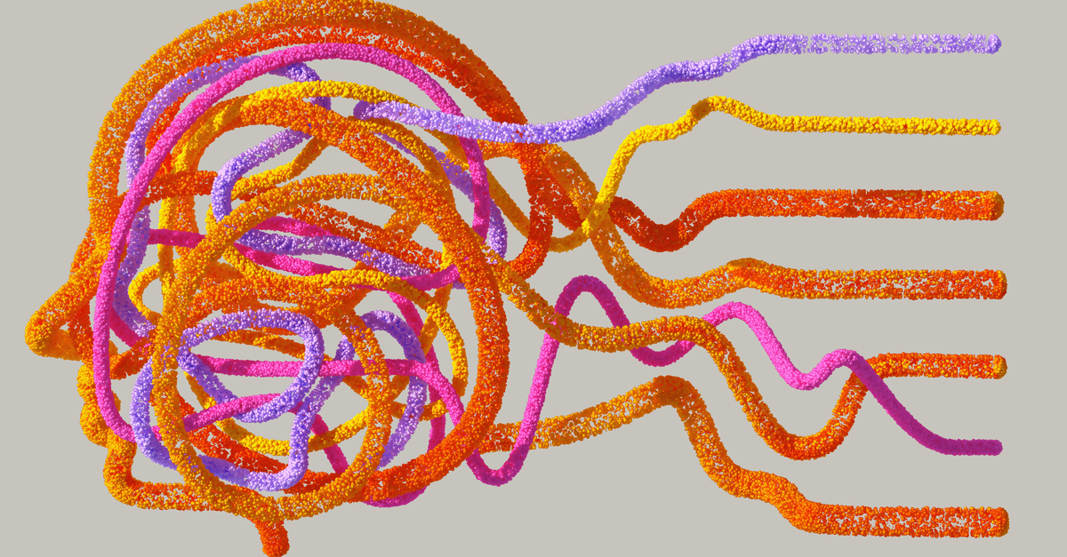
Photo: Google DeepMind / Pexels
On July 6, 1924, a German psychiatrist at the University of Jena named Hans Berger attached silver foil electrodes to the scalp of a seventeen-year-old boy recovering from neurosurgery, connected them to a galvanometer, and watched the needle jitter. What he recorded was the first-ever electroencephalogram (EEG) — a readout of the human brain's electrical activity. Berger was so uncertain of his findings that he told almost no one for five years. When he finally published in 1929, his peers laughed. When they stopped laughing, the history of neuroscience changed permanently.
The Strange Motive Behind the Machine
Hans Berger was not, by training or temperament, an engineer. He was a psychiatrist with a private obsession: he believed in telepathy. His conviction was not casual. In 1892, as a young artillery cadet on maneuvers, Berger was thrown from a horse in front of a cannon. Uninjured but shaken, he later received a telegram from his father asking if he was all right — his sister had felt a sudden, overwhelming dread at the moment of his fall. Berger spent the rest of his life trying to find the physical mechanism behind that perceived transmission of feeling between minds.
Source: How Hans Berger's quest for telepathy spurred modern brain science — Science News
His reasoning, applied to decades of reading: if the mind could transmit information, it must radiate some form of energy. Psychic energy, he believed, was probably electromagnetic and would show up as measurable electrical fluctuations on the skull. So he set about building instruments sensitive enough to detect those fluctuations. He was chasing ghosts, and he found neurons instead.
The irony is that Berger never proved telepathy existed. But the instrument he built trying to find evidence of it became one of the most important diagnostic tools in clinical medicine. Like many of history's great instruments, the EEG emerged from a question that turned out to be wrong — answered by a machine that turned out to be right.
Source: Berger and the Breakthrough: A Centennial Celebration of the Human Electroencephalogram — PubMed
How the EEG Works
The brain runs on electrochemistry. Each neuron (nerve cell) maintains an electrical charge across its membrane by pumping ions — sodium, potassium, calcium — in and out of the cell. When a neuron fires, that charge collapses in a wave called an action potential, which lasts about one millisecond. A single neuron firing produces a signal too small to detect through the skull. But the brain contains roughly 86 billion neurons, and when large populations of them fire in synchrony — as they do during sleep, seizures, attention, or emotion — the summed electrical field is strong enough to be detected on the scalp's surface.
Source: Normal Sleep EEG: Overview, Stage I Sleep, Stage II Sleep — Medscape
Think of it like a sports stadium. One person clapping is inaudible from outside the building. Fifty thousand people clapping together can be heard a block away. EEG electrodes are the microphone pressed against the stadium wall — they pick up the collective rhythm, not individual voices.
The voltage differences detected at the scalp are tiny, measured in microvolts (millionths of a volt). A modern EEG amplifier boosts those signals roughly a million times before displaying them as waveforms on screen. Different brain states produce characteristic wave shapes: the slow, rolling delta waves of deep sleep; the faster, irregular theta waves of light sleep; the calm alpha waves of a relaxed, eyes-closed awake state; and the rapid, low-amplitude beta waves of active thinking.
Source: Aserinsky Discovers REM Sleep — EBSCO Research Starters
What the Waveforms Actually Tell a Clinician
Each EEG waveform is characterized by two numbers: frequency (how many oscillations per second, measured in hertz) and amplitude (how large those oscillations are, measured in microvolts). Neurologists have learned to read specific patterns as signatures of specific brain states.
| Wave type | Frequency | Associated state |
|---|---|---|
| Delta (δ) | 0.5–4 Hz | Deep sleep, severe brain injury |
| Theta (θ) | 4–8 Hz | Drowsiness, meditation, memory encoding |
| Alpha (α) | 8–13 Hz | Relaxed wakefulness, eyes closed |
| Beta (β) | 13–30 Hz | Active thinking, alertness, anxiety |
| Gamma (γ) | 30–100 Hz | High-level cognition, sensory binding |
Epileptic seizures produce distinctive spike-and-wave patterns — sharp, sudden deflections that stand out from background activity the way a thunderclap stands out from ambient noise. This is why EEG became, and remains, the gold-standard tool for diagnosing epilepsy.
The People Who Brought the EEG to the World
Berger published his first paper in 1929 under the deliberately modest title "Über das Elektrenkephalogramm des Menschen" (On the Electroencephalogram of Man). The reception in Germany was disbelief. His methodology seemed sloppy; the signals seemed too small to be real; his telepathy research colored colleagues' view of his judgment.
The turnaround came from Cambridge. In 1934, the physiologist Edgar Douglas Adrian and his colleague B.H.C. Matthews replicated Berger's results using superior amplifiers, confirmed every major finding, and published their own validation. Adrian had already won the 1932 Nobel Prize in Physiology or Medicine for his work on nerve impulses. His endorsement was unchallengeable. The scientific community accepted the EEG almost overnight.
Source: Hans Berger and the Electroencephalogram — Neurology, American Academy of Neurology
Adrian and Matthews did more than confirm — they extended. They mapped the alpha rhythm's origins to the visual cortex, they showed EEG changed with attention, and they introduced the instrument to British neurology. From Cambridge the technique spread to the United States, where the Boston City Hospital established one of the first clinical EEG labs in the late 1930s.
Back in Jena, Berger's final years were tragic. The Nazis had dismissed him from his professorship in 1938, citing his age, though political pressure played a role. Isolated, stripped of the laboratory that was his life, and suffering from depression, Hans Berger died by suicide on June 1, 1941 — less than a decade after his invention had been proven valid and was beginning to save lives.
Source: Hans Berger — Wikipedia
How the EEG Changed Everything We Know About Sleep
In 1953, twenty-nine years after Berger's first recording, a graduate student named Eugene Aserinsky was monitoring sleeping subjects in the laboratory of sleep researcher Nathaniel Kleitman at the University of Chicago. Aserinsky — who had borrowed a room from the physiology department and initially recorded his own eight-year-old son — noticed something odd: at predictable intervals across the night, sleeping subjects' eyes began making rapid, coordinated movements. Their EEG looked almost like a waking brain. Their breathing and heart rate shifted.
Aserinsky and Kleitman published the finding in Science that same year, naming it rapid eye movement (REM) sleep. It was arguably the most important discovery in sleep science of the twentieth century. It told us that sleep is not a single passive state but a cycling architecture of distinct stages, each with its own EEG signature and its own physiological role.
Source: Kleitman & Aserinsky (1953): REM Sleep Discovery — Optimised Life
Without the EEG, REM sleep could not have been defined. The technique allowed researchers to divide sleep into NREM stages 1 through 3 plus REM, map how people cycle through them roughly every 90–100 minutes, and understand how disruptions to that cycle produce cognitive and emotional damage. The entire modern science of sleep medicine — every sleep study, every diagnosis of sleep apnea, insomnia, or narcolepsy — runs through EEG.
Source: The power of direct observation: discovery of REM sleep — Nature Reviews Neuroscience
A Century of Progress: From Two Electrodes to 256 Channels
Berger's original setup used two electrodes — one on the forehead, one at the back of the skull — connected to a galvanometer. The resulting signal was so coarse it could barely distinguish wake from sleep. Modern clinical EEG uses the international 10-20 system: 19 to 21 electrodes arranged in a standardized grid across the scalp, referenced by the distances between skull landmarks. High-density research EEGs use 64, 128, or 256 electrodes, providing enough spatial resolution to localize activity to specific brain regions.
Source: Early History of Electroencephalography and Establishment of the Field — ASET
The biggest practical limitation of traditional EEG is the gel. Electrodes must make reliable electrical contact with the scalp, which traditionally meant applying conductive gel under each electrode cup — a process that takes 20 to 45 minutes and leaves the subject's hair matted. Dry-electrode systems developed from the 2010s onward use spring-loaded pins or foam contacts to achieve acceptable contact without gel, enabling quicker setup and wearable form factors. Consumer EEG headsets from companies like Muse (InteraXon) use dry electrodes with 4–7 channels for meditation feedback and sleep tracking, though their spatial resolution is a fraction of clinical systems.
The other fundamental limitation is spatial. EEG has outstanding temporal resolution — it can track neural events that occur in milliseconds — but it is imprecise about where in the brain those events originate. Think of it as excellent at telling you when the stadium goes wild but poor at telling you which section of seats caused the noise. Functional MRI (fMRI) offers much better spatial resolution but is slow (seconds per scan) and requires the subject to lie motionless in a large magnet. Researchers frequently combine EEG and fMRI to get the best of both.
EEG and the Brain-Computer Interface Revolution
The modern interest in brain-computer interfaces (BCIs) has given EEG a second wind it might not have needed. BCIs aim to translate brain signals into control commands for computers, prosthetics, or other devices without requiring muscle movement — a life-changing prospect for people with paralysis from spinal cord injuries, ALS, or stroke.
Source: Editorial: 100 years of electrophysiology in psychiatry — PMC / NCBI
EEG-based BCIs work by having users modulate their own brain rhythms — for example, imagining moving their left hand reliably suppresses the mu wave (a rhythm near 10 Hz) over the right motor cortex. A classifier algorithm learns to detect that suppression and translates it into a command. The approach is non-invasive and inexpensive compared to implanted electrodes, which makes it attractive for widespread clinical deployment.
Implanted approaches like those developed by Neuralink and Synchron record directly from cortical neurons, producing cleaner, higher-resolution signals than surface EEG can achieve. But EEG remains the only modality that is cheap, portable, and completely non-invasive — properties that matter enormously when the goal is getting technology out of research labs and into the hands of patients.
A Timeline of EEG Milestones
| Year | Event |
|---|---|
| 1875 | Richard Caton records electrical currents from the brains of rabbits and monkeys |
| 1924 | Hans Berger records first human EEG at University of Jena |
| 1929 | Berger publishes first EEG paper after five years of doubt |
| 1934 | Adrian and Matthews confirm Berger's results; scientific community accepts EEG |
| 1938 | EEG used clinically to diagnose epilepsy for the first time |
| 1941 | Hans Berger dies by suicide under Nazi persecution |
| 1953 | Aserinsky and Kleitman discover REM sleep using EEG |
| 1958 | International 10-20 electrode placement system standardized |
| 1970s | Computerized EEG (CEEG) enables digital recording and analysis |
| 1990s | High-density 256-channel systems introduced for research |
| 2000s | Dry-electrode consumer EEG headsets enter market |
| 2020s | EEG integrated with machine learning for real-time BCI control |
Source: Berger Studies the Human Electroencephalogram — EBSCO Research Starters
Brain-Computer Interfaces: From EEG to Neuralink (2024–2026)
The EEG-based BCI section above describes the field's non-invasive tier. But 2024 marked an inflection point, when implanted intracortical devices moved from laboratory milestones into the first sustained human trials — and the contrast with scalp EEG became impossible to ignore.
The bandwidth gap is the core issue. A typical research EEG with 64 channels records population-averaged signals carrying roughly 2–5 bits per second of usable control information. Intracortical electrode arrays — implanted directly in the motor cortex — record from individual neurons, yielding information rates roughly 1,000 times higher. That gap matters practically: EEG can let a paralyzed person move a cursor or select letters; intracortical arrays can let them control a robotic arm with ten degrees of freedom simultaneously.
Synchron's Stentrode: the vascular shortcut
Synchron's Stentrode device takes a middle path between surface EEG and open-brain surgery. Rather than drilling through the skull, surgeons thread a stent-like electrode array through the jugular vein and into a blood vessel adjacent to the motor cortex. The FDA granted Breakthrough Device Designation to the Stentrode in August 2020. On July 6, 2022, a team at Mount Sinai West Medical Center in New York performed the first US implant — in an ALS patient who was discharged 48 hours later. The patient enrolled in Synchron's COMMAND trial, which is studying whether endovascular BCI can reliably restore digital device control in people with severe paralysis without the risks of open craniotomy.
Source: Synchron Announces First Human U.S. Brain-Computer Interface Implant — Business Wire, July 2022
Source: Synchron's Stentrode Receives Breakthrough Device Designation from FDA — Business Wire, August 2020
Neuralink's PRIME study and the thread retraction problem
Neuralink's approach is more invasive: a neurosurgical robot drills through the skull and inserts 64 flexible polymer threads — each thinner than a human hair — carrying a total of 1,024 electrodes into the motor cortex. On January 28, 2024, Noland Arbaugh, a 29-year-old quadriplegic, became the first person implanted in Neuralink's PRIME early feasibility study, with the procedure performed at Barrow Neurological Institute in Phoenix. Within weeks, approximately 85 percent of the threads had retracted from the brain tissue, reducing the active electrode count to roughly 15 percent of the original. Neuralink addressed the performance loss through algorithm updates rather than re-surgery — demonstrating that signal-processing software can partially compensate for hardware degradation. A second participant, known publicly as Alex, was implanted in July 2024 and avoided the thread retraction issue seen in the first case.
Source: Noland Arbaugh — Wikipedia
Source: Neuralink PRIME Study: What We've Learned So Far — The Mancunion, November 2024
The University of Pittsburgh's arm-control work
While Neuralink and Synchron dominate media coverage, the most detailed published data on high-bandwidth intracortical BCI comes from the Rehab Neural Engineering Lab at the University of Pittsburgh, led by Andrew Schwartz and Michael Boninger. Their work has demonstrated simultaneous proportional control over ten degrees of freedom in a robotic arm — three-dimensional translation, three-dimensional orientation, and four-dimensional hand shape — using signals recorded from arrays implanted in the motor cortex of paralyzed participants. This work established what the intracortical bandwidth advantage actually buys: not just cursor control, but the near-natural manipulation of a physical limb.
Cognitive liberty: the ethical frontier
As intracortical BCIs produce richer and richer neural data, ethicists and legal scholars have begun debating what they call cognitive liberty — the right to mental privacy and to freedom from non-consensual neural monitoring or modification. Current BCI implants record motor signals for device control. But the same hardware could, in principle, passively record cognitive activity far beyond movement intent. The question is not purely hypothetical: corporations own the implanted devices, control the data pipelines, and are not currently covered by any US law specifically protecting neural data. Chile became the first country to add neurorights to its constitution in 2021. The US has no equivalent framework. As BCIs move from clinical trials into commercial products, the gap between what the technology can do and what the law restricts it from doing will widen.
Source: Neuralink Beyond First Human 2026: Updates — Axis Intelligence
What People Get Wrong About EEG
The most common misconception is that EEG can "read thoughts." It cannot. It reads population-level electrical rhythms — the average behavior of millions of neurons, filtered through skull, scalp, and gel. Picking out the content of a specific thought from an EEG trace is not possible with any current technology, and for basic physical reasons, may never be. The spatial blur introduced by the skull (which is a poor conductor) destroys the fine-grained spatial information that would be needed to decode what, specifically, someone is thinking.
What EEG can detect is the neural correlates of cognitive states — whether someone is paying attention, drowsy, imagining movement, or experiencing a seizure. Those are categories of brain state, not the content of thought.
A second misconception concerns EEG therapy. Several consumer products market EEG biofeedback (neurofeedback) as a treatment for ADHD, anxiety, or peak performance. The evidence for these consumer applications is mixed. Clinical neurofeedback for epilepsy has a longer track record, but most neurofeedback claims made in direct-to-consumer wellness products go substantially beyond what peer-reviewed evidence supports.
Where EEG Stands Today
A century after Berger's first recording, EEG is performed approximately 2 million times per year in the United States alone, making it one of the most common neurological procedures. Its core clinical applications have not changed: it remains the primary diagnostic tool for epilepsy, the monitoring standard during brain surgery and intensive care, and the definitive method for diagnosing brain death.
What has changed is everything around it. Machine learning classifiers can now read EEG traces and detect seizures with accuracy approaching that of expert neurologists, enabling 24-hour automated monitoring for patients in intensive care. Ambulatory EEG systems — small wireless recorders patients wear at home for days — have replaced the requirement to catch epileptic activity in a hospital. Neonatal EEG monitors brain development in premature infants in ways that no imaging technology can match.
Source: Fiftieth Anniversary of the Discovery of Rapid Eye Movement (REM) Sleep — Journal of Neuropsychiatry
Berger's search for telepathy was wrong. His search for a physical correlate of mental activity was right. The instrument he built to prove the first claim became the foundational tool for exploring the second — and a hundred years later, it is still doing the job.
Q: What does an EEG measure? An EEG measures the summed electrical activity of large populations of neurons, detected as voltage fluctuations at the scalp surface. Individual neurons firing produce signals too small to detect; the EEG captures the synchronized activity of millions of cells acting together.
Q: How is an EEG different from an MRI or CT scan? MRI and CT produce structural images of the brain's anatomy. EEG records real-time electrical activity — it tells you what the brain is doing moment to moment, not what it looks like. EEG has millisecond temporal resolution; MRI has millimeter spatial resolution. They answer different questions.
Q: Can EEG diagnose Alzheimer's disease? EEG changes are documented in Alzheimer's disease — slowing of background rhythms and reduced alpha power are common findings — but it is not used as a standalone diagnostic. Alzheimer's diagnosis still relies on clinical assessment, neuropsychological testing, and in some cases biomarkers (PET imaging, cerebrospinal fluid).
Q: Why did Berger wait five years to publish his EEG results? Berger was deeply uncertain. He knew his telepathy motivations would invite ridicule, and he was unsure whether the signals he was recording were true brain activity or electrical artifacts from scalp muscle movement. He spent five years running controls, testing different electrode placements, and trying to eliminate alternative explanations before he felt confident enough to submit.
Q: Is the EEG still used today or has it been replaced by newer technology? EEG is performed roughly 2 million times annually in the US and remains irreplaceable for epilepsy diagnosis, intraoperative monitoring, and brain death determination. No other clinical tool matches its combination of millisecond time resolution, low cost, portability, and safety. Newer technologies complement it; none have replaced it.
Q: What are the limitations of EEG as a brain-computer interface? Surface EEG provides coarse spatial resolution because the skull and scalp electrically blur signals from different brain regions. It cannot resolve activity from sources deeper than about 3 centimeters from the surface. This limits the number of independent control signals it can provide for BCI applications. Implanted electrodes overcome this but require surgery.
The Takeaway
Hans Berger wanted to find the physics of telepathy and found the physics of thought instead. The EEG he built in pursuit of a scientifically invalid idea became the lens through which modern neurology sees the brain — diagnosing epilepsy, defining the architecture of sleep, and now forming the non-invasive half of the brain-computer interface field. The instrument is a hundred years old. It has survived every scan and imaging breakthrough that was supposed to make it obsolete. In clinical medicine and neuroscience research alike, the squiggly lines Berger first doubted for five years are still the clearest window we have into the brain's electrical life.
Read next:
Weekly digest
Stay ahead of the curve
Get the best science, AI, and tech stories delivered to your inbox every week. No spam — unsubscribe any time.
About the Author
SciHub101 is an independent science and technology publication founded and edited by Buğra Sözeri, a software engineer and app developer based in Türkiye. Articles are drafted against peer-reviewed literature and primary sources, fact-checked, and edited by a human before publication. Where we compare products, anything built by our own team is labelled as such — see the editorial guidelines and About page for how corrections and disclosures work.
Related Articles

The Fiber Optic Cable: How Glass Replaced Copper
Charles Kao's 1966 paper proved pure glass could carry light across continents. Corning proved it in 1970. Today 1.5 million km of fiber carries 95% of global internet traffic.

The Electric Toothbrush: From Corded Swiss Prototype to AI-Powered Dental Coach — SciHub101
How Philippe-Guy Woog's 1954 Broxodent, a glue-gun toy frog idea, and University of Washington research built the $4 billion electric toothbrush market — and what the evidence says it does.

The Science of Sleep: What Happens in the Brain During REM Sleep
Sleep is not a passive shutdown but an active state of brain and body reorganization. During an 8-hour sleep cycle, humans alternate between non-REM (NREM) sleep—characterized by slow theta waves, reduced muscle tone, and energy conservation—and REM (rapid eye movement) sleep, when the brain shows activity patterns similar to wakefulness, the eyes move rapidly beneath closed eyelids, and skeletal muscles are paralyzed except for the diaphragm. REM sleep makes up approximately 20–25 percent of sleep in adults and is the stage where most vivid dreaming occurs. The neuroscience of REM reveals that memory consolidation (particularly emotional and procedural memories), creativity processing, and amygdala-driven emotion regulation all occur during this stage. The glymphatic system—a network of fluid pathways that clear metabolic waste from the brain during sleep—becomes most active during deep NREM sleep, not REM. This article explains the neuroscience of REM sleep, the discovery of the glymphatic system, and why sleep is essential to brain health.
The Science of Déjà Vu: What Actually Causes It
Roughly two-thirds of the general population reports experiencing déjà vu at some point in their lives — that disorienting conviction that a present moment has been experienced before, accompanied by the simultaneous knowledge that it cannot have been. The experience lasts a few seconds, then dissolves. For most people it is occasional and benign. For patients with temporal lobe epilepsy, it can be a reliable seizure precursor. The fact that déjà vu can be reproduced reliably in clinical settings — both by electrical stimulation of specific brain regions and by certain drug compounds — tells neuroscientists where to look for the mechanism.

Why Déjà Vu Happens — The Brain Science
Why does déjà vu happen? Neuroscience explains the memory mismatch, dual-processing, and temporal lobe theories behind the 'already seen' feeling.

Why We Forget Our Dreams — The Brain Science
Most adults forget 95% of dreams within minutes. The neuroscience: low norepinephrine, MCH neurons, weak hippocampal coupling, and how to remember more.