Blood Types Explained — ABO and Rh Systems
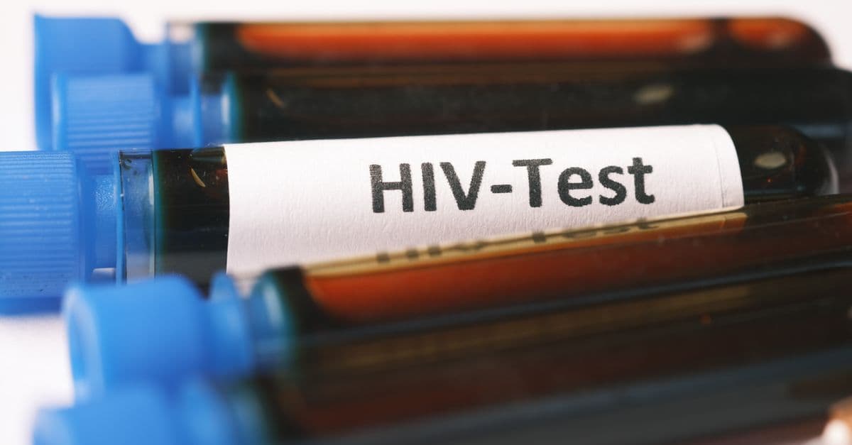
Photo: Towfiqu barbhuiya / Pexels
Every drop of human blood carries a molecular ID badge on the surface of its red blood cells. These badges — sugar and protein markers called antigens — determine your blood type. Knowing yours can save your life in a transfusion, and understanding how it's inherited reveals beautiful classical genetics in action.
What Is a Blood Type?
A blood type is defined by which antigens sit on the outer membrane of your red blood cells (RBCs), and which antibodies float in your plasma. If foreign antigens enter your bloodstream — say, from an incompatible transfusion — your antibodies trigger clumping (agglutination), which can be fatal.
The two most clinically important systems are ABO and Rh, but humans actually have over 40 recognized blood group systems (Duffy, Kell, MNS, Kidd, Lewis, and more).
The ABO System
Discovered by Austrian physician Karl Landsteiner in 1901 (Nobel Prize 1930), the ABO system has four types:
| Type | Antigen on RBC | Antibody in Plasma |
|---|---|---|
| A | A | anti-B |
| B | B | anti-A |
| AB | A and B | none |
| O | none | anti-A and anti-B |
- Type O has no ABO antigens, so it doesn't trigger immune reactions in any ABO recipient — universal donor (for red cells, and especially O-negative).
- Type AB has no ABO antibodies, so it can receive any ABO red cells — universal recipient.
Before Landsteiner, transfusions were a deadly lottery. His discovery cut transfusion mortality by orders of magnitude.
The Rh System
In 1940, Landsteiner and Alexander Wiener discovered the Rh factor (named after the rhesus monkey, in which they first identified it). The clinically important antigen is RhD:
- Rh-positive — has the D antigen (~85% of people of European descent, ~99% of East Asians).
- Rh-negative — lacks the D antigen.
Why Rh Matters in Pregnancy
If an Rh-negative mother carries an Rh-positive fetus, fetal blood cells can leak into maternal circulation (especially at birth). The mother's immune system makes anti-D antibodies. In a subsequent Rh+ pregnancy, those antibodies cross the placenta and attack fetal RBCs — hemolytic disease of the newborn (HDN), historically a major cause of fetal death.
The solution: Rho(D) immune globulin ("RhoGAM"), introduced in 1968. Given to Rh- mothers at ~28 weeks and post-delivery, it prevents sensitization. HDN deaths in developed countries dropped by >95%.
Other Blood Group Systems
While ABO and Rh dominate transfusion practice, others matter in specific cases:
- Duffy (Fy) — Duffy-negative RBCs resist Plasmodium vivax malaria; common in sub-Saharan Africa.
- Kell (K) — anti-K antibodies can also cause HDN.
- MNS — rarely involved in transfusion reactions.
- Kidd (Jk) — implicated in delayed hemolytic reactions.
- Lewis, Lutheran, Diego — niche but documented.
Today the International Society of Blood Transfusion (ISBT) recognizes 45+ systems and hundreds of antigens.
Inheritance — Classical Genetics
ABO inheritance is a classic example of codominance and recessivity:
- Alleles: I^A, I^B, i (where i = O).
- I^A and I^B are codominant; i is recessive.
Possible genotypes:
| Genotype | Phenotype |
|---|---|
| I^A I^A or I^A i | Type A |
| I^B I^B or I^B i | Type B |
| I^A I^B | Type AB |
| ii | Type O |
Punnett example: Parent 1 (I^A i) × Parent 2 (I^B i) →
- 25% I^A I^B (AB)
- 25% I^A i (A)
- 25% I^B i (B)
- 25% ii (O)
One pair of parents can produce children of all four ABO types — a fact that has resolved (and occasionally created) many paternity disputes.
Rh is simpler: D is dominant over d. Two Rh+ parents (Dd × Dd) can have an Rh- child (dd) with 25% probability.
Distribution by Population
Blood type frequencies vary significantly by ancestry:
| Group | O+ | A+ | B+ | AB+ | O- | A- | B- | AB- |
|---|---|---|---|---|---|---|---|---|
| US general | 37% | 36% | 9% | 3% | 7% | 6% | 1.5% | 0.6% |
| East Asian | 40% | 28% | 24% | 7% | <1% | <1% | <1% | <0.1% |
| Sub-Saharan African | 49% | 27% | 20% | 4% | low Rh- overall | |||
| Indigenous American | up to 100% O+ in some isolated groups |
This variation reflects evolutionary history, founder effects, and possibly selective pressure (e.g., disease resistance).
Transfusion Compatibility — Red Cells
| Recipient \ Donor | O- | O+ | A- | A+ | B- | B+ | AB- | AB+ |
|---|---|---|---|---|---|---|---|---|
| O- | ✅ | ❌ | ❌ | ❌ | ❌ | ❌ | ❌ | ❌ |
| O+ | ✅ | ✅ | ❌ | ❌ | ❌ | ❌ | ❌ | ❌ |
| A- | ✅ | ❌ | ✅ | ❌ | ❌ | ❌ | ❌ | ❌ |
| A+ | ✅ | ✅ | ✅ | ✅ | ❌ | ❌ | ❌ | ❌ |
| B- | ✅ | ❌ | ❌ | ❌ | ✅ | ❌ | ❌ | ❌ |
| B+ | ✅ | ✅ | ❌ | ❌ | ✅ | ✅ | ❌ | ❌ |
| AB- | ✅ | ❌ | ✅ | ❌ | ✅ | ❌ | ✅ | ❌ |
| AB+ | ✅ | ✅ | ✅ | ✅ | ✅ | ✅ | ✅ | ✅ |
Plasma compatibility runs inversely: AB plasma is universal donor; O plasma is universal recipient.
Can Blood Type Be Changed?
Naturally, no — your ABO type is fixed by genes. But there are fascinating edge cases and engineering breakthroughs:
- Bombay phenotype (hh) — Lacks the H antigen precursor to A and B. Tests as O but cannot receive O blood from normal donors. Extremely rare (~1 in 250,000, more common in India).
- Bone marrow transplant — Can change a recipient's blood type to that of the donor over months.
- Enzyme conversion — In a landmark 2019 study, Withers et al. at the University of British Columbia discovered gut bacterial enzymes that efficiently strip A and B antigens off red cells, converting them to functional type O. Clinical trials are ongoing (2024–2026). If approved, this could solve chronic O- shortages.
How Blood Type Is Tested
Laboratory determination of blood type relies on a principle discovered by Landsteiner himself: agglutination. When incompatible blood is mixed, antigens on red cells bind to antibodies in plasma, causing visible clumping.
Forward typing — the standard method — adds patient serum to reagent red cells of known types (A, B, and O). If the patient has anti-A antibodies, agglutination occurs with A and AB cells. If anti-B, then with B and AB. A patient with both anti-A and anti-B is type O; type AB shows no agglutination.
Reverse typing adds patient red cells to reagent serum of known types. This cross-check confirms results and catches rare variants like Bombay phenotype. Gel card technology and automated analyzers now dominate labs, reading agglutination patterns electronically and providing results in under 5 minutes.
Source: International Society of Blood Transfusion (2024) — Standardized methodology for blood group serology in diagnostic laboratories.
Molecular typing has become increasingly important for transfusion medicine in developed countries. DNA sequencing identifies blood group alleles directly, bypassing serologic testing. This is especially valuable for rare blood types, immune compromised patients, and prenatal diagnostics where serum antibodies haven't yet formed.
Blood Banking, Storage, and the Cold Chain
Once collected, blood enters a strict logistical system. Whole blood stored at 1–6°C (refrigerated) lasts 35–42 days, depending on the anticoagulant used (citrate-phosphate-dextrose, CPDA, or CPDA-1). Red blood cell concentrates (RBCs separated from plasma) also last 35–42 days refrigerated, but up to 42 days if additive solutions (like ADSOL or Nutricel) are added post-collection.
Platelets must be stored at room temperature (20–24°C) with continuous agitation to prevent aggregation — but this short shelf life of only 5–7 days creates chronic supply challenges. Fresh frozen plasma (FFP) and cryoprecipitate are frozen at −18°C or lower and can be stored for 1–2 years.
The cold chain — maintaining temperature control from donor through hospital transfusion service — is critical. A single temperature excursion can compromise a unit's safety (risk of bacterial growth). Blood banks use alarm-equipped refrigerators and emergency backup power. In developing countries, lack of reliable refrigeration is a major barrier to safe transfusion.
Source: AABB (American Association of Blood Banks) (2023) — Technical Manual, 20th edition. Storage and shelf-life standards.
Wastage remains high: in the US, approximately 2–5% of collected blood is discarded annually due to expiration before transfusion, outdated inventory, or failed testing. This creates both economic and ethical pressure to develop extended-storage solutions and artificial blood substitutes.
Blood Type and Disease Susceptibility
Beyond transfusion compatibility, blood type influences disease risk in subtle but measurable ways. The Duffy antigen (particularly absence of Duffy-negative phenotype) is the primary genetic determinant of resistance to Plasmodium vivax malaria. Duffy-negative individuals predominate in sub-Saharan Africa, where P. vivax historically had limited transmission opportunity — an example of evolutionary selection for disease resistance written into the blood type system.
ABO blood type has also shown associations with various infectious and non-infectious diseases:
- Gastric cancer: Individuals with type A blood have modestly elevated risk (about 20% higher than type O) in some populations. The mechanism is unclear but may relate to inflammatory responses or H. pylori colonization patterns.
- Type 1 diabetes: Type O individuals show slightly increased risk in some study cohorts, though the effect is small and confounded by other genetic factors.
- Venous thromboembolism (VTE): Blood types A, B, and AB are associated with increased VTE risk compared to type O. Type O individuals have lower von Willebrand factor (vWF) and factor VIII levels, both clotting factors. This protective effect is one reason type O is overrepresented among blood donors — they're also preferred for plasma export because their plasma lacks ABO antibodies.
These associations are typically relative risk increases of 10–30%, not absolute causation. A person with type A blood is not destined for gastric cancer; rather, the population-level signal reflects complex gene-environment interactions.
Source: Franchini, M. & Lippi, G. (2016). The molecular basis of blood groups and platelet alloimmunization. Transfusion Medicine Reviews, 30(3), 107–116.
Hemolytic Transfusion Reactions and Acute Compatibility Crises
While compatibility tables show who can receive what, the biological reality of incompatible transfusion is dramatic and sometimes fatal. When incompatible blood enters a recipient's bloodstream, antibodies bind to antigens on the donor's red cells, triggering a cascade of destruction called a hemolytic transfusion reaction.
Acute hemolytic transfusion reactions (occurring within minutes to hours) manifest as fever, chills, back pain, and hemoglobinuria (dark urine — a sign that freed hemoglobin is in the urine). In severe cases, the immune attack is violent: donated RBCs are lysed (burst open) by complement fixation, releasing hemoglobin into the bloodstream. Hemoglobin precipitates in kidney tubules, causing acute kidney injury. The cascade of clotting activation can trigger disseminated intravascular coagulation (DIC) — a life-threatening condition where blood clots form throughout the circulation simultaneously, depleting clotting factors and causing severe bleeding.
Mortality from acute hemolytic transfusion reactions was historically 15–30%, but modern transfusion medicine has driven this down to <1% in developed countries through:
- Type and cross-matching before transfusion — verifying compatibility before blood is transfused
- Immediate cessation of transfusion if a reaction is suspected
- Supportive care — aggressive hydration to protect kidneys, treatment of DIC, management of hyperkalemia (dangerous potassium rise from lysed RBCs)
Delayed hemolytic transfusion reactions occur days to weeks after transfusion when a patient's immune system, previously sensitized by an earlier transfusion, produces antibodies against an antigen on donor RBCs. These are usually less severe than acute reactions but can still cause significant hemolysis and fever. Patients who receive frequent transfusions (for chronic anemia, leukemia, or sickle cell disease) are at highest risk because each transfusion exposes them to new antigens and opportunities for alloimmunization (developing antibodies against donor antigens).
Source: Heddle, N. M., et al. (2015). "Pathophysiology of febrile nonhemolytic transfusion reactions." Current Opinion in Hematology, 22(6): 536–542. — Mechanisms underlying various transfusion reaction types.
The practical consequence: multi-transfused patients accumulate antibodies to minor antigens and can become extraordinarily difficult to transfuse. A patient might be compatible with standard O-negative blood when tested, but after 20 transfusions develop antibodies to Duffy, Kell, and MNS antigens, requiring weeks-long searches for blood that lacks all these antigens. For patients with critical anemia who need transfusion now, these delays can be life-threatening.
Rare Blood Types and the Global Supply Crisis
While O- donors get the spotlight, the rarest blood types present a clinical paradox. AB- individuals (~0.6% of US population) are universal recipients — they can receive any RBC type — yet AB plasma is a universal donor, creating strategic value. But Rh-null — the true "golden blood" — poses both scientific fascination and logistical nightmare.
Rh-null individuals lack all Rh antigens and can donate to anyone with rare Rh variants who is sensitized. There are fewer than 50 known Rh-null individuals alive, mostly clustered in small populations or known through blood bank registries. Some, like Maureen Crowley (pseudonym, often cited in blood bank literature), spent decades as the "world's most needed blood donor," traveling for phlebotomy appointments.
Other ultra-rare types include U-negative (lacking the U antigen, predominantly African descent) and Diego-negative (found in some East Asian and Indigenous populations). Modern multi-transfused patients with rare antibodies can face months of searching for compatible units.
To address this, blood banks maintain rare-blood registries and biobanks that catalog and preserve rare units. The International Rare Donor Panel, coordinated through ISBT, connects blood services globally. A patient in New York might receive rare blood collected in South Africa, shipped via specialized couriers.
Future Directions — Engineered Blood and Alternatives
The demand for transfusable blood far exceeds supply. The 2024 WHO estimates about 108 million blood donations globally, yet demand exceeds this in most developed nations. Research into alternatives has accelerated:
Artificial blood and blood substitutes:
- Oxygen carriers based on perfluorocarbons or hemoglobin-based oxygen carriers (HBOCs) can temporarily deliver oxygen but don't replace the full function of RBCs. Trials in the 1990s–2010s found safety concerns (renal effects, hypertension) and limited benefit over transfusion, slowing development.
- Microencapsulated red cells: Research teams (notably at MIT and University of Tokyo) have created artificial RBC-like particles using polymer shells and synthetic cytoplasm. Still experimental.
- Bioreactor-cultured RBCs: In-vitro red cell production using erythroid progenitor cells scaled in bioreactors. Cynergistek and similar biotech firms are pursuing this. One major hurdle: cost. Laboratory-produced RBCs currently cost $300–$500 per unit versus $100–$200 for donated blood.
Enzymatic antigen stripping: The 2019 Withers study was a landmark because enzyme-treated type A or B blood becomes functionally type O (lacking A/B antigens) while retaining full RBC function. Phase 1 clinical trials (2023–2024) are testing patient tolerance. If successful, this could dramatically reduce O- shortages by converting excess A and B units.
Source: Withers, S. G. et al. (2019). An enzymatic pathway in the human gut microbiome that converts A to universal O type blood. Nature Microbiology, 4(11), 1869–1876.
Gene editing: CRISPR-based approaches to delete A and B antigens in donor cells before expansion are being explored. This would theoretically allow any blood type to become O. Challenges include regulatory approval for gene-edited therapeutics and manufacturing scale-up.
The next decade will likely see a shift from purely donation-based transfusion services to a hybrid model: traditional donation for common types, supplemented by manufactured or converted units for rare and high-demand types.
The takeaway
Your blood type is a genetic passport written on every red cell, determining transfusion compatibility and hinting at disease risk. ABO and Rh systems are just two of over 40 recognized blood group systems, reflecting the incredible complexity of our immune identity. While Landsteiner's discovery transformed transfusion medicine a century ago, the next frontier — enzymatic conversion, cultured blood, and personalized rare-type registries — promises to solve chronic supply crises and make transfusion even safer.
Frequently Asked Questions
Q: What is the rarest blood type?
A: AB- is rarest globally (~0.6%). But "Rh-null" — lacking all Rh antigens — is the true unicorn, found in fewer than 50 known living people worldwide. It's called "golden blood" because it can be donated to anyone with rare Rh variants.
Q: Can two O parents have a non-O child?
A: Almost never. Both must contribute i alleles, so the child should be O. Extreme exceptions (cis-AB phenotype, chimerism) exist but are vanishingly rare.
Q: Does blood type affect personality?
A: No. The Japanese "ketsueki-gata" personality theory has been repeatedly debunked in controlled studies. It's pseudoscience akin to astrology.
Q: Does blood type affect COVID-19 or disease risk?
A: Some studies (Ellinghaus et al., 2020, NEJM) found a modest association between type A and severe COVID, and type O with slight protection. Effects are small and not clinically actionable.
Q: Why is O- the "universal donor"?
A: O- red cells carry no A, B, or D antigens, so no common ABO/Rh antibody will attack them. It's the default for emergency trauma transfusion when type is unknown.
Q: Can I donate to a family member directly?
A: Yes if compatible, but "directed donation" is no safer than the regular blood supply in developed countries — and family donors can actually pose risk for graft-versus-host disease, so blood is irradiated first.
Read next:
- How the Immune System Recognizes Threats
- How Does the Human Heart Actually Work
- Insulin and Diabetes Explained
Sources
- Landsteiner, K. (1901). Über Agglutinationserscheinungen normalen menschlichen Blutes.
- American Red Cross — Blood Type Distribution Data, 2024.
- Withers, S. G. et al. (2019). An enzymatic pathway in the human gut microbiome that converts A to universal O type blood. Nature Microbiology.
- International Society of Blood Transfusion — Red Cell Immunogenetics Working Party.
- Ellinghaus, D. et al. (2020). Genomewide Association Study of Severe Covid-19 with Respiratory Failure. NEJM.
Follow new articles
Add SciHub101 to your feed reader to see new science and technology stories.
About the Author
SciHub101 is an independent science and technology publication founded and edited by Buğra Sözeri, a software engineer and app developer based in Türkiye. AI tools assist with drafting and research. Source documentation in the existing archive is uneven, and articles are being reviewed and corrected. The Editorial Team byline identifies editorial responsibility; it does not certify specialist or independent human review. Where we compare products, anything built by our own team is labelled as such — see the editorial guidelines and About page for how corrections and disclosures work.
Related Articles

Proteins and Amino Acids — Life's Building Blocks
Proteins are chains of 20 amino acids that fold into 3D shapes powering every cell. Learn structure levels, the genetic code, folding, and AlphaFold.

How Hearing Works — From Sound to Brain
How the ear turns air pressure waves into the experience of sound — outer, middle, and inner ear anatomy, cochlear mechanics, and hearing loss.
Why Do We Have Blood Types? The Science and Evolution
Blood types are inherited molecular markers on red blood cells. The ABO polymorphism has been kept alive in primates for at least 20 million years, and infections such as malaria, cholera, and norovirus are the leading explanation for why.
Blood Type Classification: Understanding Transfusions Explained
How Karl Landsteiner discovered ABO blood groups enabling safe blood transfusions
Complete Biology Guide 2026 — How Life Works
The complete guide to biology in 2026: cells and genetics, evolution, the human body, neuroscience, disease, immune systems, and the future of genetic engineering.