How the Heart Works — Anatomy and Function
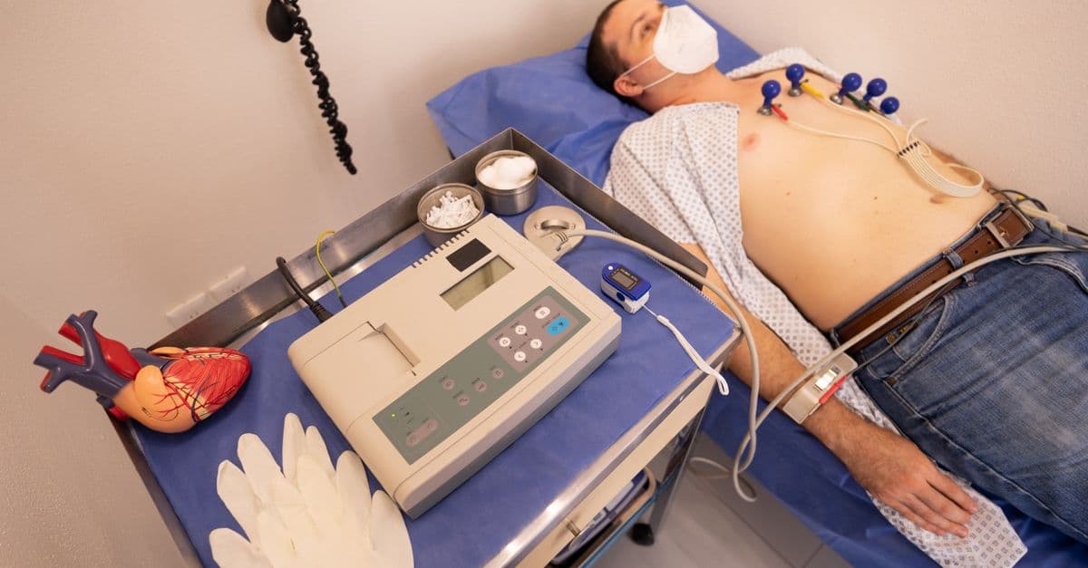
Photo: Los Muertos Crew / Pexels
The human heart beats roughly 100,000 times a day, moving about 7,500 liters of blood through 100,000 km of vessels — and it does this without conscious instruction, from the fourth week of embryonic development until the moment you die. Understanding how this fist-sized muscular pump works is one of the most useful pieces of biology you can learn, because cardiovascular disease remains the single largest cause of death worldwide (about 17.9 million people per year, according to the WHO).
This article walks through the anatomy, the electrical wiring, the cardiac cycle, and the most common things that go wrong — plus the modern treatments that have transformed cardiology over the past 50 years.
Four Chambers, Two Circuits
The heart is really two pumps stacked side by side, sharing a wall (the septum).
- Right side (right atrium + right ventricle) handles the pulmonary circulation: it receives deoxygenated blood from the body via the superior and inferior vena cavae and pumps it through the pulmonary arteries to the lungs.
- Left side (left atrium + left ventricle) handles the systemic circulation: it receives freshly oxygenated blood from the lungs via the pulmonary veins and pumps it through the aorta to every tissue in the body.
The left ventricle does dramatically more work than the right — it must generate enough pressure to push blood from your chest all the way down to your toes and back. Its wall is roughly three times thicker than the right ventricle's as a result.
The Valves
Four valves keep blood flowing in one direction only:
| Valve | Location | Function |
|---|---|---|
| Tricuspid | Right atrium → right ventricle | Prevents backflow into atrium during ventricular contraction |
| Pulmonary | Right ventricle → pulmonary artery | Prevents backflow into ventricle after ejection |
| Mitral (bicuspid) | Left atrium → left ventricle | Prevents backflow during ventricular contraction |
| Aortic | Left ventricle → aorta | Prevents backflow into ventricle during diastole |
The "lub-dub" you hear through a stethoscope is the valves snapping shut: the lub (S1) is the mitral and tricuspid closing as the ventricles start to squeeze, and the dub (S2) is the aortic and pulmonary closing as the ventricles relax.
The Cardiac Cycle: Systole and Diastole
Each heartbeat is divided into two phases:
- Diastole — the ventricles relax and fill with blood. The atria contract near the end of diastole to top up the ventricles (the "atrial kick," responsible for about 20–30% of ventricular filling).
- Systole — the ventricles contract, valves close, pressure spikes, and blood is ejected into the pulmonary artery and aorta.
At rest, systole takes roughly 0.3 seconds and diastole about 0.5 seconds at a heart rate of 70 bpm. As heart rate climbs (during exercise), diastole shortens disproportionately — which is why severely tachycardic hearts can fail to fill adequately.
Stroke volume is the amount of blood ejected per beat (~70 mL at rest in a healthy adult). Cardiac output is stroke volume × heart rate, normally ~5 L/min at rest, and capable of rising to 20–25 L/min in trained athletes during peak exercise.
The Electrical Conduction System
The heart has its own pacemaker — it does not need the brain to tell it when to beat. The wave of electrical depolarization travels in a precisely timed sequence:
- SA (sinoatrial) node — the natural pacemaker, in the upper right atrium, fires 60–100 times per minute at rest. Initiates each beat.
- AV (atrioventricular) node — delays the signal by ~0.1 seconds so the atria finish contracting before the ventricles start.
- Bundle of His — carries the signal into the interventricular septum.
- Left and right bundle branches — split the signal toward each ventricle.
- Purkinje fibers — distribute the signal rapidly through the ventricular muscle, so the entire ventricle contracts almost simultaneously.
An electrocardiogram (ECG/EKG) records this electrical activity from the skin:
- P wave — atrial depolarization (atria contracting).
- QRS complex — ventricular depolarization (the big spike — ventricles contracting).
- T wave — ventricular repolarization (ventricles resetting).
The PR interval, QRS width, and QT interval are the most clinically watched measurements. Abnormalities here flag everything from blocks to dangerous arrhythmias.
Heart Rate Regulation
The autonomic nervous system continuously fine-tunes heart rate and contractility:
- Sympathetic stimulation (fight-or-flight, via norepinephrine on β1 receptors) speeds the heart up, increases contractility, and dilates coronary arteries.
- Parasympathetic stimulation (rest-and-digest, via the vagus nerve and acetylcholine) slows the heart down.
Hormones like adrenaline (from the adrenal medulla) and thyroid hormones also influence rate. Long-term blood pressure regulation involves the kidneys via the renin-angiotensin-aldosterone system — see our companion article on how the kidneys work for that loop.
The Coronary Arteries
The heart muscle itself needs blood. The left main, left anterior descending (LAD), left circumflex, and right coronary artery branch off the aorta just above the aortic valve. The LAD supplies most of the left ventricle and septum — a blockage here is the classic "widow-maker." Coronary blood flow happens primarily during diastole, which is one reason tachycardia is dangerous in coronary disease.
What Goes Wrong: Common Heart Conditions
Atherosclerosis and myocardial infarction (heart attack). Cholesterol-laden plaques build up in the coronary arteries over decades. When a plaque ruptures, a clot forms and blocks flow — the downstream muscle starts dying within minutes. Time-to-treatment is everything. See our article on cholesterol and lipoproteins for the molecular backstory.
Arrhythmias. Disordered electrical activity. Atrial fibrillation (AFib) is the most common — chaotic atrial activity that raises stroke risk roughly 5-fold because blood pools in the atria and can clot. Ventricular fibrillation, by contrast, is immediately lethal without defibrillation.
Heart failure. The heart can't pump adequately. Split into systolic (reduced ejection fraction — the muscle is weak) and diastolic (preserved ejection fraction — the muscle is stiff and can't fill). Both end the same way: fluid backs up, lungs fill, exercise tolerance collapses.
Valvular disease. Aortic stenosis (the aortic valve calcifies and narrows, common over 75) and mitral regurgitation (the mitral valve leaks) are the two biggest in older adults.
Modern Treatments
Cardiology has been revolutionized in the past 40 years:
| Treatment | What it does | Used for |
|---|---|---|
| Statins | Lower LDL cholesterol via HMG-CoA reductase | Prevent atherosclerosis |
| Beta blockers | Slow heart rate, reduce contractility | Hypertension, heart failure, post-MI |
| ACE inhibitors / ARBs | Block RAAS, lower afterload | Hypertension, heart failure |
| PCI / stents | Mechanically open blocked coronaries | Acute MI, severe angina |
| Pacemakers | Pace the heart when its own conduction fails | Heart block, severe bradycardia |
| ICDs | Shock the heart out of lethal arrhythmias | Survivors of cardiac arrest, severe HF |
| TAVR | Replace aortic valve via catheter (no open surgery) | Aortic stenosis |
| LVAD | Mechanical pump assisting the left ventricle | Bridge to transplant or destination therapy |
| Heart transplant | Replace the heart entirely | End-stage heart failure |
The development of door-to-balloon time protocols (getting a heart attack patient's blocked artery open within 90 minutes of hospital arrival) alone has cut MI mortality dramatically since the 1990s.
The Frank-Starling Mechanism: How the Heart Self-Regulates
One of the most elegant properties of the heart is its ability to self-regulate: when more blood returns to the heart (e.g., during blood donation recovery or pregnancy), the heart automatically beats harder without any external instruction. This property, the Frank-Starling mechanism or length-tension relationship, arises from the structure of cardiac muscle itself.
Cardiac myocytes contain overlapping thick filaments (myosin) and thin filaments (actin). When the ventricle stretches slightly (from increased venous return), the sarcomeres are stretched to an optimal overlap length where myosin and actin interact most effectively. The result: increased contractility proportional to stretch. This mechanism is fundamental: it ensures the left and right ventricles pump equal amounts of blood each beat, even though they have different resting lengths. Overstretching the sarcomeres (as in dilated cardiomyopathy) moves past the optimal length, and contractility declines.
The Frank-Starling mechanism explains why pregnancy, which increases blood volume by 30–50%, causes the heart to adapt by increasing stroke volume without requiring new neural signals. It also explains why acute severe mitral regurgitation (the mitral valve leaks, overdistending the left ventricle beyond optimal length) causes acute heart failure: the mechanism works only within a physiological range.
Source: Circulation Journal (2023) — Frank-Starling Mechanism and Cardiac Physiology in Health and Disease.
Risk Factors and Prevention
Cardiovascular disease is predominantly preventable. Modifiable risk factors account for roughly 80% of cardiac risk:
- Hypertension (high blood pressure) — the single strongest modifiable risk factor. Every 20 mmHg increase in systolic BP above 115 mmHg doubles cardiovascular risk. Control via DASH diet, exercise, weight loss, and antihypertensive drugs reduces mortality 15–20%.
- Smoking — triples heart attack risk. Quitting restores normal risk within 12 months.
- LDL cholesterol — causal link proven by statin trials. Lowering LDL from 100 to 70 mg/dL via statins reduces cardiac events ~30%. PCSK9 inhibitors lower LDL further.
- Diabetes — doubles coronary risk via inflammation, endothelial dysfunction, and autonomic neuropathy. Tight glycaemic control reduces complications.
- Obesity and sedentary lifestyle — weight gain increases afterload (the pressure the left ventricle must overcome to eject blood); exercise improves multiple risk factors simultaneously.
- Poor diet — high saturated fat and sodium intake drives both hypertension and atherosclerosis.
- Chronic stress and depression — linked to increased cortisol, inflammation, and sympathetic tone; both increase cardiac risk.
Non-modifiable factors include age (risk rises steeply after 60), male sex (women have lower risk pre-menopause due to oestrogen cardioprotection), and family history (genetic variants in lipid metabolism, inflammation, and blood pressure regulation contribute).
A 2023 study in Circulation found that adherence to seven simple lifestyle factors (not smoking, regular exercise, healthy diet, moderate alcohol, normal weight, normal blood pressure, normal glucose) reduced cardiovascular mortality by approximately 75% compared to those meeting none, across all socioeconomic and ethnic groups.
Source: American Heart Association (2024) — Cardiovascular Risk Factor Modification Guidelines.
Cardiac Biomarkers and Diagnosis
Modern cardiology relies on biomarkers — measurable indicators of cardiac damage or dysfunction — to diagnose and risk-stratify patients.
Troponin (cardiac isoforms cTnI and cTnT) is the most specific marker of myocardial infarction. Detectable in blood within 3–4 hours of chest pain onset, peaking at 24–48 hours. High-sensitivity troponin assays can rule out MI within 1–3 hours, dramatically improving emergency department efficiency. Importantly, troponin elevation is not specific to MI — it rises in sepsis, heart failure, PE, and renal failure, so clinical context matters.
B-type natriuretic peptide (BNP) and its more stable form NT-proBNP are released by ventricles under stretch (volume overload). Values >100 pg/mL raise suspicion for heart failure; <35 pg/mL essentially rules it out. BNP-guided therapy in heart failure reduces hospitalisations.
D-dimer — elevated in pulmonary embolism (clot in lung vessels), though nonspecific. Low D-dimer effectively excludes PE in low-risk patients, sparing unnecessary imaging.
Lipoprotein(a) — an LDL-like particle, genetically determined. Elevated in ~20% of population; accounts for 5–10% of cardiovascular disease population risk. Not modifiable by standard statins, but new therapies targeting Lp(a) synthesis are emerging.
Exercise and Cardiac Adaptation
Regular aerobic exercise is one of the most powerful interventions for cardiac health. The heart is a muscle; it adapts to training.
Physiological hypertrophy — benign left ventricular thickening from chronic exercise — increases stroke volume and cardiac efficiency. Athletes achieve cardiac outputs of 20–25 L/min at maximal exercise, 4–5 times resting cardiac output, compared to 15–20 L/min in untrained individuals.
Autonomic remodelling — endurance training lowers resting heart rate via increased parasympathetic tone (vagal conditioning). Elite athletes may have resting heart rates of 40–50 bpm without pathology.
Vascular adaptations — capillary density in skeletal muscle increases; endothelial function (nitric oxide production, vasodilatory capacity) improves.
Cardioprotective signalling — exercise triggers anti-inflammatory and antioxidant pathways, reduces sympathetic tone, and improves glucose metabolism. These changes persist even on rest days, creating a cumulative protective effect.
Guidelines recommend 150 minutes of moderate aerobic activity weekly or 75 minutes of vigorous activity for cardiovascular health. Even brief bouts of activity (brisk 3-minute walks) accumulate benefits. See our article on the science of exercise for detailed guidance.
Frequently Asked Questions
Q: Can the heart really beat outside the body?
A: Yes, briefly. The sinoatrial node fires autonomously as long as cells have oxygen and metabolic substrates. Isolated heart preparations in research labs (Langendorff perfusion) keep hearts beating for hours. This autonomous activity demonstrates that the heart doesn't rely on the brain for its rhythm — the brain only modulates it.
Q: Why does the left ventricle wall get thick in athletes?
A: Chronic exercise causes physiological hypertrophy — the muscle adapts to higher workload by adding sarcomeres in parallel, thickening the wall while maintaining or improving function. This adaptation is benign and reversible when training stops, unlike pathological hypertrophy from chronic hypertension (where thickening occurs with fibrosis and diastolic dysfunction).
Q: What does ejection fraction (EF) actually measure?
A: The percentage of blood in the left ventricle ejected per beat. Normal is 55–70%. Below 40% is reduced ejection fraction (HFrEF, systolic heart failure); 40–49% is borderline; preserved EF (HFpEF) maintains EF >50% but has stiff, non-compliant ventricles that can't fill adequately, still causing heart failure symptoms.
Q: How long can you live without a heartbeat?
A: Brain damage begins within 4–6 minutes of cardiac arrest without blood flow. Cardiopulmonary resuscitation (CPR) maintains some perfusion, extending the window. Defibrillation (electrical shock) within 3–5 minutes of ventricular fibrillation triples or quadruples survival; beyond 10 minutes, survival is rare without special techniques like extracorporeal CPR.
Q: Are heart transplants common and what is long-term survival?
A: Approximately 3,500–4,000 transplants are performed annually in the US, limited by donor supply (only ~3,000 donor hearts available for ~4,000 candidates yearly). One-year survival is now ~90% with modern immunosuppression; median survival post-transplant is approximately 13 years. Lifetime risk of chronic rejection (gradual scarring of the graft) remains the long-term limitation.
Q: Does caffeine cause arrhythmias in people without heart disease?
A: For most people, no. Large cohort studies (including >400,000 person-years of follow-up) have not found a meaningful link between moderate caffeine intake and atrial fibrillation in the general population. However, individuals with existing arrhythmias or certain genetic variants may be sensitive; personal tolerance varies and should guide consumption.
Sex Differences in Heart Disease and Cardiology
Heart disease affects men and women differently, yet this truth is often overlooked in clinical practice. Younger women have substantially lower coronary risk than men, a phenomenon attributed to oestrogen's cardioprotective effects (lower LDL, higher HDL, improved endothelial function). However, after menopause, women's risk rises steeply, eventually exceeding men's in very advanced age.
Atrial fibrillation follows a different trajectory: women develop AFib later but at higher lifetime risk due to longer life expectancy. Women with AFib have higher stroke risk (CHA₂DS₂-VASc scoring is more aggressive for women).
Symptoms diverge: Women experiencing heart attacks report atypical symptoms more often — dyspnea (shortness of breath), fatigue, nausea, jaw pain — rather than classic substernal chest pain. This atypical presentation leads to delayed diagnosis; women experience longer door-to-balloon times in acute MI.
Heart failure phenotypes differ: women are overrepresented in heart failure with preserved ejection fraction (HFpEF), often driven by diastolic dysfunction and hypertension. Men have proportionally more systolic heart failure from coronary disease.
Treatment responses vary: women tolerate certain beta blockers differently (more symptomatic side effects); ACE inhibitors show larger blood pressure reductions in women. Hormone replacement therapy, once thought protective, was found (by the Women's Health Initiative trial, 2002) to increase coronary risk — a finding that reshaped clinical practice.
The implications: one-size-fits-all cardiology is suboptimal. Sex-specific risk scores, symptom recognition training, and sex-specific pharmacology are increasingly recognized as necessary.
Source: Mehta, L. S., et al. (2021). Acute Myocardial Infarction in Women and Sex-Based Differences in Presentation and Outcomes. Circulation, 143(8). — Comprehensive review of sex differences in cardiovascular disease.
The Aging Heart and Senescent Cardiac Cells
Cardiac aging is a distinct pathophysiological process, separating normal senescence from disease. Even in the absence of hypertension, diabetes, or coronary disease, the aging heart exhibits specific changes:
Diastolic dysfunction becomes increasingly prevalent with age. The ventricular wall becomes stiffer (increased fibrosis, collagen cross-linking), impairs relaxation, and fills poorly. Older adults compensate via increased atrial contractility and tachycardia during exertion; however, when atrial fibrillation develops, loss of the atrial kick causes acute heart failure in previously asymptomatic individuals.
Senescent cardiac cells accumulate with age due to telomere shortening and diminished capacity for autophagic clearance of damaged proteins. These cells secrete pro-inflammatory cytokines (IL-6, TNF-α), creating local inflammation. The senescent cell burden correlates with heart failure risk.
Mitochondrial dysfunction increases with age — aged cardiac mitochondria produce more reactive oxygen species and generate less ATP efficiently. This reduces the energy available for contraction.
Conduction system aging increases susceptibility to arrhythmias. The sinoatrial node loses pacemaker cells; conduction velocity slows; refractory periods lengthen. Atrial fibrillation incidence rises from ~1% at age 60 to ~10–15% by age 85.
Exercise capacity declines with age even in healthy individuals. Maximum achievable cardiac output drops 20–30% per decade after age 30, primarily due to reduced maximum achievable heart rate (max HR = 220 - age, roughly) and reduced contractility.
Emerging therapies targeting aging include senolytics — drugs that selectively eliminate senescent cells — which have shown promise in animal models and are entering clinical trials in heart failure.
Cardiac Stem Cells and Regenerative Medicine
Unlike skeletal muscle, mature cardiac muscle cannot regenerate. After myocardial infarction, dead myocytes are replaced by fibrosis (scarring), permanently reducing contractile function. This has spurred interest in cardiac stem cell therapy.
Bone marrow-derived stem cells were initially hoped to differentiate into cardiomyocytes and replace lost tissue. Trials like REPAIR-AMI (2006) showed modest improvements in ejection fraction. However, the benefit-to-effort ratio proved suboptimal, and enthusiasm waned. The mechanism appears to be paracrine: stem cells secrete cytokines that reduce inflammation and apoptosis, rather than differentiating into new muscle.
Induced pluripotent stem cells (iPSCs) are reprogrammed from mature cell types (skin, blood) to an embryonic state, then differentiated into cardiomyocytes in vitro. iPSC-derived cardiomyocytes can be generated in large numbers, genetically corrected if needed, and potentially transplanted. Early animal studies are promising, but translation to humans faces challenges: engraftment efficiency is low (~1–10% of transplanted cells survive), arrhythmia risk is high (young myocytes don't connect smoothly to mature tissue), and immune rejection or tumorigenicity remains a concern.
Gene therapy targeting heart failure via viral vectors is emerging. Restoring calcium handling (SERCA2a gene therapy) or improving myofilament function shows promise in preclinical and early clinical studies. The challenge: targeting the entire heart requires systemic delivery, ensuring safe tropism to cardiac tissue.
Most progress to date involves supportive technologies: ventricular assist devices (LVADs) are increasingly used as destination therapy, not just bridge to transplant, allowing decades of prolonged life for end-stage patients.
The Heart's Role in Systemic Physiology
The heart is not merely a pump—it's an endocrine organ secreting hormones that regulate blood pressure, fluid balance, and metabolism:
Natriuretic peptides (ANP from atria, BNP from ventricles) are released when the heart stretches. They promote sodium and water excretion by the kidneys, reduce sympathetic tone, and dilate blood vessels — collectively reducing cardiac preload and afterload. This is a negative feedback loop: when the heart is overstretched, it secretes factors that reduce its workload.
Endothelins are powerful vasoconstrictors secreted by endothelial cells in response to wall stress. Dysfunctional endothelin signaling contributes to pulmonary hypertension.
Cardiac troponins, beyond their role in contraction, appear to have signaling functions in hypertrophy and apoptosis pathways, making them potential therapeutic targets.
These endocrine functions mean heart disease is never purely cardiac—it's systemic, affecting kidneys, lungs, brain, and metabolism.
The Takeaway
The heart is a triumph of evolutionary engineering: a self-pacing, self-regulating pump operating continuously for decades. Understanding its anatomy, electrophysiology, and physiology reveals why it fails in specific predictable ways and illuminates how modern medicine repairs it. The three biggest killers—atherosclerotic myocardial infarction, arrhythmias, and heart failure—now have evidence-based treatments; 80% of the remaining preventable risk comes from lifestyle factors. Cardiovascular disease is fundamentally preventable, yet remains the leading cause of death globally because prevention is underutilized. Knowing how the heart works should motivate action: diet, exercise, stress management, and regular monitoring are not merely recommendations—they are the most powerful medicines available.
Related Reading
Weekly digest
Stay ahead of the curve
Get the best science, AI, and tech stories delivered to your inbox every week. No spam — unsubscribe any time.
About the Author
SciHub101 is an independent science and technology publication founded and edited by Buğra Sözeri, a software engineer and app developer based in Türkiye. Articles are drafted against peer-reviewed literature and primary sources, fact-checked, and edited by a human before publication. Where we compare products, anything built by our own team is labelled as such — see the editorial guidelines and About page for how corrections and disclosures work.
Related Articles

How Color Perception Works — Biology of Color
How human eyes and brain construct color from light. Cones, trichromacy, color blindness, animal vision, and why color isn't a property of objects.

How Pregnancy Works — The Biology of New Life
How pregnancy works: fertilization, implantation, placenta, the three trimesters, labor, complications, and how modern medicine has changed outcomes.
How the Human Heart Pumps Blood: A Complete Guide
Your heart beats about 100,000 times a day without a single conscious thought. Here's the remarkable engineering behind it.
How Does the Human Heart Actually Work?
Your heart beats 100,000 times a day, pumping ~5 litres of blood every minute. Here's the elegant biology of the most reliable mechanical organ in your body.
How Lungs Work — Breathing and Gas Exchange
How the lungs move air, exchange oxygen and CO2, and what goes wrong in asthma, COPD, pneumonia, and at high altitude. A clear, evidence-based explanation...

How the Human Heart Works: The Cardiac Cycle Explained
The heart beats more than 100,000 times per day without a single conscious command, regulated by an electrical system so precise that it can be measured from electrodes on the skin. Here is how that system works, from the first spark in the SA node to the pressure wave that reaches your fingertips.