How Do Octopuses Change Color? The Science of Chromatophores and Camouflage
Photo: yu zhang / Pexels
An octopus approaches a patch of gravel on the seafloor and, in less than one-third of a second, its entire body transforms from pale gray to an intricate mosaic of dark spots, bumps, and textured ridges that replicate the appearance of the rocks beneath it. The transformation is not a slow wash of color—it is a complete reengineering of the skin's visual properties. Most startling, the octopus performing this feat is colorblind. Its eyes contain only one type of photoreceptor; it cannot distinguish the wavelengths it is reproducing with such precision. The system that accomplishes this is neither a paint job nor a passive material property. It is an active, brain-controlled display panel built from millions of individually addressable cells arranged in three distinct biological layers, operating on timescales that rival human reflexes. Understanding how it works requires unpacking each layer separately—and then confronting one of biology's deepest puzzles: how a colorblind animal perceives color at all.
The Three-Layer Skin System: A Biological Display Panel
The skin of an octopus, cuttlefish, or squid is not a uniform organ. It is a stratified structure with three functionally distinct layers, each employing a different physical mechanism to generate color and light-manipulating properties. The brain does not control all three layers equally; some respond to motor commands on the timescale of tens of milliseconds, while others shift colors over seconds through osmotic pressure changes. Understanding their interaction is the key to understanding how octopus camouflage actually works.
Chromatophores — specialized pigment-containing cells consisting of an elastic sac filled with granules of concentrated pigment (yellow, orange, red, or brown), surrounded by a ring of radial muscle cells arranged around the sac's perimeter like spokes of a wheel. Each chromatophore is directly innervated by its own motor neuron from the central brain, allowing individual control. When the muscles contract, they pull the sac outward like a balloon inflating, expanding it from an essentially invisible pinpoint to a disk up to one millimeter in diameter, instantly revealing the color inside. When the muscles relax, the elastic properties of the sac itself cause it to snap back to its small, concealed state, making the color vanish. This expansion and retraction cycle takes less than 200 milliseconds.
The biophysics here is elegant and economical. The chromatophore does not generate color; it stores it, kept invisible by being kept small. The pigment itself—yellow through brown—reflects light across a limited range of wavelengths, and by controlling which chromatophores are expanded at any given moment, the brain can compose a spatial pattern of color across the entire body surface. A common cuttlefish has roughly 200,000 to 300,000 chromatophores, each independently addressable by a dedicated neuron. This is a pixel array controlled by living biology.
Source: Hanlon, R.T. and Messenger, J.B. (1996), Cephalopod Behaviour, Cambridge University Press — the foundational reference on cephalopod skin structure and neuronal control of chromatophores.
The speed is critical. Under predation or defensive threat, an octopus must change its appearance quickly enough that a striking predator cannot track visual landmarks on its skin. The 200-millisecond timescale is fast enough to defeat most predator reaction times. In experiments, researchers have shown that octopuses can complete a full color and pattern changeover in the time it takes a fish predator to strike.
Iridophores — cells that do not contain pigment but instead contain stacks of thin, crystalline protein plates—typically made of a molecule called guanine—separated by layers of cytoplasm or water. These structures do not reflect color through pigment; they reflect it through constructive and destructive interference of light waves. When two light waves reflect off two different layers of the stack, and the difference in their path lengths equals an integer multiple of the light's wavelength, the reflected waves reinforce each other, and that wavelength is strongly reflected. All other wavelengths cancel out. This process, called thin-film interference, is the same mechanism that makes soap bubbles iridescent and oil slicks shimmer.
The advantage of iridophores is that they can reflect a much wider range of colors than chromatophores—including metallic blues, greens, and silvers that pigment alone cannot produce. The disadvantage is speed. Chromatophores respond to motor commands within milliseconds. Iridophores respond to changes in osmotic pressure around the cell, a slower process involving water movement into and out of the cytoplasm surrounding the plates. Changing iridophore color takes seconds, not tens of milliseconds. This slower timescale suggests iridophores are not primarily for rapid escape, but rather for the fine-tuning of camouflage once the octopus has settled in place, and for social signaling displays that do not need to be instantaneous.
Leucophores — cells packed with spherical protein granules (made of purine compounds, often guanine or hypoxanthine) that are small enough to scatter all wavelengths of visible light equally, producing broadband white reflection. Leucophores serve as a reflective backing layer, ensuring that colors from the chromatophore and iridophore layers above appear bright and vivid rather than muddy. Without leucophores, the body would absorb much of the light hitting the chromatophores, dulling the reflected color. With them, the skin becomes a high-contrast display.
The three-layer system functions as a biological analog to the LCD displays found in screens. The leucophores act as the backlight, providing brightness. The iridophores and chromatophores act as the color-filtering layers, modulating what wavelengths of that light are allowed through. The difference is that this system is not electronic—it is biological, controlled by the nervous system, and responsive to sensory input in real time.
| Layer | Cell type | Mechanism | Color range | Response speed |
|---|---|---|---|---|
| Top | Chromatophores | Muscle-controlled pigment expansion | Yellow to brown | <200 ms |
| Middle | Iridophores | Crystalline plate spacing (interference) | Blue, green, iridescent | Seconds |
| Bottom | Leucophores | Broadband light scattering | White reflection | Fixed |
How the Brain Commands the Skin: The Visual-Motor Loop
The neural control system that orchestrates this display is direct and efficient. Photons from the environment hit the octopus's eyes, where they are absorbed by photoreceptor cells in the retina. The signal is then processed in the optic lobes, a massive neural structure that comprises roughly one-quarter of the octopus's entire brain. These lobes extract certain features from the visual scene: not a pixel-by-pixel image, but a set of abstract visual properties that predict what the substrate looks like.
The specific features extracted are still being actively researched, but they include at minimum: mean brightness (average reflectance), spatial frequency (coarseness of texture), and local contrast (edges and transitions). The optic lobes also receive information about body orientation and position, allowing the brain to map the spatial distribution of the visual features being detected across different regions of the body.
This information is then fed to motor systems that control the chromatophore muscles. Each chromatophore muscle group is directly wired to a motor neuron originating from the brain. When the brain sends a signal down that neuron, the muscle contracts, the chromatophore expands, and the pigment becomes visible. The entire pathway is monosynaptic or disynaptic—one or two neural synapses between the sensory input and the motor output—meaning there is almost no delay or filtering. The system is as direct as the connection between your hand touching a hot stove and your hand pulling away.
Source: Nilsson, D.E. et al. (2012), "A Conserved Vertebrate Genetic Toolkit for Vision," Nature Neuroscience, Vol. 15 — comparative study showing that despite structural differences, cephalopod and vertebrate optic systems rely on conserved genes and functional principles for visual processing.
What makes this neurobiologically remarkable is that the octopus does not need to consciously "decide" to change its color to match the background. The system is largely automatic. Place an octopus on a checkerboard pattern, and within seconds, the chromatophore pattern will begin matching the geometry of the squares. The visual system is computing the statistics of the background, and the motor system is translating those statistics into chromatophore positions, without any apparent deliberation.
The Colorblind Paradox: Seeing Color Without Color Vision
Here is the puzzle that has troubled cephalopod researchers for over a century: the octopus and cuttlefish can match colors they cannot perceive.
An octopus eye, like all cephalopod eyes, contains only one type of photoreceptor in the retina: a rod-like structure sensitive to blue light. The visual pigment present is a variant of rhodopsin tuned to wavelengths around 490 nanometers (blue-green). Compare this to the human eye, which contains three types of cone cells: sensitive to short (blue), medium (green), and long (red) wavelengths. Color vision in humans—the ability to discriminate between wavelengths and perceive hue—arises from comparing the response levels across these three receptor types. A red object activates the long-wavelength cones more than the short-wavelength ones; a blue object shows the opposite pattern. The brain compares these signals and generates the sensation of red or blue.
An animal with only one type of photoreceptor cannot perform this comparison. By the standard definition, the octopus should be colorblind—unable to distinguish red from green, blue from yellow. And by standard behavioral tests of color discrimination, it is. Octopuses do not distinguish colors in the way humans do.
Yet they match colors with remarkable precision. Place an octopus on a red substrate, and within minutes, the animal displays predominantly orange and reddish chromatophores. Place it on a yellow substrate, and the display shifts toward yellow. Place it on a bright blue background, and the octopus rearranges its chromatophores and iridophores to approximate blue. The matching is not perfect—octopuses match substrates better when they have had time to observe them under stable lighting—but it is good enough to fool predators and human observers alike.
This paradox catalyzed decades of research, and the leading hypothesis comes from work by Christopher Stubbs and colleagues at the University of California Santa Barbara and the Scripps Institution of Oceanography. Their proposal is that cephalopods exploit chromatic aberration in their own optical system.
Chromatic aberration is a flaw (or in this case, a repurposed feature) of all simple lens systems. Different wavelengths of light bend (refract) at slightly different angles when passing through a lens, causing them to focus at slightly different distances behind the lens. A lens focusing blue light will bring it to a point 100 micrometers behind the lens; red light, refracted less, might focus 150 micrometers behind. This is why old camera lenses produce colored fringes around bright objects.
The octopus's pupil is not round. It is a horizontal slit in some species and a highly irregular, bumpy shape in others. The pupil shape can also be adjusted by the octopus through muscular control. Stubbs and colleagues proposed that by controlling pupil shape and accommodation (adjusting the focus of the lens), the octopus could essentially sample the scene at multiple focal depths simultaneously. Different depths would receive different chromatic filtering—shorter wavelengths (blues) in focus at one depth, longer wavelengths (reds) in focus at another. By comparing the brightness of the image at different focal settings, or by the distribution of blur around focused points, the octopus could extract spectral information encoded as spatial or brightness variation rather than as a multi-channel color code.
Source: Stubbs, A.L. and Stubbs, C.W. (2016), "Spectral Discrimination in Color Blind Animals via Chromatic Aberration and Pupil Shape," Proceedings of the National Academy of Sciences, Vol. 113 — presents the chromatic aberration hypothesis and argues that pupil shape modulation enables wavelength sampling.
This hypothesis is compelling but not yet definitively proven. Cuttlefish can match colors even in complete darkness (tested by researchers, who observed camouflage pattern changes with lights completely off), suggesting that either tactile information about substrate texture is sufficient to trigger appropriate color patterns, or the pattern-generation system has sufficient redundancy that multiple sensory modalities can drive similar outputs. The darkness experiment also suggests that the color matching may not require visual color perception at all—it may be more about matching the statistical texture of the background using whatever information is available (visual, tactile, or both).
Recent research has also identified opsin genes (the genes encoding light-sensitive proteins) in octopus skin itself. If the skin can express light-sensitive proteins, there is the possibility of peripheral light detection—the skin itself "seeing" to some degree and triggering local chromatophore responses. This would explain rapid, local pattern responses to light changes without requiring the central brain to process all visual information first.
Papillae and Three-Dimensional Camouflage
Chromatophores and iridophores handle color and pattern. But a cuttlefish or octopus camouflage display also includes a three-dimensional component: the texture and bumpy topography of the skin surface.
Papillae — muscular, bumpy projections from the skin surface that can be individually raised, retracted, or held at intermediate heights, allowing the animal to change the skin's macroscopic texture in real time. A flat skin looks flat; a skin covered in raised papillae looks bumpy, rocky, or spiky.
An octopus matching a smooth sandy seafloor will flatten its papillae and display uniform, fine-textured chromatophore patterns. An octopus matching a rocky reef will raise its papillae and pair that topographic change with a coarser, more mottled chromatophore pattern. An octopus trying to mimic a seaweed-covered substrate might raise papillae into elongated, waving shapes. This three-dimensional component is not incidental—it is necessary for complete camouflage. An animal that perfectly matched background color but did not match texture would cast shadows that revealed its outline.
The papillae are directly wired to the brain as well. The same visual system computing chromatophore patterns also computes papilla heights and positions. In some cuttlefish species, papillae can be raised or lowered within one second, much slower than chromatophore expansion but still rapid enough to contribute to escape responses.
Source: Hanlon, R.T., Forsythe, R.B., and Joneschild, D.E. (1999), "Crypsis, Conspicuousness, Mimicry and Polyphenism as Aspects of Adaptive Coloration in Cephalopods," Biological Journal of the Linnean Society, Vol. 88 — describes the integration of papilla, chromatophore, and iridophore control in cuttlefish camouflage displays and documents individual papilla-raising speeds.
Signaling, Not Just Camouflage
While camouflage is often presented as the primary function of octopus color change, the system is also used extensively for social signaling, threat displays, and mating communication. An octopus confronted by a rival or a predator does not always try to hide. Instead, it often performs a display—darkening its body, raising all papillae into pronounced spikes, and sometimes flashing high-contrast patterns in rapid sequences.
These displays serve multiple functions. They can startle a predator, making the octopus appear larger or more dangerous than it actually is. They can also signal aggression or dominance to a rival octopus. Males competing for mating access to a female engage in dramatic color displays, flashing patterns across their bodies in rapid sequences. The intensity and speed of the display can predict the outcome of the contest—a male displaying intensely tends to win access to the female, even against a larger male that displays less vigorously.
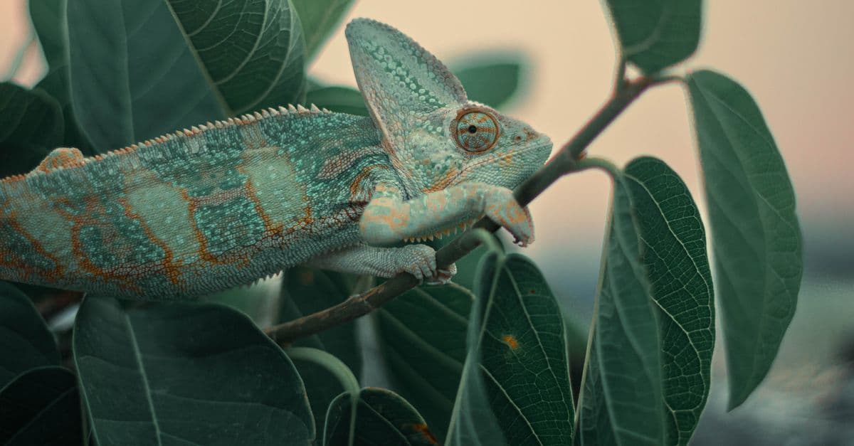
This observation parallels what researchers have found with chameleons: the most visually impressive color-change ability in an animal does not necessarily mean the color change primarily serves camouflage. Chameleons, as covered in the science of animal camouflage, use color change primarily for social signaling, not hiding. The same principle may apply more to octopuses than is often acknowledged. An animal with the ability to match its background has options—it can blend in when it wants to, but it can also stand out dramatically when signaling is more adaptive than hiding.
How Fast Can the System Really Respond?
The chromatophore system can expand and retract in under 200 milliseconds. But the complete camouflage response—including changes to iridophores and papillae—takes longer. In laboratory studies, a cuttlefish placed on a new background will show initial chromatophore changes within 200-400 milliseconds, a slower refinement phase over the next 10-30 seconds as iridophores adjust and papillae reach their final heights, and then further fine-tuning over minutes.
This multiphasic response suggests a layered strategy. The fast chromatophore changes provide immediate, gross-level camouflage—enough to break up the outline and match major brightness features. The slower iridophore and papilla adjustments fine-tune the match, providing details and three-dimensional accuracy. For an animal being pursued by a predator, reaching 70% camouflage match in 200 milliseconds is better than achieving 95% match in 30 seconds. For an animal settling into a hunting position, the slower refinement phase is worth the wait.
The speed of chromatophore response also appears to be tuned to the ecology of different species. Species hunted by visually-acute predators (like certain fish and cephalopod predators) show faster chromatophore response times than species living in deeper water or in environments with slower-moving predators. This is consistent with the idea that the system has been shaped by predation pressure: in a high-threat environment, the selective advantage of millisecond-level speed is high; in a low-threat environment, other priorities (energy efficiency, precise color matching) can trade off against speed.
Comparison With Chameleon Color Change: Two Solutions to One Problem
The octopus color-change system is often compared to that of chameleons, but the two systems are nearly opposite in their fundamental mechanisms. Understanding the differences illuminates what each system is optimized for.
Chameleons do not use chromatophores as a primary mechanism—or rather, they do have some chromatophore-like cells, but these are not the primary driver of the dramatic color changes that chameleons are famous for. Instead, chameleons rely primarily on iridophores: arrays of nanocrystals arranged in specific geometric patterns within the skin. When a chameleon is calm, these crystals are closely packed and reflect short wavelengths (blues and greens). When the chameleon is stressed, excited, or engaging in social signaling, skin cells expand, spacing the crystals further apart, and the reflected wavelengths shift toward reds and yellows.
The key difference is that chameleon color change is not directly controlled by motor neurons commanding individual cells. Instead, it is driven by systemic physiological changes—stress hormones (like catecholamines released during fight-or-flight responses), changes in body temperature, and changes in autonomic nervous system tone (parasympathetic versus sympathetic). These hormonal and autonomic changes alter tissue osmotic pressure and turgor across large skin regions, affecting crystal spacing throughout that region simultaneously. The change is slower (taking seconds to minutes rather than milliseconds) and coarser (affecting broad areas rather than individual pixels).
More fundamentally, chameleon color change is not primarily for camouflage. The research of Dolph Schluter, Darren Stuart-Fox, and others has shown that chameleon color change is primarily a social signal, broadcasting competitive status, readiness to mate, and alarm state. The camouflage function exists—a resting chameleon is well-matched to its foliage—but this match is a static property of resting coloration, not the dramatic displays that are the reason chameleons are famous.
An octopus system optimizes for speed and pixel-level control. A chameleon system optimizes for broadcasting large-scale internal physiological states. The octopus has invested heavily in fine-grained motor control; the chameleon has invested in honest signaling of internal state. These are different design priorities reflecting different ecological niches and evolutionary pressures.
Source: Teyssier, J., Saenko, S.V., van der Meulen, D., Marshall, J., and Oberwinkler, J. (2015), "Photonic Crystals Cause Active Colour Change in Chameleons," Nature Communications, Vol. 6 — identified iridophore crystal structures as the primary mechanism of chameleon color change, replacing the older chromatophore hypothesis.
Energy Cost and the Nervous System Burden
Operating hundreds of thousands of individually-controlled chromatophores is metabolically expensive. Each chromatophore requires a dedicated motor neuron, and motor neurons consume ATP continuously, whether firing or silent (there is a baseline metabolic cost to maintaining ion gradients). The octopus neural system must devote substantial neural tissue to the control of color display. The brachial lobes of the octopus brain—the structures directly controlling the arms and skin—are among the largest neural centers in the animal.
Yet cephalopods have chosen to bear this cost. The speed and fine-grained control afforded by direct motor neuron control of millions of cells must provide selective advantage sufficient to justify the metabolic investment. In environments with high predation pressure and prey with acute vision (like certain fish species), the ability to change camouflage faster than a predator can strike appears to be worth the cost.
The system also reveals something about cephalopod neural organization more broadly. Cephalopods have evolved a nervous system where a significant fraction of the total neurons are dedicated to real-time sensorimotor control—controlling the arms, the skin, the suckers—with relatively less emphasis on central processing and learning compared to vertebrates. This distributed architecture is part of the explanation for why octopuses have such high neural counts (500 million neurons) while being shorter-lived and smaller-bodied than many vertebrates with fewer neurons. Much of that neural tissue is dedicated to control systems for a highly complex, flexible body.
Paper Thin: The Physiological Limits of the System
One constraint on the chromatophore system is mechanical. Each chromatophore is an elastic sac with muscle fibers pulling on its perimeter. The sac is roughly 0.5 to 1 millimeter in diameter when maximally expanded, and it is packed densely—hundreds of thousands to millions per animal. The skin itself is typically only a few millimeters thick. The chromatophore layer occupies only the topmost portion of the skin; below it are iridophores, leucophores, and connective tissue.
This means there is a physical limit to how densely packed these cells can be. You cannot have infinitely fine control if you only have a few hundred thousand pixels to work with. Octopuses have solved this by using pattern generation algorithms that work on relatively coarse texture scales—their camouflage patterns are typically composed of features in the range of 1-5 millimeters in size. They can match fine details through iridophore and papilla adjustments at slower timescales, but for the rapid 200-millisecond response, the resolution is limited.
This is still sufficient to fool most predators and prey, but it means that a sufficiently high-resolution background pattern—finer than the chromatophore spacing—cannot be perfectly matched by the octopus. In practice, octopuses are observed rearranging their body position and orientation when settling into camouflage, suggesting that they actively seek out background substrates that match the spatial scale of their available chromatophore control.
The Takeaway
The octopus color-change system is not a single mechanism but an integrated biological display panel composed of three layers of cells (chromatophores, iridophores, leucophores), a three-dimensional textural component (papillae), and a direct neural control system linking the animal's visual perception to the motor neurons commanding each display element. The system is optimized for speed and fine-grained control in environments where predators are visually acute and the advantage of millisecond-level camouflage changes justifies the metabolic and neural cost. The paradox of colorblind color matching reveals that color perception and color production are not as tightly linked as our intuition suggests—the octopus likely detects color through indirect means (chromatic aberration, peripheral opsin expression) rather than through conventional color vision, and it uses abstract visual features (brightness, texture, spatial frequency) as sufficient drivers for appropriate color and pattern responses. The system is evidence that evolution solves problems with the tools available; the octopus does not have three types of photoreceptors, so it built a different kind of visual system that works around that constraint. The result is a camouflage system so effective that it prompted human researchers to ask how an animal could possibly achieve what it was achieving—and the answers revealed principles of neural control, visual processing, and biomechanical design that apply far beyond cephalopods.
Frequently Asked Questions
Q: How do octopuses control individual chromatophores if they are colorblind?
A: Each chromatophore has its own dedicated motor neuron from the brain. The brain does not need to "see" each chromatophore individually—it computes the visual properties of the background (brightness, texture, spatial frequency), then sends motor commands to chromatophore populations based on those abstract features. Which specific chromatophores to expand is determined by pre-wired neural circuits and learned patterns, not by conscious color vision.
Q: Can octopuses match colors they have never seen before?
A: Yes, to a degree. Octopuses placed on novel substrates with colors and patterns they have never encountered will begin generating matching patterns within seconds. This suggests the pattern-generation system is not a lookup table of memorized patterns but a dynamic algorithm that computes appropriate responses to visual features in real time.
Q: How many chromatophores does an octopus have?
A: It varies by species and body size. A common cuttlefish has roughly 200,000 to 300,000 chromatophores. A large octopus likely has similar numbers, though exact counts for living octopuses are difficult to establish. Each chromatophore is individually innervated, meaning the brain has potentially hundreds of thousands of independent pixels to control.
Q: Is chromatophore control the only way cephalopods change color?
A: No. Iridophores and leucophores also contribute to color and light manipulation. Iridophores respond more slowly (over seconds) and allow production of structural colors like iridescent blues and silvers. Chromatophores are fastest and most directly controlled, making them primary for rapid camouflage changes. Papillae add a three-dimensional texture component. The three systems work together, with different speeds suited to different functions.
Q: How does the octopus brain manage motor control of millions of cells simultaneously?
A: The octopus brain uses a hierarchical control strategy. The optic lobes compute summary statistics of the visual scene (not pixel-by-pixel details), and these summary properties are translated into high-level commands to motor systems. Local neural circuits in the skin and nervous system then expand those high-level commands into specific chromatophore positions. This distributed processing means the central brain does not need to compute every detail—it sets general parameters and delegates fine details to local circuits.
Q: Do octopuses use color change only for camouflage?
A: No. Octopuses also use rapid color changes for threat displays, courtship, and signaling dominance during contests with rivals. A fleeing or fighting octopus may darken dramatically and raise all papillae into spikes, making itself appear larger and more threatening. Males competing for mates perform elaborate rapid displays. Color change serves multiple functions depending on the behavioral context.
Read next:
Follow new articles
Add SciHub101 to your feed reader to see new science and technology stories.
About the Author
SciHub101 is an independent science and technology publication founded and edited by Buğra Sözeri, a software engineer and app developer based in Türkiye. AI tools assist with drafting and research. Source documentation in the existing archive is uneven, and articles are being reviewed and corrected. The Editorial Team byline identifies editorial responsibility; it does not certify specialist or independent human review. Where we compare products, anything built by our own team is labelled as such — see the editorial guidelines and About page for how corrections and disclosures work.
Related Articles
How Animals Were Domesticated: Genetics, Behavior, and the Domestication Syndrome
Dogs diverged from wolves at least 15,000 years ago — possibly 40,000 years ago — and the transformation involved not just behavioral tameness but a cascade of physical changes no one selected for: floppy ears, piebald coats, shortened snouts, reduced brain volume. The same cluster of traits appears in domesticated cattle, pigs, horses, and foxes bred in a 60-year Russian experiment. This pattern has a name, a mechanistic explanation, and implications for understanding a transition that reshaped the human species.
Why Whales Sing Underwater
Why whales sing underwater: the SOFAR channel, low-frequency physics, Payne and McVay's 1971 discovery, and what remains unproven about whale language.
How Octopuses Are So Intelligent: The Animal That Thinks With Its Arms
An octopus has roughly 500 million neurons — about as many as a dog — but two-thirds of those neurons are not in its brain. They are distributed across eight arms, each of which can taste, touch, decide, and act without ever consulting the central brain. This is the story of how intelligence can evolve in a completely different shape.
How Octopus Intelligence Works: The Alien Mind Living in the Sea
Octopuses have nine brains, blue blood, and three hearts — and their intelligence evolved completely independently from ours.

Why Do We Get Itchy? The Biology of Itch
Why do we feel itchy? The neuroscience of pruritus — dedicated itch neurons, histamine, the itch-scratch cycle, and why itching is contagious.
How Animal Camouflage Works: Crypsis, Mimicry, and the Biology of Invisibility
A cuttlefish can reproduce the texture of a gravel bed on its skin — in complete darkness. Chameleons change color not primarily to hide, but to communicate. A stick insect's body plan is so precise that its eggs mimic seeds, tricking ants into carrying them to new locations. Camouflage is not a coat of paint; it is a complete biological system built by evolution over hundreds of millions of years.