How Mantis Shrimp See So Many Colors
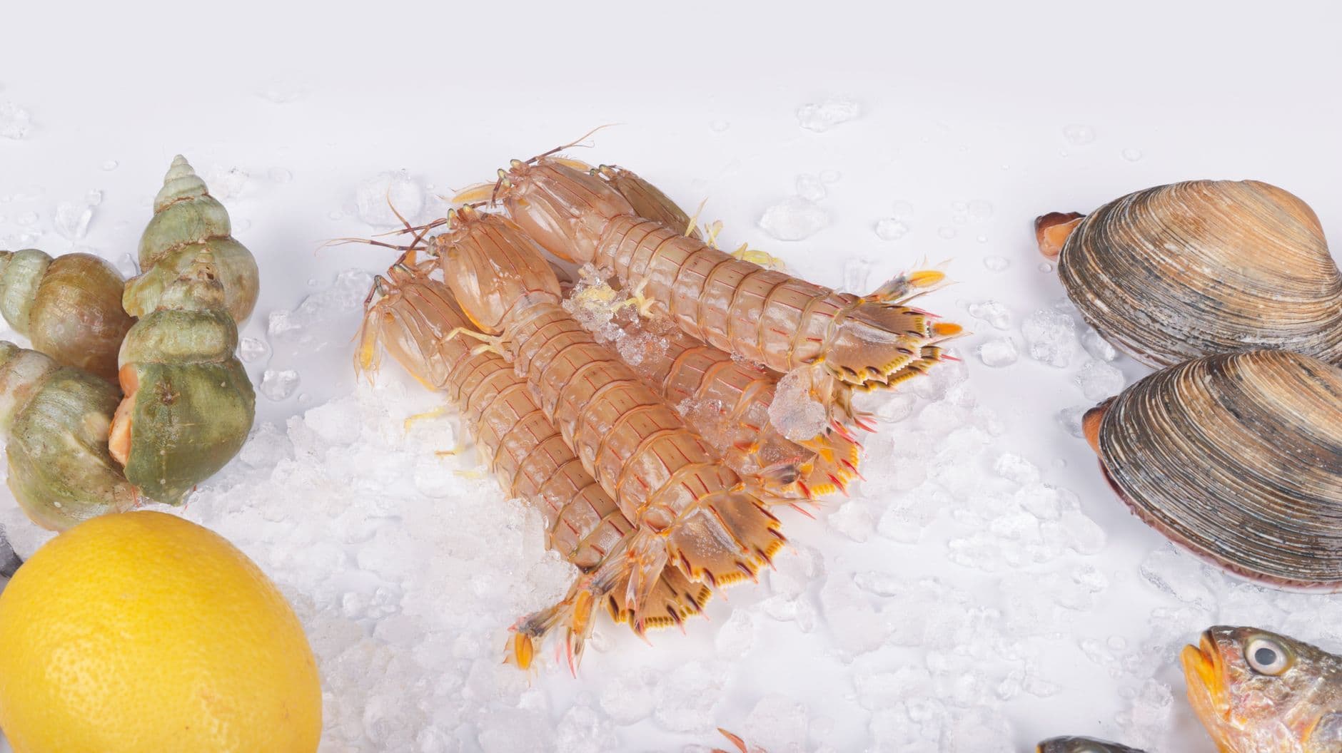
Photo: Change C.C / Pexels
Mantis shrimp carry up to 16 photoreceptor types in their compound eyes, more than any other animal studied, compared to three cone types in a typical human retina. For decades that number fed a popular claim that mantis shrimp must see colors humans cannot even imagine. Behavioral testing published in Science in 2014 overturned that assumption: mantis shrimp actually discriminate between similar colors worse than humans do, and figuring out why rewrote how vision scientists think about color processing itself.
An eye built like nothing else in nature
The peacock mantis shrimp (Odontodactylus scyllarus) belongs to a group of roughly 450 stomatopod species, all marine crustaceans, and its compound eyes are unlike anything else in the animal kingdom. Each eye sits on its own mobile stalk capable of rotating independently through a wide range of motion, and each eye is itself divided into three optical regions, giving a single eye its own trinocular depth perception without needing input from its twin.
Running across the middle of each eye is a specialized strip called the midband, a band of modified ommatidial rows that houses most of the eye's exotic sensory hardware. Six of those rows are tuned to different regions of the color spectrum, stretching from ultraviolet down through the visible range, while additional midband rows are dedicated entirely to detecting polarized light rather than color.
That split matters. A human retina devotes essentially all of its photoreceptors to color and brightness. A mantis shrimp devotes an entire separate visual channel to a property of light, polarization, that the human eye cannot register at all. Stomatopods are the only animal group confirmed to detect circularly polarized light using a biological mechanism, a finding first reported by researchers at the University of Queensland and the University of Bristol (Chiou et al., 2008, Current Biology).
Not every mantis shrimp sees the same way
The 16-receptor, six-row midband described above belongs to the most visually elaborate stomatopods, not to the group as a whole. Roughly 400 to 450 stomatopod species are currently recognized, spread across shallow tropical and subtropical seas, on reef flats, in rubble fields, seagrass beds, and sandy burrow systems, and their eyes vary considerably in how many midband rows they carry (Cronin & Marshall, various; summarized in Cronin et al., 2014, Journal of Experimental Biology).
Species like Neogonodactylus oerstedii and Pseudosquilla ciliata carry the full six-row midband, the version responsible for the extreme photoreceptor counts most coverage of mantis shrimp vision describes. Other species, including Squilla empusa and Alima pacifica, have a stripped-down two-row midband instead. Some of these simpler-eyed species appear to rely on a single visual pigment across their entire retina, peaking at around 507 nanometers, which would leave them with very limited color discrimination of any kind, comparison-based or otherwise.
Phylogenetic work suggests the two-row midband is a derived, simplified state rather than an ancestral one, meaning some stomatopod lineages evolved away from elaborate color-and-polarization processing rather than toward it. Researchers studying the underlying optic lobes have found that the neural circuitry associated with fine color and polarization analysis is present only in six-row species, not in the pared-down two-row ones, which suggests the reduction in eye hardware is matched by a reduction in the brain tissue that would otherwise process it. In other words, "mantis shrimp vision" as popularly described, the 12-to-16-channel, polarization-sensing version, is really the high end of a spectrum that runs from nearly color-blind burrow dwellers to the reef-patrolling peacock mantis shrimp most articles picture.
Why 16 receptor types do not mean better color vision
Human trichromatic color vision runs on comparison. The brain reads out the ratio of signal strength across just three cone types, tuned roughly to red, green, and blue wavelengths, and computes perceived color from those ratios. It is computationally expensive but precise: humans can typically distinguish wavelengths differing by around 5 nanometers.
A team led by Hanne Thoen, working with Justin Marshall's lab at the University of Queensland, tested whether mantis shrimp used a similar comparison strategy. They trained live mantis shrimp to associate specific colored targets with a food reward, then tested how finely the animals could tell two nearby wavelengths apart. The results, published in Science in 2014, were the opposite of what a comparison-based system would predict: mantis shrimp could only reliably discriminate colors that differed by roughly 15 to 25 nanometers, a coarser resolution than human color vision despite having four to five times as many receptor types (Thoen et al., 2014, Science).
Color discrimination — the ability to tell two similar wavelengths of light apart, tested behaviorally by seeing how close in wavelength two colors can be while an animal still reliably tells them apart.
| Visual system | Photoreceptor types | Discrimination limit | Strategy |
|---|---|---|---|
| Human eye | 3 cone types | ~5 nm wavelength difference | Neural comparison of ratios across receptors |
| Mantis shrimp eye | Up to 16 receptor types | ~15–25 nm wavelength difference | Each receptor reports its own channel with little cross-comparison |
| Most mammals (dogs, cats) | 2 cone types (dichromatic) | Coarser than human trichromacy | Limited comparison across two channels |
| Butterflies (e.g., Papilio swallowtails) | Up to 6 spectral receptor types | Finer than mantis shrimp in tested ranges | Comparison-based, closer to human strategy |
Mantis shrimp have far more receptor types than any of the animals above, yet their discrimination is the coarsest of the group, evidence that receptor count and discrimination fineness are not the same thing.
The Thoen team's explanation is that mantis shrimp are not running a comparison system at all. Instead, each of the twelve color-sensitive channels appears to report its own signal with minimal cross-processing between receptor types, a strategy the researchers described as closer to direct color recognition than to comparative discrimination.
Inside the color-discrimination experiment
The 2014 finding did not come from measuring photoreceptor absorbance in a lab and inferring what the animal must perceive. It came from behavioral testing on live, freely moving mantis shrimp, which is a slower and more demanding kind of evidence but a far more direct one, since it tests what the animal can actually act on rather than what its hardware could theoretically support.
Thoen and colleagues worked with Haptosquilla trispinosa, a stomatopod species with the full six-row midband, at Lizard Island Research Station on the Great Barrier Reef. Each animal was allowed to leave its burrow and was trained to approach one of two colored light sources for a food reward, learning to treat one specific wavelength as the "correct" choice. Once an animal reliably chose the trained color, researchers presented it with that same trained wavelength alongside a second wavelength shifted progressively closer, testing whether the animal could still tell the two apart.
Some animals were trained on light at 470 nanometers, in the blue range, and tested against wavelengths shifting toward longer, greener light; others were trained at 570 nanometers, in the yellow-green range, and tested against shorter wavelengths shifting toward blue. The gap between the trained color and the test color started wide, at up to 100 nanometers apart, easily distinguishable by nearly any visual system, and was narrowed step by step until the animal's choices dropped to chance level.
That threshold, the smallest wavelength gap an individual mantis shrimp still reliably told apart, is what produced the 15-to-25-nanometer figure. It is a considerably coarser threshold than the roughly 5-nanometer gap humans can typically resolve, and the gap held even though the mantis shrimp's receptor count vastly outnumbers the human eye's three cone types. Researchers have since proposed, based partly on this behavioral pattern, that stomatopods may instead be running a form of time-based signal comparison, one that scans a scene with rapid eye movements and compares receptor outputs across time rather than across simultaneous channels, a mechanism still being tested in follow-up work (Thoen et al., 2014, Science; Cronin et al., Journal of Experimental Biology).
Why would an animal trade precision for speed?
A comparison-based system needs the brain to weigh signals from multiple receptor types against each other before it can label a color, which takes processing time. A direct-channel system, where each receptor type reports something closer to "yes, this wavelength band is present," can generate a usable answer faster, at the cost of fine gradation between similar shades. Researchers describe this as trading resolution for reaction speed.
The direct-channel theory, in plain terms
Picture a mail sorting facility. One approach uses a single scale, weighing every letter and calculating a precise price based on the ratio of weight to size, slow but exact. Another approach uses sixteen separate chutes, one per rough weight class, and a letter simply falls into whichever chute matches closely enough. The second system sorts faster but cannot tell a letter that is 2 grams over the chute's threshold from one that is 20 grams over. That is roughly the tradeoff Thoen's team proposes for mantis shrimp vision: many parallel, coarse channels instead of one finely calibrated comparison system.
This does not mean mantis shrimp see a duller world. It means they may process visual information the way a barcode scanner processes color-coded labels rather than the way a paint-matching machine blends a custom shade, fast categorical recognition rather than fine gradient discrimination. The functional payoff, researchers hypothesize, is speed: recognizing a predator, a rival, or a patch of coral fast enough to react before it matters, rather than lingering on a precise hue.
Can mantis shrimp really see ultraviolet and polarized light at the same time?
Yes. Several midband rows are tuned into the ultraviolet range, below about 400 nanometers, alongside the rows tuned to visible wavelengths, and separate midband rows detect linear and circular polarization independent of color entirely. A mantis shrimp's visual system can, in principle, register wavelength, ultraviolet content, and polarization state from the same scene simultaneously.
The polarization channel nobody else has
Circular polarization vision is the more genuinely unique part of the mantis shrimp visual system, and it is separately documented from the color-discrimination story above. Linearly polarized light vibrates in a single plane; circularly polarized light rotates as it travels, like a corkscrew. Several insects and cephalopods can detect linear polarization, useful for orientation and navigation, but stomatopods remain the only confirmed animal group that also detects circular polarization.
The mechanism relies on a specific photoreceptor cell, known as R8, sitting in rows 5 and 6 of the midband. Its microvillar structure functions as what researchers call an achromatic quarter-wave retarder, converting circularly polarized light into linearly polarized light that the underlying photoreceptors can then register, functioning across nearly the entire visible spectrum rather than a single wavelength the way most manufactured optical quarter-wave plates do (Chiou et al., 2008, Current Biology).
Researchers led by the Marshall lab later documented that some stomatopod species reflect circularly polarized light from patches on their bodies and appear to use it as a signal, proposed as a possible private communication channel invisible to most predators and rivals that lack the same detection hardware (Gagnon et al., 2015, Current Biology). That is still an active area of study rather than settled fact, but the detection capability itself, unlike the exact communicative purpose, is well documented across multiple research groups.
How the R8 cell physically twists light into a new state
The circular-polarization trick described above depends on a specific piece of cellular geometry, not on any exotic chemistry. Photoreceptor cells build their light-catching membrane into microvilli, tiny finger-like folds packed with visual pigment, and in most eyes, including the R1-through-R7 cells that sit beneath R8 in the mantis shrimp midband, those microvilli all point in the same one or two directions across a given cell.
R8 breaks that pattern deliberately. Its microvilli are oriented at roughly 45 degrees relative to the microvilli of the R1–R7 cells stacked underneath it, and the cell itself is unusually long, around 150 micrometers, which gives incoming light enough travel distance through the birefringent (light-splitting) microvillar material to shift phase by exactly a quarter wavelength (Roberts et al., 2009, Nature Photonics). That quarter-wavelength shift is precisely what is needed to convert circularly polarized light, light whose electric field corkscrews as it travels, back into ordinary linearly polarized light, the kind every photoreceptor already knows how to register.
The 45-degree offset is also what lets the system tell left-handed circular polarization from right-handed circular polarization, not just detect that circular polarization is present. Because R8 sits at an angle to R1–R7, clockwise-rotating light gets converted into linear light aligned one way, and counterclockwise-rotating light gets converted into linear light aligned the other way, so the receptors underneath can distinguish the two by which axis lights up (Chiou et al., 2008, Current Biology).
Why does this work across so many colors at once?
A manufactured quarter-wave plate, the kind used in polarizing camera filters and 3D-movie glasses, is typically tuned to one wavelength and drifts out of alignment for others. The mantis shrimp's version stays accurate across nearly the entire visible spectrum because its performance depends on cell length and microvillar geometry rather than a fixed refractive property tied to one wavelength, an engineering solution that took human optics decades longer to approximate with stacked multi-layer films.
This distinction between linear and circular detection is not a minor technicality. Linear polarization patterns occur naturally and abundantly, scattered by the atmosphere and reflected off water and wet surfaces, which is why linear polarization vision shows up independently in several unrelated animal lineages for navigation and glare reduction. Circular polarization, by contrast, is rare in nature outside of a handful of reflective structures on stomatopod bodies themselves, which is part of why researchers suspect the ability evolved specifically as a signal that only other stomatopods, carrying the matching R8 hardware, can read.
What this means for camera and sensor design
The mantis shrimp's polarization-sensing structures have drawn direct interest from optical engineers because the biological quarter-wave retarder works efficiently across a broad spectral range, something conventional manufactured polarization optics struggle to do without stacking multiple wavelength-specific components. Research teams have used the mantis shrimp eye as a template for compact polarization camera designs, including exploratory work on detecting the way certain tissue types, including some cancerous tissue, alter the polarization state of reflected light, an approach that would not require the bulky optical stacks traditional polarimeters use.
This line of research remains largely at the prototype and proof-of-concept stage rather than deployed clinical technology, and readers should not treat it as an available diagnostic tool. What makes it noteworthy for a biology explainer is more basic: a structure that evolved to help a shrimp find prey and rivals on a reef became, independently, a template engineers borrowed for a human medical imaging problem. That kind of direct transfer from animal sensory hardware to applied optical engineering is uncommon enough to be worth flagging on its own.
What people get wrong about mantis shrimp vision
The most common misconception is the direct inversion of what the 2014 research actually found. Because mantis shrimp have more photoreceptor types than any other studied animal, the assumption spread quickly, including across popular science coverage, that they must experience unimaginably rich or "supercharged" color vision. Behavioral testing shows close to the opposite: their fine color discrimination underperforms a human's in the wavelength ranges tested.
A second, related misconception is treating "sees more colors" and "has more photoreceptor types" as interchangeable. Photoreceptor count sets the raw hardware ceiling for what wavelengths an eye can register at all, but discrimination, the ability to tell two similar wavelengths apart, depends on how the nervous system processes and compares those signals downstream. Mantis shrimp have the broader hardware but appear to skip much of the downstream comparison step that gives human vision its fine gradation.
A third point worth separating out clearly: the coarse color discrimination finding and the circular polarization detection ability are two different, independently documented capabilities. One (color) turned out to be less impressive than the initial hype suggested. The other (polarization) remains a genuinely rare capability, confirmed in multiple studies, that almost no other animal on the planet shares.
The takeaway
Mantis shrimp do not have "better color vision" than humans in the straightforward sense that twelve to sixteen photoreceptor types once suggested. Behavioral research from Justin Marshall's lab, published in Science in 2014, found their fine color discrimination is actually coarser than a human's, evidence that they likely trade discrimination precision for a faster, more direct form of color recognition. What remains genuinely exceptional, and well documented by separate research, is their capacity to detect circularly polarized light, a dimension of vision essentially no other animal group can access, which makes a coral reef informationally richer to a mantis shrimp than it could ever appear to a human observer looking at the identical patch of coral.
Read next:
FAQ
Q: How many types of color receptors do mantis shrimp have compared to humans?
Mantis shrimp have up to 16 photoreceptor types in the midband region of their compound eyes, compared to three cone types in the human retina. Behavioral research shows this larger receptor count does not translate into finer color discrimination overall.
Q: Do mantis shrimp actually see more colors than humans?
Not in terms of fine discrimination. Behavioral experiments published in Science in 2014 by Thoen, How, Chiou, and Marshall found mantis shrimp could only reliably tell apart wavelengths differing by roughly 15 to 25 nanometers, coarser than human color discrimination of around 5 nanometers, suggesting their many receptors prioritize fast recognition over fine comparison.
Q: Can mantis shrimp see something humans cannot see at all?
Yes. Mantis shrimp can detect circularly polarized light, a property of light invisible to the human eye and to nearly every other studied animal, using a specialized photoreceptor structure that functions as a natural quarter-wave retarder across most of the visible spectrum.
Q: Why do mantis shrimp have eyes on independently moving stalks?
Each eye sits on a mobile stalk and is itself split into three optical zones, giving a single eye trinocular depth perception on its own. This likely helps mantis shrimp judge distance precisely when striking fast-moving or well-camouflaged prey.
Q: Has mantis shrimp vision influenced real technology?
Yes, at the research stage. Their polarization-sensing eye structures have inspired prototype work on compact bioinspired polarization cameras, including exploratory research into detecting tissue changes associated with cancer, without the bulky optics conventional polarimeters require. This remains experimental research rather than deployed medical technology.
Q: How fast can a mantis shrimp's claw strike?
Research from Sheila Patek's lab measured the peacock mantis shrimp's raptorial appendage accelerating at roughly 10,400 times the acceleration of gravity, generating peak strike forces up to about 1,501 newtons, fast enough to create cavitation bubbles that collapse with their own separate force spike.
Q: What physically lets an R8 photoreceptor detect circular polarization?
The R8 cell's microvilli sit at roughly 45 degrees to the microvilli of the receptor cells beneath it, and the cell is unusually long, around 150 micrometers. That combination shifts incoming light's phase by exactly a quarter wavelength, converting circularly polarized light into linearly polarized light the underlying receptors can already register, while the 45-degree offset lets the system tell clockwise-rotating light from counterclockwise-rotating light.
Q: Is linear polarization vision the same thing as circular polarization vision?
No. Linear polarization detection, which occurs in bees, some fish, and cuttlefish, only registers light vibrating in a fixed plane and is common enough in nature to support navigation and glare reduction. Circular polarization detection requires the extra 45-degree-offset R8 mechanism, is rare outside stomatopods, and appears to function more as a private signaling channel than a navigation tool.
Follow new articles
Add SciHub101 to your feed reader to see new science and technology stories.
About the Author
SciHub101 is an independent science and technology publication founded and edited by Buğra Sözeri, a software engineer and app developer based in Türkiye. AI tools assist with drafting and research. Source documentation in the existing archive is uneven, and articles are being reviewed and corrected. The Editorial Team byline identifies editorial responsibility; it does not certify specialist or independent human review. Where we compare products, anything built by our own team is labelled as such — see the editorial guidelines and About page for how corrections and disclosures work.
Related Articles


The Longest-Living Animals Explained: Biology of Extreme Longevity
A Greenland shark alive today may have been born before the Thirty Years' War ended in 1648. An ocean quahog clam called Ming was 507 years old when researchers accidentally killed it by opening its shell. A bowhead whale carries harpoon fragments from weapons manufactured in the 1800s, embedded in its blubber for over a century. Extreme longevity in animals is not accidental — it is the product of specific biological mechanisms that researchers are now mapping in molecular detail.
How Bioluminescence Works in Animals: The Chemistry of Living Light
More than 76% of deep-sea animals produce their own light. Fireflies flash in coordinated sequences. A jellyfish discovered off the coast of Friday Harbor, Washington, gave biologists a tool that transformed cell biology, earned three scientists a Nobel Prize, and now glows inside cancer cells in laboratories around the world. Bioluminescence — light produced by a living organism — is one of evolution's most useful accidents, and its chemistry is surprisingly simple.
How Sharks Sense the World: Electroreception, Smell, and the Lateral Line
Sharks read faint electric fields, track scent plumes one nostril at a time, and feel water motion along their flanks. Some popular claims about those senses, from the mile-away drop of blood to the all-seeing predator, do not survive the data.