The X-Ray: Röntgen's Accidental Discovery That Transformed Medicine
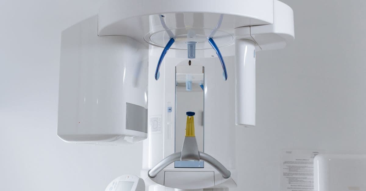
Photo: cottonbro studio / Pexels
On the evening of November 8, 1895, a 50-year-old German physicist named Wilhelm Conrad Röntgen was working alone in his darkened laboratory at the University of Würzburg, experimenting with a Crookes tube — a glass vacuum tube that produced a beam of electrons. He had covered the tube with black cardboard to block any light. Across the room, a piece of paper coated with barium platinocyanide began to glow. There was no light source that could explain it. Röntgen had just discovered X-rays: a previously unknown form of radiation that could pass through solid objects and make the invisible visible. He spent the next seven weeks in almost complete secrecy, barely leaving his laboratory, before telling the world what he had found.
The Story Behind the Discovery
The Crookes tube — named for British physicist William Crookes, who developed it in the 1870s — is a sealed glass tube from which most of the air has been pumped out, with metal electrodes at each end. When a high voltage is applied, the remaining gas ionizes and electrons (then called "cathode rays") stream from the negative electrode (cathode) to the positive electrode (anode). These electrons travel at a significant fraction of the speed of light inside the tube.
What Röntgen was studying in November 1895 was whether cathode rays could penetrate the glass wall of the tube and travel through air. Several researchers had made similar investigations; Philipp Lenard had built tubes with thin aluminum windows to allow cathode rays to escape. Röntgen was repeating related experiments with a different tube design when he noticed the glow from the barium platinocyanide screen.
Barium platinocyanide — a fluorescent compound that glows green when struck by certain forms of radiation or ultraviolet light. Röntgen used paper coated with it as a fluorescence detector. It glowed in response to X-rays even when placed several feet away from the Crookes tube and with no optical connection to it.
The critical observation was distance. Cathode rays — the electron beam inside the tube — could not travel more than a few centimeters through air before being absorbed. The glow from the barium platinocyanide screen was occurring at a distance of about one meter. Whatever was causing the screen to glow was not cathode rays.
Source: LITFL (Life in the Fast Lane) — "November 8, 1895: X-rays discovered" — the detailed sequence of Röntgen's November 8 discovery and the following weeks of secret experimentation.
Röntgen picked up the screen and carried it around the room. The glow continued at every distance from the tube, through walls, through books, through his hand — but with his hand, something uncanny happened. He could see the shadow of the bones inside his hand projected on the screen. He was looking at his own skeleton.
The physicist kept his discovery to himself for seven weeks while he systematically documented its properties. He determined that X-rays:
- Traveled in straight lines
- Could not be deflected by magnetic fields (unlike cathode rays)
- Penetrated most materials but were partially blocked by dense ones
- Were not reflected or refracted in the same way as light
- Caused fluorescent materials to glow
- Darkened photographic plates
He named them "X-rays" — the mathematical convention for an unknown quantity — because he did not know what they were. He was right to be cautious. The nature of X-rays would not be definitively established until Max von Laue demonstrated in 1912 that they were electromagnetic waves, like light but at much higher frequencies and shorter wavelengths.
The First X-Ray Photograph
On December 22, 1895, Röntgen asked his wife, Anna Bertha, to hold her left hand over a photographic plate for fifteen minutes while he exposed it to X-rays. The resulting image — showing the bones of her hand and her wedding ring in crisp detail against the soft tissue shadow — was the world's first radiograph of a human body.
Source: PBS NewsHour — "'I Have Seen My Death': How the World Discovered the X-Ray" — documents Anna Bertha's reaction and the December 22, 1895 photograph.
Anna Bertha reportedly looked at the image and said, "I have seen my death." The phrase captures something genuine about the psychological impact of X-ray images on the people who first encountered them. Seeing a skeleton photograph of a living person disturbed a cultural boundary between the living and the dead that had never before been visually transgressed.
Röntgen submitted his paper, "Über eine neue Art von Strahlen" (On a New Kind of Rays), to the Würzburg Physical-Medical Society on December 28, 1895. He included the photograph of his wife's hand as an illustration. He also mailed copies of the paper with copies of X-ray photographs to several leading physicists in Europe on January 1, 1896.
The response was immediate and global. Within weeks, physicians across Europe and North America were using X-rays to locate bullets in soldiers' bodies, find bone fractures, and diagnose tuberculosis by its effect on lung tissue. Within months, X-ray photography studios had opened in major cities. The public was simultaneously thrilled and unnerved.
How X-Rays Actually Work
An X-ray is a photon — a particle of electromagnetic radiation — at a much higher energy than visible light. Visible light has photon energies of roughly 1.8 to 3.1 electron volts (eV). Medical X-rays have photon energies of 20,000 to 150,000 eV (20–150 keV). This high energy is what allows X-rays to penetrate matter rather than being absorbed or reflected at the surface.
Photon — a quantum of electromagnetic energy. The energy of a photon is proportional to its frequency and inversely proportional to its wavelength. X-rays have much higher frequency and much shorter wavelength than visible light.
Production: The X-Ray Tube
Modern X-ray tubes work on the same principle as Röntgen's Crookes tube, but with deliberate engineering instead of accidental observation. Electrons are emitted from a heated tungsten cathode and accelerated through a high voltage (typically 20–150 kilovolts) toward a tungsten anode. When the high-speed electrons strike the tungsten anode, two things happen:
Bremsstrahlung radiation ("braking radiation"): The electrons decelerate abruptly and release their kinetic energy as X-ray photons. Think of it like a car hitting a wall — the energy of motion has to go somewhere, and in this case it goes into radiation.
Characteristic X-rays: Some electrons knock inner-shell electrons out of tungsten atoms. When electrons from outer shells fall in to fill those vacancies, they release X-ray photons at specific energies characteristic of tungsten's atomic structure.
The result is a spectrum of X-ray energies, controllable by adjusting the accelerating voltage and the current. Higher voltage produces higher-energy X-rays that penetrate denser tissue; lower voltage produces lower-energy X-rays that show better contrast between soft tissues.
Why Some Tissues Show and Others Don't
X-rays interact with matter primarily through photoelectric absorption: a material's atoms absorb X-ray photons and re-emit lower-energy photons in random directions. Dense materials with high atomic numbers absorb X-rays strongly; light materials with low atomic numbers absorb them weakly.
Bone contains calcium (atomic number 20) in its crystalline mineral matrix. Calcium absorbs X-rays much more strongly than the carbon, hydrogen, and oxygen that make up soft tissue. This is why bones appear white (high absorption = fewer X-rays reaching the detector) and soft tissue appears grey to black in a standard X-ray image. Air-filled spaces (lungs) absorb almost nothing and appear black.
Lead (atomic number 82) absorbs X-rays even more strongly than calcium, which is why lead aprons protect patients and radiographers from scattered radiation.
Source: University of Michigan Medical School / Radiology Key — "Radiation and Its Discovery" — covers the physics of X-ray production and tissue interaction.
Contrast Agents
For soft tissue structures that are nearly invisible on standard X-rays — the gastrointestinal tract, blood vessels, the urinary system — radiologists use contrast agents: substances with high X-ray absorption that can be introduced into the body. Barium sulfate is used to coat the GI tract for fluoroscopic examination. Iodine-based compounds are injected into the bloodstream to visualize blood vessels (angiography) and organs. The principle is the same as natural tissue contrast, but artificially enhanced.
The People Involved
Wilhelm Conrad Röntgen (1845–1923)
Röntgen was a careful, methodical physicist with a reputation for meticulous experimental work but no particular fame before November 1895. He was born in Lennep, Prussia (now part of Remscheid, Germany), and had a somewhat difficult early career — he was dismissed from his first university position at Zurich on a technicality related to his lack of an appropriate diploma. He eventually held chairs at Würzburg and Munich.
Röntgen's response to his discovery says a great deal about his character. He declined to patent the X-ray or any of the apparatus associated with it, believing that scientific discoveries should be freely available for humanity's benefit. He gave away the prize money from the Nobel award to the University of Würzburg. He refused speaking fees and made himself difficult to interview. He was deeply uncomfortable with the celebrity his discovery brought.
Source: History.com — "German scientist discovers X-rays, November 8, 1895" — documents Röntgen's discovery and his explicit refusal to patent.
When World War I devastated Germany's economy, Röntgen donated his remaining savings to the German war effort and died in 1923 in financial difficulty. His refusal to patent an invention that generated enormous commercial wealth for others stands as one of the more striking examples of scientific altruism on record.
The Nobel Committee awarded Röntgen the first Nobel Prize in Physics in 1901 — the first Nobel Prize ever awarded in that category. The citation was simply "for the discovery of the remarkable rays subsequently named after him."
Thomas Edison and Clarence Dally
Within weeks of Röntgen's announcement in January 1896, Thomas Edison began investigating X-rays for potential commercial application. He developed a fluoroscope — a device that used a fluorescent screen to produce real-time X-ray images rather than static photographs — and exhibited it at the 1901 Pan-American Exposition. The fluoroscope became an important medical and industrial imaging tool.
To develop his fluoroscope, Edison employed a glassblower named Clarence Madison Dally, who worked extensively with X-ray tubes in Edison's West Orange laboratory. Dally was exposed to extreme doses of X-ray radiation over several years without any protective measures — because the dangers of radiation were not yet understood.
Source: Wikipedia — "Clarence Madison Dally" — documents Dally's work for Edison, his radiation injuries, and his death.
Dally developed severe radiation burns on his hands, which progressed to radiation-induced cancer despite multiple amputations. His left hand was amputated, then his right hand, then his left arm at the elbow, then his right arm at the shoulder. He died in October 1904 at age 33. His death is widely cited as the first recorded death from X-ray exposure.
Edison, reportedly shaken by Dally's death, ceased his X-ray work entirely. He reportedly told a reporter years later: "Don't talk to me about X-rays. I am afraid of them."
Wilhelm His Jr. and Early Clinical Radiology
The physician who arguably did most to establish clinical radiology as a medical specialty was not a single person but a cohort of early adopters — physicians who recognized within days of Röntgen's January 1896 paper that X-rays could locate fractures, foreign objects, and pathological changes in tissue with a speed and certainty that physical examination alone could never achieve. By early 1896, X-ray units were operating in hospitals in Vienna, Glasgow, and Boston.
The challenge was controlling exposure: without dosimetry — measurement of radiation dose — both patients and operators received arbitrarily large doses. Many early radiologists developed radiation-induced skin cancers and cataracts. The full scope of radiation's biological hazards was not established until the 1920s and 1930s, and not systematically regulated until after World War II.
Timeline: From Crookes Tube to CT Scanner
| Year | Event |
|---|---|
| 1875–1880 | William Crookes develops the Crookes tube — the instrument Röntgen would use |
| November 8, 1895 | Röntgen observes fluorescence at distance from covered Crookes tube — discovery of X-rays |
| December 22, 1895 | First radiograph of a human body: Anna Bertha Röntgen's hand |
| December 28, 1895 | Röntgen submits "Über eine neue Art von Strahlen" to Würzburg Physical-Medical Society |
| January 1, 1896 | Röntgen mails paper and X-ray photographs to leading European physicists |
| January 1896 | X-ray apparatus in clinical use in Vienna, Glasgow, and US within weeks |
| 1896 | Edison develops fluoroscope; Clarence Dally begins X-ray work |
| 1901 | Röntgen awarded first Nobel Prize in Physics |
| 1904 | Clarence Dally dies of radiation-induced cancer — first known X-ray fatality |
| 1912 | Max von Laue demonstrates X-rays are electromagnetic waves (Nobel Prize 1914) |
| 1913 | William Coolidge develops hot cathode X-ray tube — more controllable, safer |
| 1920s–30s | Radiation safety standards begin to develop; lead shielding adopted |
| 1948 | Image intensifier developed — real-time fluoroscopy with dramatically lower dose |
| 1972 | Godfrey Hounsfield and Allan Cormack independently develop computed tomography (CT) — Nobel Prize 1979 |
| 1980s | Digital radiography begins replacing film-based X-ray |
| 1990s | Computed radiography (CR) and direct digital radiography (DR) become clinical standard |
| 2000s | Cone-beam CT enters dental and orthopedic practice |
| 2020s | AI-assisted image interpretation becomes routine in radiology |
Röntgen's Refusal to Patent
The decision not to patent his discovery is unusual enough to deserve close examination. Röntgen explicitly stated that he believed the results of scientific research should belong to humanity, not to individuals. He had seen the difficulties that arose when scientific discoveries were locked up in commercial patents, and he was determined not to create such a barrier to the medical use of his finding.
The practical consequence of this decision was that within months of his January 1896 publication, dozens of manufacturers worldwide were producing X-ray tubes and fluorescent screens without paying any royalties. This is why X-ray diagnosis spread so rapidly: there was no patent holder controlling access to the technology.
Source: historytools.org — "The Accidental Discovery That Revolutionized Medicine: How X-Rays Were Found" — covers Röntgen's explicit refusal to patent and his reasoning.
The irony is that Röntgen himself died in modest financial circumstances in 1923, while X-ray equipment companies built fortunes on technology he had made freely available. Whether this represents a failure or a success depends on how you weight personal benefit against public good.
Unintended Consequences
The Radiation Martyr Problem
The first generation of radiologists worked without protective equipment and with no understanding of the cumulative biological damage they were accumulating. The initial response to X-rays was sensationalist enthusiasm: X-ray shoes were sold in shoe stores to check for proper fit. X-ray demonstrations were public entertainments. Some entrepreneurs offered X-ray hair removal services.
The biological effects of radiation exposure — DNA damage, cancer induction, teratogenesis — are probabilistic. A single X-ray exposure has a very low probability of causing cancer; repeated high exposures have a substantially higher probability. The relationship was not established until the 1920s, when radiologists began dying of cancers at rates dramatically higher than the general population.
The 1928 International Congress of Radiology established the first international radiation safety standards, introducing the concept of a maximum permissible dose. Modern standards, based on decades of epidemiological data from radiation workers and atomic bomb survivors, set annual dose limits for radiation workers at 20 millisieverts (mSv) per year, with a lifetime limit of 100 mSv in any five-year period.
Source: AuntMinnieEurope — "Was Clarence Dally a radiology martyr or victim?" — places Dally's death in the context of early radiation safety history.
The Atomic Age and Radiation Fear
The same physics that makes X-rays medically useful — high-energy photons that ionize atoms — makes radioactive fallout from nuclear weapons deeply dangerous. The nuclear attacks on Hiroshima and Nagasaki in 1945 produced X-ray-like gamma radiation in addition to visible light, heat, and blast. The subsequent study of survivors (the Life Span Study, ongoing since 1950) remains the primary epidemiological dataset from which radiation risk at low doses is extrapolated.
Public anxiety about radiation, often imprecise about the distinction between diagnostic X-rays and fallout, has complicated evidence-based communication about radiation health risks ever since. A chest X-ray delivers approximately 0.02 mSv — equivalent to about 2.4 days of natural background radiation from soil, air, and cosmic rays. A full-body CT scan delivers approximately 10 mSv — equivalent to about 3 years of natural background. These are not zero risks, but they are orders of magnitude smaller than the risk of undiagnosed disease.
CT, MRI, and the Imaging Revolution
The X-ray itself is a two-dimensional shadow: a three-dimensional object projected onto a flat detector. Structures overlap and obscure each other. Computed tomography (CT), developed independently by Godfrey Hounsfield at EMI and Allan Cormack at Tufts University in the early 1970s (Nobel Prize 1979), solved this by taking hundreds of X-ray images from different angles and using a computer to reconstruct a three-dimensional density map of the scanned volume. The result is a three-dimensional X-ray image in which any cross-section can be viewed without interference from overlapping structures.
CT is now among the most diagnostically powerful tools in medicine. Over 80 million CT scans are performed annually in the United States alone, primarily for trauma evaluation, cancer staging, and vascular disease assessment.
Source: iData Research — "Over 75 Million CT Scans Are Performed Each Year and Growing" — global CT scan volume statistics.
Magnetic resonance imaging (MRI), developed in the 1970s and 1980s using nuclear magnetic resonance, uses no X-rays at all — it uses magnetic fields and radio waves to image soft tissue. It is in many ways a complementary technology: excellent where X-rays are weak (soft tissue contrast, no ionizing radiation), limited where X-rays excel (speed, availability, bone detail). The two technologies coexist and are often used in sequence for complex diagnostic problems.
Where X-Ray Technology Stands Today
The glass and film technology of Röntgen's era has been entirely replaced in modern radiology. Digital detector panels — flat-panel detectors using amorphous silicon with a scintillator layer — convert X-ray photons directly to digital signals. Images appear on workstation screens within seconds of exposure, can be adjusted for contrast and brightness digitally, transmitted anywhere in the world instantly, and stored indefinitely without degradation.
Artificial intelligence has entered radiological interpretation. Deep learning systems trained on millions of annotated chest X-rays can detect pneumonia, tuberculosis, and lung nodules with accuracy comparable to experienced radiologists, and they can process images in seconds. FDA-cleared AI tools for X-ray interpretation were available in clinical use by 2023. The AI does not replace the radiologist — it flags potential findings for human review and provides a first pass on high-volume screening applications.
Airport security X-ray systems use the same ionizing radiation physics to examine baggage, but at much lower energies than medical imaging, and with dual-energy technology that allows the system to differentiate organic materials (which might be explosives) from metals by their different X-ray absorption at two different energies.
Source: ReadMyMRI — "How Many Medical Imaging Scans Are Done Per Year?" — US imaging volumes including CT and X-ray.
Röntgen's original observation — that a Crookes tube could make a fluorescent screen glow across a dark room — has spawned a diagnostic technology used in virtually every hospital and clinic on Earth. The barium platinocyanide screen in his Würzburg laboratory has been replaced by flat-panel digital detectors; the photographic plates by storage phosphor systems and direct digital capture; the hand-made Crookes tubes by precision-engineered rotating-anode X-ray tubes producing controlled spectra at kilowatt power levels. The physics is identical. The engineering is unrecognizable.
FAQs
Q: How did Röntgen discover X-rays? A: On November 8, 1895, Röntgen noticed that a barium platinocyanide-coated screen in his darkened laboratory glowed when he activated a nearby Crookes tube, even though the tube was covered with black cardboard and no light could reach the screen. The distance and the screen's behavior showed the effect was not caused by cathode rays. He spent seven weeks investigating the phenomenon in secret before publishing his findings.
Q: Why didn't Röntgen patent the X-ray? A: Röntgen believed that scientific discoveries should be freely available to all of humanity. He explicitly declined to patent his discovery or any of the apparatus associated with it, accepting no royalties or commercial benefit from the technology's rapid adoption. He donated his Nobel Prize money to the University of Würzburg.
Q: What is the connection between X-rays and the first Nobel Prize? A: Röntgen received the first Nobel Prize in Physics, awarded in 1901, for his discovery of X-rays. The Nobel Prizes were first awarded in 1901, the year the Nobel Foundation was established under the terms of Alfred Nobel's 1895 will.
Q: Who was Clarence Dally and why does he matter? A: Clarence Madison Dally was a glassblower employed by Thomas Edison to develop X-ray fluoroscope technology. Dally worked with X-ray tubes without protection for years and developed progressive radiation-induced cancer. He had both arms amputated before dying at age 33 in 1904. He is generally cited as the first person known to have died from X-ray radiation exposure, and his death is credited with making Edison end his X-ray research.
Q: How dangerous is a medical X-ray? A: A chest X-ray delivers approximately 0.02 millisieverts (mSv) of radiation dose — equivalent to about 2.4 days of natural background radiation from the environment. A full-body CT scan delivers approximately 10 mSv — comparable to about 3 years of background radiation. These doses carry a very small but non-zero increase in lifetime cancer risk. Medical X-rays are recommended when the diagnostic benefit outweighs this small risk; they are not recommended for screening in asymptomatic people without specific indications.
Q: What is computed tomography (CT) and how does it differ from a standard X-ray? A: A standard X-ray is a two-dimensional shadow projection — a three-dimensional body projected onto a flat detector, with structures overlapping each other. CT takes hundreds of X-ray images from different angles around the patient and uses a computer to reconstruct a three-dimensional density map. Any cross-section — axial, coronal, sagittal, or any oblique plane — can then be displayed. CT provides vastly more diagnostic information for complex anatomy, at the cost of higher radiation dose and greater equipment complexity.
Q: How are X-rays used outside medicine? A: X-rays are used in airport and shipping security screening (dual-energy systems that distinguish organic from metallic materials); non-destructive testing of industrial components (detecting cracks, voids, and flaws in metal castings and welds); X-ray crystallography (determining the atomic structure of molecules — the technique that revealed the double helix of DNA in 1953); food safety inspection (detecting bone fragments and foreign objects in packaged food); and art authentication (examining the underdrawings and paint layers in paintings).
Q: Did anyone else discover X-rays independently? A: Several researchers had observed unusual fogging of photographic plates near Crookes tubes before 1895, including Arthur Goodspeed and William Jennings at the University of Pennsylvania in 1890, but they did not investigate the cause. Röntgen's contribution was not just observing the effect but systematically documenting its properties, identifying it as a distinct phenomenon, and recognizing its significance. He is the credited discoverer.
The Takeaway
Röntgen's seven weeks of secret work after November 8, 1895 — systematic, careful, refusing to announce an incompletely understood result — set a standard for scientific rigor that his successors in radiology often failed to match. The early enthusiasm for X-rays, untethered from any understanding of their biological hazards, killed Clarence Dally and damaged dozens of pioneering radiologists before the medical community accepted that ionizing radiation required measurement, limits, and protection. The technology that Röntgen gave freely to the world without a patent has since generated enormous medical value and some medical harm — and the ongoing work of radiation safety, AI-assisted diagnosis, and dose reduction is the continuing attempt to maximize the first and minimize the second.
Read next:
Follow new articles
Add SciHub101 to your feed reader to see new science and technology stories.
About the Author
SciHub101 is an independent science and technology publication founded and edited by Buğra Sözeri, a software engineer and app developer based in Türkiye. AI tools assist with drafting and research. Source documentation in the existing archive is uneven, and articles are being reviewed and corrected. The Editorial Team byline identifies editorial responsibility; it does not certify specialist or independent human review. Where we compare products, anything built by our own team is labelled as such — see the editorial guidelines and About page for how corrections and disclosures work.
Related Articles

Vaccination: The Invention That Has Saved More Lives Than Any Other
Vaccination began with cowpox, a boy named James Phipps, and Edward Jenner in 1796. Discover how the science of immunization works and why it remains our most powerful public health tool.

The Vacuum Cleaner: From Puffing Billy to Roomba
The vacuum cleaner's full history: Booth's 1901 horse-drawn machine, Spangler's 1907 portable upright, Hoover, Dyson's 5,127 prototypes, and iRobot's Roomba in 2002.
Eijkman's Chickens: How Beriberi Led to Vitamins
Christiaan Eijkman's chickens in Java developed beriberi on white rice and recovered on brown, starting the path to vitamin B1 and a 1929 Nobel Prize.
The Invention of MRI
How Lauterbur's 1973 Nature paper and Mansfield's math turned nuclear magnetic resonance into the scanner that sees inside living tissue without radiation.
Anesthesia: The Invention That Made Modern Surgery Possible
How William Morton's 1846 ether demonstration at Mass General transformed surgery from conscious agony into painless procedure — and why we still don't fully understand it.
The Lithium-Ion Battery: How Three Scientists Powered the Wireless Age
The lithium-ion battery's full history: Whittingham's 1976 Exxon work, Goodenough's LiCoO2 breakthrough, Yoshino's 1985 prototype, the 2019 Nobel Prize, and today.