The History of DNA: From Miescher to the Double Helix
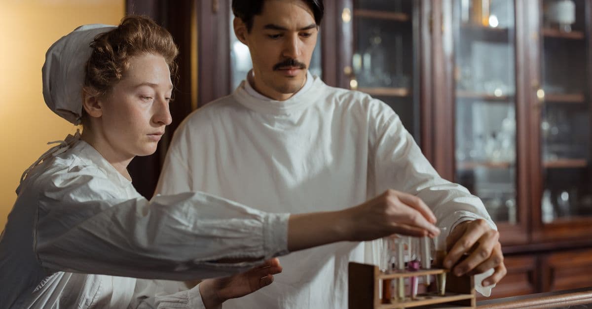
Photo: cottonbro studio / Pexels
One molecule carries the instructions for nearly every living thing on Earth. It helps determine whether a cell becomes a bacterium or a blue whale, whether eyes are blue or brown, and whether some inherited diseases run in a family. The publication of DNA's double-helix structure on 25 April 1953 is one of the most celebrated moments in science. But the road there was 84 years long, and it was built by many people whose names rarely appear in the short version of the story.
This article traces that road in order: the first isolation of DNA, the decades when scientists thought it was too simple to matter, the experiments that proved it carries genes, the chemical and X-ray clues that revealed its shape, and the long argument about who deserves credit. If you want to know how DNA works rather than how it was found, our explainer on how DNA works covers the biology.
Key takeaways
- Friedrich Miescher isolated a phosphorus-rich substance he called "nuclein" from white blood cells in 1869; his paper appeared in 1871. It was DNA, though its function was unknown.
- From the 1910s, Phoebus Levene's "tetranucleotide" model portrayed DNA as a simple repeating molecule, which helped persuade scientists that proteins, not DNA, carried genes.
- Avery, MacLeod and McCarty showed in February 1944 that purified DNA could transform one pneumococcus strain into another, the first strong evidence that DNA is genetic material.
- Erwin Chargaff's 1950 measurements showed that adenine matches thymine and guanine matches cytosine, a key clue to base pairing.
- Rosalind Franklin and Raymond Gosling's May 1952 X-ray image, Photo 51, was shown to James Watson in January 1953 without Franklin's knowledge; a 2023 Nature reassessment argues she was an equal contributor, not merely a victim.
Timeline of the DNA discovery
| Year | Event | Key people |
|---|---|---|
| 1869 | Nuclein isolated from white blood cells in Tübingen | Friedrich Miescher |
| 1871 | Miescher's nuclein paper published after his supervisor repeated the work | Miescher, Felix Hoppe-Seyler |
| 1909 | Ribose identified as a component of nucleic acids | Phoebus Levene, Walter Jacobs |
| c. 1910s | Tetranucleotide hypothesis proposes a simple, repeating DNA | Phoebus Levene |
| 1928 | Bacterial "transformation" discovered in pneumococcus | Frederick Griffith |
| 1929 | Deoxyribose identified as DNA's sugar | Phoebus Levene |
| 1944 | DNA identified as the transforming principle | Oswald Avery, Colin MacLeod, Maclyn McCarty |
| 1950 | Base-ratio rules (A = T, G = C) published | Erwin Chargaff |
| 1952 | Hershey–Chase experiment with bacteriophage | Alfred Hershey, Martha Chase |
| May 1952 | Photo 51 recorded at King's College London | Rosalind Franklin, Raymond Gosling |
| January 1953 | Wilkins shows Photo 51 to Watson | Maurice Wilkins, James Watson |
| 25 April 1953 | Double-helix papers published in Nature | Watson and Crick; Wilkins and colleagues; Franklin and Gosling |
| April 1958 | Franklin dies, aged 37 | Rosalind Franklin |
| 1962 | Nobel Prize in Physiology or Medicine | Watson, Crick, Wilkins |
| 2023 | Nature comment reassesses Franklin's role | Matthew Cobb, Nathaniel Comfort |
Miescher finds "nuclein" (1869)
In the winter of 1869, a young Swiss physician named Friedrich Miescher was working in the laboratory of the biochemist Felix Hoppe-Seyler at the University of Tübingen, in rooms set up in the kitchen of the town's medieval castle. Miescher wanted to understand the chemistry of the cell, and he chose white blood cells because they were easy to get: he collected them from pus on used surgical bandages from a nearby clinic.
After digesting away the protein-rich parts of the cells and isolating their nuclei, he obtained a substance unlike any protein then known. It was acidic, rich in phosphorus and resistant to the enzymes that broke down proteins. He named it nuclein, because it came from the nucleus.
Hoppe-Seyler was cautious. He repeated the experiments himself before allowing the work to be published, so Miescher's paper, written in 1869, appeared only in 1871. Miescher later moved to Basel and switched to salmon sperm, whose heads are almost entirely nucleus, which made purification much easier. According to the historian of science Ralf Dahm, writing in EMBO Reports in 2010, Miescher came surprisingly close to the modern idea: he speculated that hereditary information might be encoded in large, complex molecules through differences in their chemical structure. But he ultimately rejected nuclein as the key to heredity, partly because of a misidentification in chicken eggs, and he died in 1895 still doubtful.
What Miescher had isolated was a mix of what we now call DNA, along with associated proteins. His work established that the nucleus contains a distinct chemical substance. The question of what it was for would take another 75 years.
Levene and the "boring molecule" problem
In the early 20th century, the Russian-born American biochemist Phoebus Levene, working at the Rockefeller Institute in New York, worked out much of nucleic acid chemistry. With Walter Jacobs he identified ribose as a component of nucleic acids in 1909, and in 1929 he identified deoxyribose, the sugar that gives deoxyribonucleic acid its name. He also showed that the basic unit of a nucleic acid is a nucleotide: a phosphate group, a sugar and a base, linked in that order. That part of his work was correct and has lasted.
Levene also proposed the tetranucleotide hypothesis: the idea that DNA was made of the four bases, adenine (A), guanine (G), cytosine (C) and thymine (T), in equal amounts, repeated in a fixed short unit. If that were true, DNA would be as monotonous as a wallpaper pattern, unable to carry the variety of information heredity seemed to require.
The hypothesis was partly a product of the crude analytical methods of the time, which could not distinguish small differences in base composition. But it was very influential. Chromosomes were known to contain both DNA and protein. Proteins are built from 20 different amino acids and come in enormous variety, so through the 1920s and 1930s most scientists assumed that genes must be made of protein and that DNA was a structural scaffold. This "protein paradigm" is a good example of how a reasonable-sounding assumption can slow a field for decades.
Griffith's transforming principle (1928)
The first crack came from an unexpected direction. In 1928, the British medical officer Frederick Griffith was studying Streptococcus pneumoniae (pneumococcus) to understand pneumonia. Some strains had a smooth sugar capsule and killed mice; others lacked the capsule and were harmless. When Griffith injected mice with heat-killed virulent bacteria mixed with live harmless bacteria, the mice died, and he recovered live virulent bacteria from them.
Something from the dead bacteria had permanently changed the live ones, and the change was inherited by their offspring. Griffith called it "transformation." He did not identify what the transforming substance was. Griffith was killed in an air raid on London in 1941, three years before the substance was identified. Our article on Griffith's transformation experiment describes the mouse experiments in detail.
Avery, MacLeod and McCarty prove DNA carries genes (1944)
At the Rockefeller Institute, the physician and bacteriologist Oswald Avery spent more than a decade trying to purify the "transforming principle." He was joined by Colin MacLeod, who arrived in 1935, and later by Maclyn McCarty, who took over from MacLeod in 1941.
Their method was a process of elimination. They grew large volumes of virulent bacteria, killed them, and stripped away component after component: capsule sugars, proteins and lipids. The purified extract still transformed harmless bacteria into virulent ones. Enzymes that digest protein or RNA did not stop it. An enzyme preparation that destroyed DNA did. Chemical analysis showed the active fraction had the composition expected of DNA.
The paper, "Studies on the Chemical Nature of the Substance Inducing Transformation of Pneumococcal Types," was published in the Journal of Experimental Medicine on 1 February 1944. It concluded that the transforming principle was a nucleic acid of the deoxyribose type.
The reception was mixed. Some biologists, including a young Erwin Chargaff, saw at once that this was revolutionary. Others objected that a tiny amount of protein contamination might be the real agent, and that bacteria might be a special case. Avery himself wrote carefully and avoided overclaiming. He never received a Nobel Prize, which many historians now consider one of the prize's notable omissions.
Chargaff's rules (1950)
Erwin Chargaff, an Austrian-born biochemist at Columbia University, later said that reading Avery's paper changed the direction of his career. If DNA carried genes, it could not be the uniform molecule Levene had described. Chargaff set out to measure the base composition of DNA from many organisms, using paper chromatography and ultraviolet spectroscopy, techniques far more precise than those available to Levene.
In 1950 he published two findings that became known as Chargaff's rules:
- Base composition varies between species but is constant across tissues of the same species. That killed the tetranucleotide hypothesis: DNA was not a uniform repeat and could, in principle, carry species-specific information.
- Within any DNA sample, the amount of adenine roughly equals the amount of thymine, and guanine roughly equals cytosine. Total purines (A + G) therefore match total pyrimidines (C + T).
Chargaff did not explain why these equalities held. In hindsight they point straight at pairing, A with T and G with C, across two strands. When Chargaff met Watson and Crick in Cambridge in 1952 he was reportedly unimpressed by their grasp of chemistry, and he remained bitter for the rest of his life that the model built partly on his data brought them the Nobel Prize.
Hershey and Chase settle the argument (1952)
Doubts about Avery's result faded quickly after 1952, when Alfred Hershey and Martha Chase at the Cold Spring Harbor Laboratory performed an elegant experiment with bacteriophage T2, a virus that infects bacteria. Phage particles consist only of protein and DNA. Hershey and Chase labeled the protein with radioactive sulfur (present in protein but not DNA) and the DNA with radioactive phosphorus (present in DNA but not in that protein).
After letting the phage infect bacteria, they used a kitchen blender to shear the empty viral coats off the cells. The radioactive phosphorus, and so the DNA, went inside the bacteria; most of the radioactive sulfur stayed outside. Yet the infected cells produced complete new phage, protein coats included. DNA alone carried the instructions. The Hershey–Chase experiment persuaded many remaining skeptics, and by 1953 most biologists accepted that genes were made of DNA. The question had shifted from what DNA did to how it was built.
X-ray crystallography: the race to the structure
X-ray crystallography works by passing X-rays through a sample with a regular repeating structure. The rays scatter off electrons and form a pattern of spots on film; from the positions and intensities of those spots, a skilled crystallographer can work backward to the arrangement of atoms. In the early 1950s, three groups were relevant to DNA.
At King's College London, the physicist Maurice Wilkins had begun X-ray studies of DNA fibers. In January 1951 the chemist and crystallographer Rosalind Franklin joined the King's biophysics unit, bringing expertise in X-ray diffraction of difficult, poorly crystalline materials from her earlier work on coal and carbon in Paris. A misunderstanding about who was in charge of the DNA project soured relations between Franklin and Wilkins from the start, and they largely worked separately.
At the Cavendish Laboratory in Cambridge, the American biologist James Watson and the British physicist Francis Crick were not officially assigned to DNA at all. They took a different approach: building physical models from known bond lengths and angles and checking them against the experimental data others produced. Their first attempt, a three-strand model shown to the King's group in late 1951, put the phosphate backbones in the middle and was quickly shown to be wrong, in part by Franklin, who pointed out it did not fit what was known about DNA's water content. After that embarrassment, the head of the Cavendish, Lawrence Bragg, told them to leave DNA to King's.
At the California Institute of Technology, the chemist Linus Pauling, already famous for the alpha-helix structure of proteins, was also working on DNA. In early 1953 he and Robert Corey proposed a three-stranded structure with the phosphates inside, which also turned out to be wrong.
Franklin's two forms and Photo 51
Franklin's key experimental insight was that DNA exists in two forms depending on humidity. In drier conditions it forms a more crystalline "A" form; in wetter conditions it forms a longer "B" form. Earlier images had mixed the two, producing confusing patterns. By controlling humidity carefully, Franklin and her PhD student Raymond Gosling obtained clean images of each.
According to King's College London, the camera for the image later called Photograph 51 was set up on Friday 2 May 1952, and the film was developed on Tuesday 6 May after a total exposure of 62 hours. It was the best of their B-form photographs. Its striking X-shaped pattern of spots is the signature of a helix, and the spacing of the layer lines gave the helix's repeat and the spacing between the stacked bases.
Franklin, however, chose to concentrate on the A form, which gave sharper data suitable for the rigorous mathematical analysis she preferred, rather than on guessing a structure from the B form. That was a deliberate methodological choice, not a failure to see what the photograph showed.
January 1953: the photograph and the report
In January 1953, with Franklin preparing to leave King's for Birkbeck College, Wilkins showed Photo 51 to Watson, who was visiting London. Franklin did not know. Gosling had passed the image to Wilkins as part of handing over the work. In his 1968 memoir The Double Helix, Watson described being stunned by it.
Around the same time, Max Perutz at Cambridge gave Watson and Crick a copy of a December 1952 report prepared for a Medical Research Council committee that included a summary of the King's group's results, among them Franklin's measurements of the unit cell of the A form. Crick, who was a crystallographer by training, recognized from those parameters that the molecule had two strands running in opposite directions.
Building the model
With those clues, Watson and Crick returned to model building in February 1953. The remaining puzzle was how the bases fit inside. Watson first tried pairing like with like (A with A, and so on), which did not fit. The American crystallographer Jerry Donohue, who shared their office, told Watson that the textbook chemical forms of the bases he was using were probably wrong. With the correct forms, Watson found that an A–T pair and a G–C pair, each held together by hydrogen bonds, had almost exactly the same shape and size. That explained Chargaff's ratios at a stroke, and it meant the two backbones could stay a constant distance apart.
According to Watson's own account, Crick announced at the Eagle pub in Cambridge, on 28 February 1953, that they had found "the secret of life." The story is famous but rests largely on Watson's memoir.
Three papers in Nature, 25 April 1953
The structure was published on 25 April 1953 in Nature as three back-to-back papers. Watson and Crick's short letter proposed the model: two right-handed helical strands running in opposite directions, with sugar-phosphate backbones on the outside and paired bases stacked inside, A with T and G with C. It contained no new experimental data of its own and acknowledged only in general terms that they had been stimulated by the unpublished results and ideas of Wilkins, Franklin and their co-workers.
The second paper, by Wilkins, Alexander Stokes and Herbert Wilson, and the third, by Franklin and Gosling, presented X-ray evidence consistent with the model. Photo 51 itself appeared in the Franklin and Gosling paper. Watson and Crick's letter closed with one of the most quoted understatements in science, noting that the pairing "immediately suggests a possible copying mechanism for the genetic material."
Rosalind Franklin and the Nobel Prize
Franklin moved to Birkbeck College in 1953, where she led important work on the structure of viruses, including tobacco mosaic virus. She died of ovarian cancer on 16 April 1958, aged 37. The Nobel Prize in Physiology or Medicine for 1962 was awarded to Watson, Crick and Wilkins "for their discoveries concerning the molecular structure of nucleic acids." The Nobel statutes do not permit posthumous nominations, and a single prize can be shared by at most three people.
For decades, Franklin's story was told mainly as one of injustice: her data used without her consent, her contribution minimized, and her character caricatured in Watson's memoir as "Rosy," a description that drew criticism from colleagues at the time.
The 2023 reassessment in Nature
On 25 April 2023, the 70th anniversary of the papers, the historians Matthew Cobb and Nathaniel Comfort published a comment in Nature based on two previously overlooked documents in the archives at Churchill College, Cambridge. One was a draft news article written in 1953 by the journalist Joan Bruce, in consultation with Franklin, intended for Time magazine but never published. The other was a letter from one of Franklin's colleagues to Crick. As summarized by the University of Manchester, where Cobb works, the documents indicate that Franklin understood the structure and was working toward it in her own way.
The draft Time article described the work as being done by two teams, Wilkins and Franklin gathering X-ray evidence and Watson and Crick working on theory, which suggests that people at the time saw it as a collaboration. Cobb and Comfort concluded that Franklin was "an equal member of a quartet who solved the double helix." They did not deny that Photo 51 was shown to Watson without her knowledge or that she faced sexism. Their argument was that telling her story only as theft understates what she achieved: she helped define the problem, produced crucial data, and verified the result.
| Interpretation | Core claim | Main evidence |
|---|---|---|
| Traditional heroic account | Watson and Crick's insight solved the structure; others supplied data | The Double Helix (1968) |
| "Stolen data" account | Franklin's data were used without consent and she was denied credit | Photo 51 and MRC report episodes; Anne Sayre's 1975 biography |
| 2023 reassessment | Franklin was an equal contributor in a shared, partly competitive effort | Bruce draft for Time and colleague's letter (Cobb and Comfort, 2023) |
Who deserves credit?
Modern historians tend to describe the double helix as a synthesis that depended on several independent lines of work:
- Miescher found the substance.
- Levene worked out its basic chemistry, even though his large-scale model was wrong.
- Griffith, Avery, MacLeod and McCarty, and Hershey and Chase, showed it carries genetic information.
- Chargaff provided the base ratios.
- Franklin and Gosling, and Wilkins, produced the X-ray data and dimensions.
- Watson and Crick combined all of it in a model that explained both the data and the chemistry.
- Donohue corrected the base chemistry at a key moment.
You can read about the two model-builders' lives and later careers in our profile of James Watson and Francis Crick.
Common misconceptions
"Watson and Crick discovered DNA." They discovered its structure. DNA had been known since 1869 and its role in heredity was largely accepted by 1953.
"Franklin did not understand her own photograph." The 2023 reassessment argues the opposite: her notebooks and the Bruce draft show she was working toward a two-chain helix and chose a rigorous route through the A-form data.
"Photo 51 alone gave away the structure." The photograph showed a helix and key dimensions, but the model also needed the unit-cell data in the MRC report, Chargaff's ratios and the correct chemical forms of the bases.
"Avery's work was immediately accepted." It was influential but contested until the Hershey–Chase experiment and the double helix itself made DNA's role hard to dispute.
The double helix's legacy
The structure immediately suggested how heredity works at the molecular level. If A always pairs with T and G with C, each strand contains the information needed to rebuild the other. Separate the two strands, and each can act as a template for a new partner. In 1958, Matthew Meselson and Franklin Stahl confirmed this "semi-conservative" copying with heavy nitrogen labeling, as described in our article on the Meselson–Stahl experiment.
Molecular biology developed quickly from there. Marshall Nirenberg and Heinrich Matthaei began cracking the genetic code in 1961, showing how triplets of bases specify amino acids. Messenger RNA and transfer RNA were identified, and Crick's "central dogma" described information flowing from DNA to RNA to protein. In 1973 Herbert Boyer and Stanley Cohen joined DNA from different sources, the start of recombinant DNA technology. The Human Genome Project, declared essentially complete in 2003, read the full sequence of human DNA, and gene-editing tools such as CRISPR now let scientists change it with precision.
FAQ
Q: Who discovered DNA? A: The Swiss physician Friedrich Miescher first isolated DNA, which he called nuclein, from white blood cells in 1869 in Tübingen; his paper was published in 1871. Watson and Crick did not discover DNA itself. In 1953 they proposed its double-helix structure, using X-ray data from Rosalind Franklin, Raymond Gosling and Maurice Wilkins and Erwin Chargaff's base ratios.
Q: Did Watson and Crick steal Rosalind Franklin's data? A: "Steal" is contested. Wilkins showed Watson Photo 51 in January 1953 without Franklin's knowledge, and Watson and Crick saw her measurements in an MRC report she had not intended for them. A 2023 Nature reassessment by Matthew Cobb and Nathaniel Comfort accepts this but argues Franklin should be seen as an equal contributor rather than only as a victim.
Q: What is Photo 51? A: Photo 51 is an X-ray diffraction image of the B form of DNA recorded at King's College London in May 1952 by Rosalind Franklin and her PhD student Raymond Gosling, after 62 hours of exposure. Its X-shaped pattern indicates a helix. It was published in Franklin and Gosling's Nature paper on 25 April 1953.
Q: Why didn't Rosalind Franklin get the Nobel Prize? A: Franklin died of ovarian cancer on 16 April 1958, aged 37. The Nobel Prize in Physiology or Medicine was awarded to Watson, Crick and Wilkins in 1962, and Nobel rules do not allow posthumous nominations. Whether she would have been included had she lived is unknowable, since a prize can be shared by at most three people.
Q: What did Chargaff's rules show? A: Published in 1950, Chargaff's rules showed that DNA base composition varies between species, ruling out a simple repeating molecule, and that adenine roughly equals thymine and guanine roughly equals cytosine in any sample. Watson and Crick's model explained the second rule through A–T and G–C base pairing across two strands.
Q: What did the Avery–MacLeod–McCarty experiment prove? A: Published in February 1944, it showed that purified DNA from virulent pneumococcus could permanently transform harmless bacteria into the virulent type, and that destroying the DNA, but not protein or RNA, stopped the effect. It was the first strong evidence that genes are made of DNA rather than protein.
Q: What is the difference between DNA, genes and chromosomes? A: DNA is the molecule, a double helix of paired bases. A gene is a stretch of DNA that encodes a protein or a functional RNA. A chromosome is a single, very long DNA molecule packaged with proteins. Humans normally have 46 chromosomes containing roughly 20,000 protein-coding genes.
Sources
- Dahm, R. (2010). From discovering to understanding: Friedrich Miescher's attempts to uncover the function of DNA. EMBO Reports.
- King's College London. The story behind Photograph 51.
- University of Manchester (2023). Rosalind Franklin's contribution to the discovery of DNA structure highlighted, on Cobb and Comfort's Nature comment of 25 April 2023.
- Avery, O. T., MacLeod, C. M. and McCarty, M. (1944). Studies on the chemical nature of the substance inducing transformation of pneumococcal types. Journal of Experimental Medicine 79(2): 137–158.
Read next:
Follow new articles
Add SciHub101 to your feed reader to see new science and technology stories.
About the Author
SciHub101 is an independent science and technology publication founded and edited by Buğra Sözeri, a software engineer and app developer based in Türkiye. AI tools assist with drafting and research. Source documentation in the existing archive is uneven, and articles are being reviewed and corrected. The Editorial Team byline identifies editorial responsibility; it does not certify specialist or independent human review. Where we compare products, anything built by our own team is labelled as such — see the editorial guidelines and About page for how corrections and disclosures work.
Related Articles
How the Scientific Revolution Changed Everything
In 144 years, European thinkers traded inherited doctrine for measurement, experiment, and proof, and built the method science still runs on.
How Surgery Became Safe: Anesthesia, Antiseptics, and the Sterile Revolution
Before 1846, surgery meant unbearable agony and a near-certain chance of fatal infection. Here's how medicine solved both problems within a generation.
Plague and Disease: How Epidemics Shaped History
The Black Death killed a third of Europe. The 1918 flu killed more people than World War I. Epidemics have redirected history more than any army.
History of Medicine: From Hippocrates to Genomics
Medicine has gone from bloodletting and prayer to gene editing in just a few centuries. Here's how we got there.
History of Mathematics: From Euclid to Turing
Mathematics is the only field where we prove things are true forever. The story of how humans discovered that power spans 2,300 years and several revolutions in thought.
History of the Atom: From Dalton to Quantum Mechanics
The atom went from a philosophical guess to a nuclear reality in about 150 years. Each model that replaced the last was stranger than anyone expected.