How Animals See the World Differently: UV, Infrared, and 16-Color Vision
Photo: Jiří Dočkal / Pexels
When a bumblebee lands on a flower, it is likely following a pattern invisible to human eyes: a UV bullseye target on the petal that guides it precisely to the nectar source. When a male painted bunting attracts a mate, the female is evaluating UV reflectance in his plumage that the human eye cannot register. When a rattlesnake strikes at a mouse in total darkness, it is using a heat-imaging system that creates a thermal map of the world rather than a light-based one. Human vision feels like the default, but it is in fact a narrow and idiosyncratic slice of the sensing possibilities that evolution has explored. Understanding animal vision means understanding that perception itself is plural — there is no single world being observed, only different visual universes, each constructed from whichever photons were most useful to a given animal's ancestors.
How Vision Works: The Physics of Photoreception
Before comparing animal visual systems, the underlying mechanism requires clarity.
Photoreceptor — a cell specialized to absorb photons of light and convert that absorption into a nerve signal. The conversion process is called phototransduction.
In vertebrates, photoreceptors in the retina come in two main types: rods, which are extremely sensitive to light but do not distinguish wavelengths, and cones, which function in bright light and respond preferentially to specific wavelength ranges. Each cone type contains a different photopigment — an opsin protein bound to a chromophore molecule (typically a derivative of vitamin A). Different opsins absorb photons most efficiently at different wavelengths; the wavelength of peak absorption defines the "color channel" that cone type represents.
Color vision — the ability to distinguish objects based on wavelength composition of light, independent of brightness. It requires at least two cone types with different spectral sensitivities; the brain infers color from the ratio of signals across different cone types.
Humans have three cone types: short-wave (peak ~420 nm, perceived as blue-violet), medium-wave (peak ~530 nm, perceived as green), and long-wave (peak ~560 nm, perceived as red-yellow). All human color experience is constructed by comparing these three channels. Animals with more or fewer cone types operate with correspondingly more or fewer dimensions of color information.
Source: Jacobs, G.H. (2009), "Evolution of colour vision in mammals," Philosophical Transactions of the Royal Society B, Vol. 364 — comprehensive review of mammalian photoreceptor evolution.
Trichromacy, Dichromacy, and Tetrachromacy: The Dimensions of Color
Dichromat — an animal with two cone types. Most mammals, including dogs, cats, and horses, are dichromats. Their color space is two-dimensional: they can distinguish blue from yellow but confuse red with green (functionally similar to human red-green colorblindness, which is also dichromatic).
Dogs have two cone types: one peaking around 430 nm (blue-violet) and one around 555 nm (yellow-green). The popular belief that dogs see in black-and-white is wrong. They see a world of blues and yellows. Red objects appear muted yellow or brown to a dog. This is not a deficiency in the context of dog ecology — detecting moving prey against natural backgrounds, which contain far more contrast in the yellow-blue dimension than in the red-green dimension, does not require red-green discrimination.
Tetrachromat — an animal with four cone types. Most birds, many reptiles, and some fish are tetrachromats, with a fourth cone type extending their sensitivity into the ultraviolet range (typically 300–400 nm). The addition of a UV channel does not merely add a "fourth color" — it expands color space to four dimensions, enabling far finer discrimination of spectral differences that would look identical to a trichromat.
Source: Hart, N.S. & Hunt, D.M. (2007), "Avian visual pigments: characteristics, spectral tuning, and evolution," American Naturalist, Vol. 169 — review of bird photopigment diversity and UV vision.
The UV sensitivity of birds has direct ecological consequences documented across many species:
Plumage assessment. Male blue tits (Cyanistes caeruleus) have a UV-reflective crown patch invisible to human eyes. Females assess male fitness partly through this UV signal — UV crown brightness correlates with parasite load and immune function. Human observers see male blue tits as essentially identical in crown color; female blue tits see a graded UV signal that encodes male quality.
Foraging. Kestrels (Falco tinnunculus) can see the UV-reflective urine trails left by voles on runways through grass. From altitude, a kestrel is following luminous UV tracks to active prey — an additional foraging channel invisible to every mammalian predator sharing its range.
Flower detection. Many flowers have UV nectar guides — patterns that, under UV light, direct pollinators toward nectar sources. Sunflowers, which appear uniformly yellow to human eyes, have concentric UV dark zones in their petals that create a visible target under UV illumination.
Mantis Shrimp: The 16-Channel Spectral Scanner
The mantis shrimp (order Stomatopoda) has become the canonical example of extreme visual complexity, and the reality is more nuanced — and more scientifically interesting — than the popular version.
Mantis shrimp have compound eyes (like insects, not like vertebrates) mounted on movable stalks. Their retinas contain 16 different types of photoreceptors: four UV types, four visible light types (covering roughly the vertebrate visual range), and eight types specialized for linear and circular polarized light. The 16 spectral types span a wavelength range from approximately 300 nm (deep UV) to 720 nm (near infrared).
The popular claim is that mantis shrimp have "better color vision" than any other animal. The research does not support this interpretation. A 2014 study by Hanne Thoen and colleagues published in Science tested mantis shrimp color discrimination using a trained behavioral paradigm and found that they performed worse than expected for animals with 16 photoreceptor types — worse, in fact, than humans on tasks requiring discrimination between colors close together in wavelength.
Source: Thoen, H.H. et al. (2014), "A different form of color vision in mantis shrimp," Science, Vol. 343 — the key study demonstrating that mantis shrimp color vision works by spectral classification, not fine discrimination.
The resolution of this apparent paradox is architectural. Mantis shrimp do not compare signals across their 16 channels the way vertebrate brains compare trichromatic signals to perceive a continuous spectrum of color. Instead, each channel acts as a threshold detector: a stimulus either exceeds the detection threshold for a given channel or it does not. The 16 channels create 16 binary "bins" that classify spectral content rapidly and coarsely.
Think of the difference between a spectral scanner and a spectral analyzer. A human eye with three channels can, through signal comparison, distinguish thousands of different hues — it analyzes the spectrum finely. A mantis shrimp eye with 16 channels classifies the spectrum into 16 labeled categories rapidly — it scans the spectrum. The mantis shrimp system trades fine discrimination for speed and unambiguity: it can classify a spectral signature faster than any comparison-based system could resolve fine differences.
This makes sense for a predator that makes rapid decisions about prey capture in complex, colorful coral reef environments. A fast, unambiguous identification system — "this is target category 7, strike" — serves better than a slow, high-resolution comparison — "let me compute the exact hue relative to background before deciding."
| Animal | Cone types | Wavelength range | Color space | Notes |
|---|---|---|---|---|
| Human | 3 | ~380–700 nm | 3D (trichromatic) | Can distinguish ~10 million colors |
| Dog | 2 | ~430–555 nm | 2D (dichromatic) | Blue-yellow; no red-green discrimination |
| Honeybee | 3 | ~300–650 nm | 3D (trichromatic, UV-shifted) | UV channel replaces red channel |
| European robin | 4+ | ~300–700 nm | 4D (tetrachromatic) | Full UV vision; sees plumage UV signals |
| Mantis shrimp | 16 | ~300–720 nm | 16-channel classification | Fast classification, not fine discrimination |
| Rattlesnake (pit organs) | 1 (photoreceptors) + pit organs | Light + 700–1000+ nm thermal | Separate light + thermal imaging | Infrared through dedicated pit organ system |
Polarized Light Vision: The Hidden Dimension
Beyond wavelength, light carries another property that some animals detect: polarization — the orientation of light wave oscillation.
Reflected sunlight off water surfaces and flat surfaces becomes partially polarized in a specific geometric pattern. Several animal groups can detect polarized light and use it for navigation, communication, and prey detection.
Mantis shrimp are exceptional here too: they detect both linear and circular polarization. Some species have circular polarization patterns in their body coloration visible only to other mantis shrimp — a private communication channel unavailable to any vertebrate predator. Since few ocean predators can detect circular polarization, this may function as a covert signaling system that cannot be eavesdropped upon.
Honeybees use polarized light from the sky as a navigational reference — the pattern of polarization across the sky dome encodes the sun's position even when the sun is obscured by cloud, providing a solar compass as long as any patch of blue sky is visible.
Source: Wehner, R. (2001), "Polarization vision — a uniform sensory capacity?," Journal of Experimental Biology, Vol. 204 — comprehensive review of polarized light detection across animal taxa.
Pit Viper Infrared: Thermal Imaging Through Biology
Pit vipers (subfamily Crotalinae, including rattlesnakes, copperheads, and water moccasins) and pythons have evolved an entirely different visual system for nocturnal hunting: infrared (heat) detection.
Pit vipers have paired pit organs — specialized sensory structures positioned between the nostril and eye on each side of the head. Each pit organ is a small, deep pit containing a thin membrane (the pit membrane) that is densely innervated by nerve fibers from the trigeminal nerve. This membrane absorbs infrared radiation from 0.75 to 30 micrometers — the thermal emission range of warm-blooded prey animals.
The pit organ works on a thermal imaging principle: the pit organ cavity is thermally insulated, so when the thin membrane absorbs infrared radiation from a warm object, the membrane temperature rises in proportion to the intensity and direction of the source. The nerve fibers detect this temperature change and encode it as a spatial heat map. The two pit organs, one on each side of the head, provide the snake with binocular thermal imaging — depth perception in the thermal domain.
Sensitivity is extraordinary: pit vipers can detect temperature differences at the pit membrane of as little as 0.003°C (0.005°F), allowing detection of a mouse at a distance of about one meter in complete darkness.
Source: Gracheva, E.O. et al. (2010), "Molecular basis of infrared detection by snakes," Nature, Vol. 464 — identification of the TRPA1 ion channel as the molecular transducer for thermal sensing in snake pit organs.
The molecular mechanism involves TRPA1 (transient receptor potential ankyrin 1) ion channels in the pit membrane neurons, expressed at unusually high levels in pit vipers. TRPA1 is activated by temperature changes — essentially a heat-activated ion channel that translates infrared absorption into neural signals. The same protein family is present in humans (where it responds to chemical irritants and noxious cold), but the pit viper version has evolved markedly higher thermal sensitivity.
The brain processes pit organ signals in the same optic tectum that processes visual signals from the eyes, creating a spatially registered overlay of thermal and visual information. In effect, the snake sees a combined image with both light-based detail and thermal-based warm-blooded-prey detection.
Python thermal sensing is structurally similar but anatomically different — pythons have pit organs arranged along the lip scales rather than in a single pair of facial pits — and evolved independently from the pit viper system. This is another case of convergent evolution: two lineages facing the same ecological challenge (nocturnal warm-blooded prey detection) arriving at functionally identical solutions through different structural implementations.
Compound Eyes: Panoramic but Low-Resolution
Insects, crustaceans, and many other invertebrates have compound eyes — visual organs consisting of many individual optical units called ommatidia, each pointing in a slightly different direction. A dragonfly's compound eye may contain up to 30,000 ommatidia covering nearly 360 degrees.
Each ommatidium produces a single pixel of information; the brain assembles these pixels into a mosaic image. Compared to vertebrate eyes, which project a single lens image onto a retina containing millions of photoreceptors, compound eyes have dramatically lower spatial resolution — a dragonfly's visual acuity is roughly 100 times poorer than a human's at the same scale.
What compound eyes sacrifice in resolution they gain in field of view and motion detection. A fly detects motion with a temporal resolution of approximately 250 frames per second (compared to human flicker fusion at about 60 Hz under photopic conditions). This is why a human hand appears to a fly as a slow, predictable swatting motion — the fly is watching in visual slow motion. This motion sensitivity, combined with near-360-degree coverage, explains why catching a fly by hand requires surprising speed and angle.
Dragonflies, which are aerial predators, have a region of higher acuity (a dorsal acute zone) in the forward-upward direction — precisely where flying prey would appear silhouetted against bright sky. This regional specialization within a compound eye provides higher resolution precisely where it matters for the dragonfly's hunting strategy.
Source: Warrant, E.J. & Nilsson, D.E. (2006), "Invertebrate Vision," Cambridge University Press — comprehensive reference on arthropod and mollusk visual systems.
The Electroreception Bonus: Seeing Electric Fields
While not strictly "vision" in the photon-detection sense, electroreception deserves mention as a parallel sensory dimension that some animals use for spatial perception in ways analogous to vision.
Sharks, rays, electric eels, and many freshwater fish have electroreceptors that detect electric fields. Sharks have ampullae of Lorenzini — jelly-filled pores distributed around the snout that detect the micro-voltage bioelectric fields produced by the muscular contractions of prey (including the heartbeat of a flatfish buried under sand). This electroreception allows sharks to detect prey that are visually and olfactorily concealed.
Q: Do mantis shrimp really have better color vision than humans?
A: Not in the way the popular claim implies. Mantis shrimp have 16 photoreceptor types, but behavioral testing shows they are worse at distinguishing similar colors than humans. Their visual system works as a fast spectral classifier — rapidly categorizing spectral signatures into 16 labeled bins — rather than as a fine-tuned color discriminator. It is a different strategy optimized for speed, not superior resolution.
Q: What UV patterns do birds actually see?
A: Many bird species have UV-reflective patches in plumage that are invisible to human eyes but visible to other birds. Female blue tits assess male UV crown brightness to gauge fitness. Kestrels use UV reflectance of vole urine trails to locate active prey from altitude. Many flowers have UV nectar guide patterns used by bee and bird pollinators.
Q: How do pit vipers see in total darkness?
A: Pit vipers have paired pit organs — thermally sensitive membrane-lined pits — that absorb infrared radiation emitted by warm-blooded prey. TRPA1 ion channels in the membrane neurons transduce temperature changes into nerve signals, which are processed in the same brain region as visual signals to create a combined thermal-visual image. Sensitivity is approximately 0.003°C.
Q: Do dogs see in black and white?
A: No. Dogs are dichromats with two cone types peaking at around 430 nm and 555 nm. They see a blue-yellow world without red-green discrimination — similar to human red-green colorblindness. They distinguish blue from yellow but not red from green.
Q: Why do some animals see UV light while humans cannot?
A: Ancestral vertebrates were tetrachromats with UV vision. Most mammals lost their UV-sensitive cone early in mammalian evolution — likely because early mammals were nocturnal, making UV sensitivity less useful, and UV-absorbing lens proteins were selected for their UV-protective properties. Birds and many reptiles retained the ancestral four-cone system including UV sensitivity.
Q: What is the widest field of vision in an animal?
A: Some animals with compound eyes approach nearly 360-degree fields of view. Among vertebrates, the woodcock (Scolopax rusticola) has eyes positioned so far back on its head that it achieves a full 360-degree visual field with both eyes open. Horses have approximately 340-degree monocular fields combined.
The Takeaway
Animal visual systems are not refinements of a single design — they are multiple independent inventions and adaptations, each shaped by the specific sensory demands of a given ecological niche. The mantis shrimp's 16-channel system is not "better" than human vision; it is optimized for different tasks. UV vision in birds is not an extra luxury; it is an ecological necessity for species where mate selection, foraging, and social signaling all operate in the UV channel. Pit viper infrared is not a peculiarity; it is a complete nocturnal prey-detection system engineered by evolution for the specific task of catching endothermic prey in darkness. Each visual universe is valid, complete, and precisely tuned — the human one included. What all of them share is that they deliver exactly enough perceptual information to solve the survival problems the animal faces, and no more.
Read next:
Follow new articles
Add SciHub101 to your feed reader to see new science and technology stories.
About the Author
SciHub101 is an independent science and technology publication founded and edited by Buğra Sözeri, a software engineer and app developer based in Türkiye. AI tools assist with drafting and research. Source documentation in the existing archive is uneven, and articles are being reviewed and corrected. The Editorial Team byline identifies editorial responsibility; it does not certify specialist or independent human review. Where we compare products, anything built by our own team is labelled as such — see the editorial guidelines and About page for how corrections and disclosures work.
Related Articles
The Deepest Sea Creatures Explained: Life at the Bottom of the Ocean
At 8,000 meters below the Pacific surface, pressure exceeds 800 atmospheres — enough to crush a submarine hull — and no sunlight has penetrated for geological eons. Yet animals live there, and not just survive: they thrive in communities as dense and diverse as any shallow reef. The hadal zone is where life tests what it can endure, and the answers have rewritten basic biology.
The Most Venomous Animals Explained: How Venom Works and Why It Evolved
The geography cone snail moves at walking pace, cannot bite or scratch, and looks harmless enough to pick up from a beach. Its venom can kill an adult human within hours, and there is no antivenom. Venom is not a single thing — it is a convergently evolved chemical toolkit, independently invented more than 100 times across the animal kingdom, that does radically different things in different species. Here is what venom actually is, how it works at the molecular level, and why the most dangerous molecules on Earth are also among the most medically promising.
What If We Could See in All Wavelengths of Light?
Human eyes detect a tiny slice of the electromagnetic spectrum — visible light. If we could see ultraviolet, infrared, microwaves, X-rays, and radio waves simultaneously, the world would become a chaotic visual feast revealing hidden information at every wavelength.
How Sharks Sense the World: Electroreception, Smell, and the Lateral Line
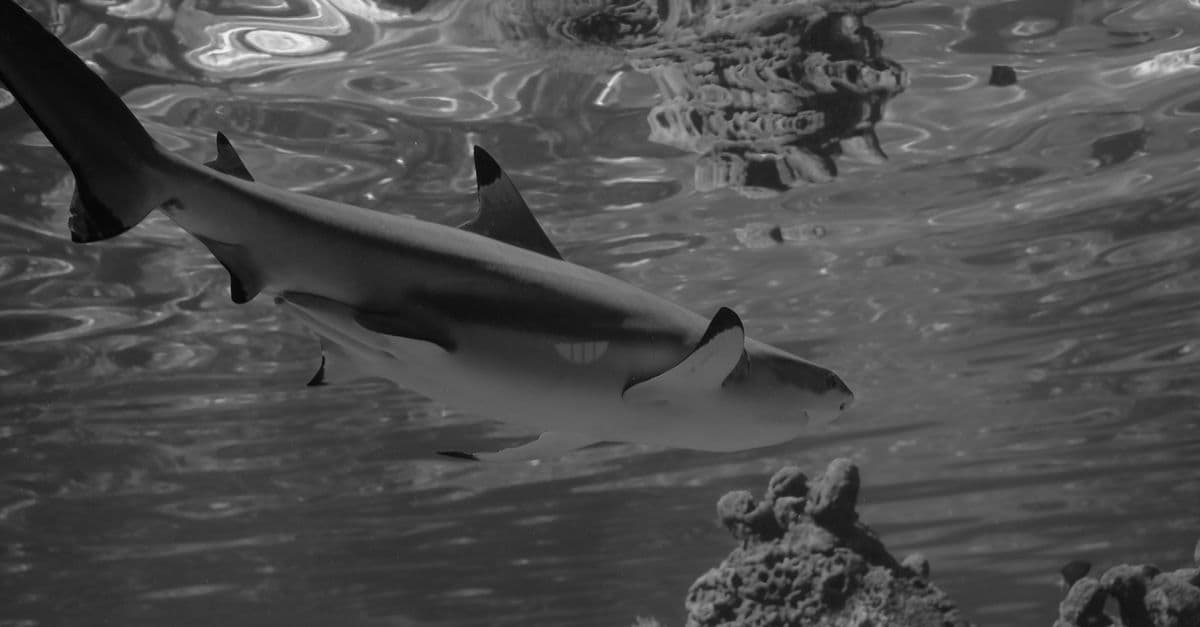
Sharks navigate a sensory world almost entirely unlike ours. They detect the bioelectric fields of a hidden flatfish buried under sand, sense water pressure waves from a struggling fish a kilometer away, and smell blood diluted to one part in ten billion. But popular culture has wildly exaggerated some of these abilities while ignoring the most extraordinary one — a sense that has no human analogue at all.
How Does the Human Eye Work? The Physics and Neuroscience of Vision
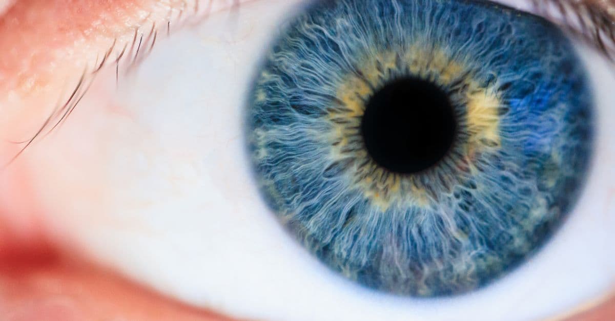
The human eye is a biological optical device, a precision camera that rivals instruments costing thousands of dollars, yet operates flawlessly for decades with zero conscious effort. Light enters through the cornea, passes through the aqueous humor, and is focused by the crystalline lens onto the retina — a thin sheet of neural tissue at the back of the eye containing two types of photoreceptors: 120 million rod cells (monochromatic, exquisitely sensitive in dim light) and 6 million cone cells (color-sensitive, active in daylight). Each photoreceptor contains millions of opsins, protein molecules that undergo a chemical transformation when struck by a photon, initiating a neural cascade that ultimately fires signals through the optic nerve to the visual cortex. The brain constructs vision from 30 discrete visual areas, each specialized for a different attribute: color, motion, depth, form. Yet we perceive a single, seamless, three-dimensional world, a construction so compelling that we believe we are seeing reality directly, unaware that the image in our visual cortex is inverted, compressed, and extensively processed.
How the Eye Works: Light, the Retina, and How the Brain Builds Sight
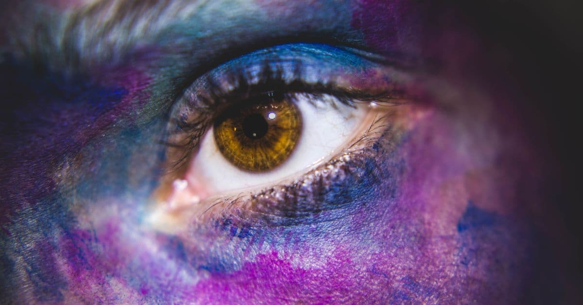
Your eye contains 126 million photoreceptors but only one million output fibers to the brain — meaning the retina compresses visual data by a factor of 126 before it leaves the eye. What you see is a model your brain built, not a photograph it took.