How Viruses Work: From Infection to Replication
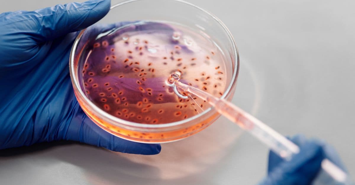
Photo: Nadezhda Moryak / Pexels
A virus is a stretch of genetic instructions in a protective shell, and it has no way to carry those instructions out on its own. It carries no ribosomes (the molecular machines that build proteins) and no way to make energy, so every virus must get inside a living cell and borrow the cell's equipment. This article follows one infection from the first contact on a cell surface to the release of new virus particles, then shows where vaccines and antiviral drugs interrupt the process and why viral DNA ended up inside our own genome.
Educational content, not medical advice. Talk to your doctor before changing any medication, treatment, or routine. Information here reflects current research but every person's situation differs.
Understanding how viruses work is the foundation of vaccine design, antiviral drug development, and a good part of modern genetics. It also clears up a common confusion. A virus is not a tiny bacterium. It is closer to a recipe card dropped into someone else's kitchen, and the kitchen does all the cooking.
Key takeaways
- Viruses are not cells: They lack metabolic machinery and can only replicate inside a host cell using the host's ribosomes and energy.
- Capsid and envelope: Viral structure consists of a protein shell (capsid) surrounding the genome, sometimes wrapped in a lipid envelope taken from the host.
- Two replication strategies: The lytic cycle produces new virions and destroys the host cell; the lysogenic or latent route keeps the viral genome quiet inside the host for a long time.
- Host specificity: Surface proteins determine which cells a virus can infect. HIV targets CD4 T cells; influenza targets respiratory epithelium.
- Interruption points: Vaccines, antibodies, and antiviral drugs each block a particular step, from attachment to release.
What is a virus, and what is it not?
A virus is a submicroscopic infectious agent made of genetic material, either DNA or RNA, enclosed in a protein coat called a capsid. Some viruses also wear an envelope, a lipid bilayer (a double layer of fat molecules) borrowed from the host cell as the virus leaves. The complete, mature particle outside a cell is called a virion. Outside a host, a virion does nothing at all. It does not eat, grow, or repair itself.
That passivity is why biologists argue about whether viruses are alive. They carry genes, they evolve by natural selection, and they reproduce, which are three hallmarks of life. But they have no metabolism of their own and cannot copy themselves without a host, which is why many textbooks place them at the edge of the living world instead of inside it. The question matters less than it sounds. Whatever label you choose, the mechanics of infection are the same.
The shell and the cargo
Capsids are built from many copies of a small number of protein subunits, which is an efficient design because a short gene can encode the subunit and the cell then makes thousands of copies. Many capsids are icosahedral, a 20-faced solid built from triangular faces. A soccer ball is a loose cousin, although a real soccer ball is a truncated icosahedron with pentagons and hexagons. Other viruses form helical rods, like a spiral staircase of protein wrapped around the genome. Bacteriophages (viruses that infect bacteria) often combine a head with a tail, like a syringe on a lander.
Genomes come in every format. A virus can store its genes as double-stranded DNA, single-stranded DNA, double-stranded RNA, or single-stranded RNA. Single-stranded RNA genomes come in two flavors. Positive-sense RNA can be read directly by ribosomes, like a finished mRNA (messenger RNA, the working copy of a gene). Negative-sense RNA is the mirror image and must be copied into positive-sense RNA before it can be read.
Seven ways to make a messenger
Virologists group viruses by how they get from genome to mRNA, because that step decides which enzymes the virus must bring with it. This scheme is called the Baltimore classification and has seven groups.
| Group | Genome | Example viruses | Key requirement |
|---|---|---|---|
| I | Double-stranded DNA | Herpesviruses, adenoviruses, poxviruses | Host or viral DNA polymerase |
| II | Single-stranded DNA | Parvoviruses | Convert to double-stranded DNA first |
| III | Double-stranded RNA | Rotaviruses | Viral RNA polymerase carried in the particle |
| IV | Positive-sense single-stranded RNA | Coronaviruses, poliovirus | Genome is translated immediately |
| V | Negative-sense single-stranded RNA | Influenza, Ebola | Carries RNA-dependent RNA polymerase |
| VI | RNA that is copied into DNA | HIV | Reverse transcriptase and integrase |
| VII | DNA that is copied through an RNA step | Hepatitis B virus | Reverse transcriptase |
The table is not trivia. Each row implies a different weak point for drugs, which comes up again below.
How big can a virus get?
Most viruses are small compared with a cell, with sizes measured in tens to hundreds of nanometers (billionths of a meter). Giant viruses break that pattern. The mimivirus, first reported in 2003, forms a roughly spherical particle about 0.75 micrometers (750 nanometers) across once its surface fibers are counted, according to the International Committee on Taxonomy of Viruses. Its genome is a single molecule of double-stranded DNA of about 1.18 million base pairs. The initial 2004 annotation predicted 911 protein-coding genes, and later transcriptome sequencing raised the count to 979 protein-coding genes.
That shift shows how much annotation improves with better data. Giant viruses carry genes for some parts of the protein-making machinery, such as tRNAs (the adapter molecules that match codons to amino acids). They still have no complete ribosome, so even the biggest virus remains dependent on a host to build its proteins.
Step one: attachment, the lock-and-key moment
Infection begins when proteins on the virion surface bind receptor molecules on a host cell. The fit is specific, like a key that opens only certain locks. That specificity explains why different viruses infect different tissues and different species.
| Virus | Viral protein | Host receptor | Main target cells |
|---|---|---|---|
| HIV | gp120 (with gp41) | CD4, plus a coreceptor such as CCR5 or CXCR4 | Helper T cells and some other immune cells |
| Influenza A | Hemagglutinin | Sialic acid on glycoproteins | Respiratory epithelium |
| SARS-CoV-2 | Spike | ACE2 | Cells of the airway, gut, and other tissues that display ACE2 |
HIV binds CD4, a protein found mainly on helper T lymphocytes, which is why the virus damages the immune system instead of, say, liver cells. For a full account of that disease and its treatment, see the HIV explainer. Influenza hemagglutinin (the "H" in H1N1) recognizes sialic acid sugars on the surface of respiratory cells. SARS-CoV-2 uses its spike protein to grab ACE2, a receptor that is present in many tissues, which helps explain why the illness it causes can reach beyond the lungs. The SARS and MERS article covers how related coronaviruses use different receptors.
The receptor is usually a normal, useful protein that the virus has learned to exploit. That is why removing the receptor is rarely a workable therapy. Cells need ACE2 and CD4 for their own jobs.
Step two: entry, getting the cargo through the wall
Once attached, the virus must get its genome past the cell membrane. Strategies fall into a few families.
Enveloped viruses fuse their membrane with a host membrane. The fusion machinery is a protein that starts in a spring-loaded shape and snaps into a new one when triggered, pulling the two membranes together like a zipper. The trigger differs by virus. HIV's trigger is receptor binding at the cell surface, so fusion happens at the plasma membrane. Influenza's trigger is acid. The cell swallows the virion into an endosome (a membrane bubble), the endosome becomes more acidic, and the lower pH makes hemagglutinin refold and drive membrane fusion, as described in a structural review of pH-dependent influenza entry.
Does SARS-CoV-2 use one entry route or two?
It can use two. In one route, the virion is taken into an endosome, where the acidic environment and an enzyme called cathepsin L help activate the spike. In the other, an enzyme on the cell surface called TMPRSS2 cuts the spike so fusion occurs at the plasma membrane. A furin-type protease also pre-processes the spike. A review in Nature Reviews Molecular Cell Biology, summarized on PubMed, lays out the roles of furin-like proteases, TMPRSS2, and cathepsin L in this multistep process. Which route dominates depends on which proteases a given cell type carries.
Non-enveloped viruses have no membrane to fuse, so they must punch through or trick the cell into taking them up. Some injectors deliver only the genome and leave the empty shell outside. Many bacteriophages do exactly this. Their tail acts like a syringe, pushes through the bacterial wall, and injects DNA. A famous 1952 experiment used this behavior to show that DNA, not protein, is the genetic material; the Hershey-Chase experiment tells that story.
Step three: uncoating and takeover
After entry, the capsid comes apart in a step called uncoating, which exposes the genome to the cell's machinery. What happens next depends on the genome type, and the Baltimore table above is the map.
Double-stranded DNA viruses such as herpesviruses and adenoviruses usually send their DNA to the nucleus. There they borrow the host's DNA polymerase and RNA polymerase to copy the genome and make mRNA. Poxviruses are the exception that proves the rule. They carry their own replication enzymes and complete much of the cycle in the cytoplasm.
RNA viruses face a problem. The cell has no enzyme that copies RNA into RNA, so the virus must bring or build one. That enzyme is the RNA-dependent RNA polymerase (RdRp). Positive-sense RNA viruses, such as coronaviruses and poliovirus, are read by host ribosomes the moment they arrive, and one of the first proteins made is the RdRp. Negative-sense viruses, such as influenza and Ebola, carry the polymerase in the particle and use it to transcribe positive-sense copies before translation can start. The influenza genome is also split into several segments, which lets two strains infecting one cell swap segments, a process called reassortment. The influenza explainer covers why that matters for pandemics.
The photocopier with no proofreader
Most RNA virus polymerases make mistakes and lack a repair step, like a photocopier that copies faithfully most of the time but never checks its work. Reviews of viral mutation rates place RNA virus mutation at about 10^-6 to 10^-4 substitutions per nucleotide per cell infection, while DNA viruses sit at 10^-8 to 10^-6, as laid out in Sanjuán and colleagues' survey of viral mutation rates.
A quick calculation shows what that means. A genome of 10,000 nucleotides copied at 10^-4 per nucleotide accumulates, on average, about one new mutation per copy. Most mutations hurt the virus, but a population producing enormous numbers of copies explores many variants, and the rare helpful ones can improve spread, evade antibodies, or resist a drug. Coronaviruses are a partial exception, since their very large RNA genomes encode an enzyme that corrects some copying errors. Even so, they still mutate far faster than DNA viruses.
Retroviruses: writing RNA into the host's own DNA
Retroviruses such as HIV reverse the usual flow of information. They carry reverse transcriptase, an enzyme that converts the RNA genome into DNA, and integrase, an enzyme that splices that DNA into a host chromosome. The integrated copy is called a provirus. From then on, every time the cell divides, the provirus is copied with it. Reverse transcriptase is error-prone, which feeds HIV's rapid evolution within a single patient.
Assembly and release: leaving the cell
The cell now holds a pool of new genomes and a pool of new viral proteins. Assembly is often self-driven. Capsid subunits snap together around genomes, much like LEGO bricks that only fit one way, and no outside instructions are needed.
Release comes in two main styles. Non-enveloped viruses commonly lyse the cell, bursting it open and spilling out the new virions. Enveloped viruses commonly bud, pushing out through the cell membrane and wrapping themselves in a piece of it. Budding can leave the cell alive for hours while it produces more virus.
Influenza adds a twist. Its hemagglutinin binds sialic acid, but sialic acid is also all over the surface of the cell the virion is trying to leave. Influenza neuraminidase cuts those sugars so the new virions are not stuck to the cell. That enzyme is the target of the drug oseltamivir, covered below.
The lytic cycle: replicate fast, destroy the cell
In the lytic cycle, the virus commandeers the cell immediately, produces new virions, and ends with the cell's destruction. The interval between infection and release is the latent period (a technical use of the word, meaning the time before new particles appear). Bacteriophage T4, which infects the gut bacterium E. coli, is the classic example. Measured values for T4 show a latent period as short as about 20 minutes, according to De Paepe and Taddei's 2006 study in PLoS Biology.
Lytic infections cause much of the damage we feel as symptoms, though not all of it. In a respiratory infection, some of the harm comes from dying cells and some from the immune response itself. The step-by-step guide to the immune response to viruses shows how the body's own defenses contribute to fever and inflammation.
The lysogenic cycle: hide and wait
In the lysogenic cycle, a virus integrates its genome into the host's DNA and goes quiet. In bacteria, the integrated phage is called a prophage and is copied along with the bacterial chromosome every time the cell divides. Bacteriophage lambda is the standard model. Its latent period in E. coli is at least about 50 minutes when it takes the lytic route, noticeably longer than T4.
Stress can flip the switch. DNA damage to a lysogenic bacterium, for instance, triggers the prophage to excise itself and start making virions, like a stowaway abandoning a sinking ship.
Latency in human viruses
Human viruses use dormancy in different ways, and the word lysogenic does not fit them all. HIV integrates a provirus into host chromosomes in a way that closely resembles a prophage. Herpesviruses take a different approach. After the first infection, herpes simplex virus establishes latency in sensory neurons, where the genome persists but few viral genes are active. Later triggers, which can include stress, fever, or immune suppression, can reactivate it. The shingles article tells the same story for the chickenpox virus, which hides in nerve cells for decades before reappearing.
Latency is why some viral infections are never fully cleared. A dormant genome is hard for the immune system to see and hard for drugs to attack, since most antivirals target active replication.
Fossil viruses in your DNA
Latency has a long-term consequence. When a retrovirus infects an egg or sperm cell and the integrated provirus is passed to offspring, the sequence becomes a permanent part of the host's lineage. These sequences are called endogenous retroviruses, or ERVs. A review of ERV envelope proteins in the placenta puts such sequences at about 8% of the human genome. Most have decayed into non-coding fragments, but a few have been recruited for host duties.
The best-known example is syncytin. The syncytin proteins come from old retroviral envelope genes, which originally helped viruses fuse with cells. In the placenta, they help cells fuse into a layer called the syncytiotrophoblast, which the same review links to fetal development, gas exchange, and tolerance between mother and fetus. The viral fusion trick survived, with a new job.
One infection, step by step in time
The steps above are easier to hold in your head as a sequence. The timeline below is a generic model, not a measurement of any one virus. Real viruses run faster or slower, and the schedule depends on the host cell and the virus.
- Contact. A virion drifts near a cell, and surface proteins bind receptors. Binding is reversible at first and becomes firm as more contacts form.
- Entry. The virion fuses at the surface or is swallowed into an endosome. The cell sees an ordinary delivery.
- Uncoating. The capsid opens and the genome is exposed.
- Early phase. The first viral genes are read. Early proteins usually switch off cell defenses and turn the cell toward viral production.
- Genome copying and late phase. Genomes are copied by the polymerase, and structural proteins, which build capsids, are made in bulk.
- Assembly. Genomes and proteins find each other and form virions.
- Release. Virions bud or burst out, and each can start the cycle in a neighboring cell.
For a phage like T4, the whole loop from injection to release can take as little as about 20 minutes under good conditions. Viruses of animals are often slower, but the logic is the same: a short quiet stretch where nothing seems to happen from outside, followed by a sudden release of a large number of particles. That quiet stretch is called the eclipse period, because no intact virion can be found in the cell even though replication is under way.
Why the early phase matters so much
The first minutes decide the outcome. A cell is not a passive victim, and it has alarms for foreign nucleic acids. Many cells detect unfamiliar RNA or DNA in the wrong place, for example double-stranded RNA in the cytoplasm, and respond by releasing interferons, signaling proteins that put neighboring cells on alert. Viruses in turn carry proteins whose job is to silence those alarms. Much of the diversity among viral genomes is not in the replication machinery at all but in these countermeasures, which is a quiet arms race that goes back as far as cells themselves.
This is also why the same virus can cause very different illnesses in different people. The speed of the cell's alarm, the state of the immune system, and the number of viral particles in the first hours all shape what follows. The overview of how the immune system recognizes threats describes the detection side of the contest.
How scientists grow and count viruses
Because viruses need living cells, they cannot be grown in a nutrient broth the way bacteria can. Laboratories grow them in cell culture, a dish of cells kept alive in a medium, or in some cases in whole organisms. One classic way to count infectious particles is the plaque assay. A thin layer of cells is covered in a gel and exposed to diluted virus. Each infectious particle kills a small patch of cells that grows into a visible clear spot, a plaque. Counting plaques at a known dilution tells you how many infectious units the sample held.
Bacteriophages are especially easy to count this way, which is one reason they were central to early molecular biology. The cultures of bacteria on a plate look like a lawn, and every phage leaves a hole in it. The approach shows a point that is easy to miss. A virus preparation contains many particles that cannot infect, such as damaged or incomplete virions, so the number of particles and the number of infectious units are not the same quantity.
Not every infection ends the same way
Infection outcomes cluster into a few patterns, which helps explain why some viral diseases are brief and others last a lifetime.
| Pattern | What happens | Typical examples |
|---|---|---|
| Acute, cleared | Fast replication, then the immune system clears the virus | Many respiratory viruses, including seasonal flu |
| Persistent | Low-level replication continues for a long time | Hepatitis B in some infected people |
| Latent | The genome sits quietly and can reactivate | Herpesviruses, including the chickenpox virus that causes shingles |
| Integrated | Viral DNA becomes part of the host chromosome | HIV; ancient retroviruses in the germline |
The pattern depends on the virus and on the host. A virus that is cleared in one person may persist in another whose immune system is weakened. The point of the table is to separate two ideas that often get mixed up. How fast a virus replicates and how long it stays in the body are different properties.
Where vaccines and drugs interrupt the cycle
Every step above is a potential target. The cleanest way to see the pattern is to line up the steps with the interventions that act on them.
| Step | What the virus does | Intervention that acts here |
|---|---|---|
| Attachment | Binds receptor | Antibodies from infection or vaccination block the binding protein |
| Entry and fusion | Fuses membranes | Antibodies; some drugs that block fusion proteins |
| Genome copying | Uses polymerase or reverse transcriptase | Polymerase inhibitors; HIV reverse transcriptase and integrase inhibitors |
| Release | Cuts free from the cell | Neuraminidase inhibitors (oseltamivir, zanamivir, peramivir) |
| Transcription of RNA | Makes viral mRNA | Baloxavir marboxil, a cap-dependent endonuclease inhibitor |
The CDC lists oseltamivir, zanamivir, and peramivir as neuraminidase inhibitors and baloxavir marboxil as a different type that interferes with viral RNA transcription. The agency advises that treatment ideally starts within 48 hours of symptom onset for the greatest benefit, with earlier treatment for people who are hospitalized or at high risk. That timing reflects the biology. Antivirals work best while the virus is still ramping up copies.
How vaccines fit in
Vaccines work before infection, not during it. They present viral proteins, or instructions for making them, to the immune system, which produces antibodies and memory cells. If the real virus later appears, those antibodies block attachment and the memory cells respond quickly. Traditional vaccines use killed or weakened virus, subunit vaccines use only selected proteins, and mRNA vaccines deliver instructions so the recipient's own cells briefly make a viral protein. The overview of how vaccines work compares the approaches, and the mRNA vaccine article covers the COVID-19 case in detail.
Why HIV needs several drugs at once
HIV has a very high mutation rate and a provirus that survives treatment, so a single drug lets resistant variants take over. Antiretroviral therapy combines drugs that hit different steps, which means a variant must acquire several unlikely mutations at once to escape. The World Health Organization describes antiretroviral therapy as stopping the virus from replicating and notes that it does not cure HIV, but allows people to live long, healthy lives. The WHO fact sheet estimates 41.0 million people were living with HIV at the end of 2025, with about 570,000 HIV-related deaths and 1.2 million new infections that year.
What people get wrong about viruses
Several beliefs about viruses are common and slightly off.
Antibiotics do not treat viral infections. Antibiotics hit structures bacteria have and viruses lack, such as bacterial cell walls and bacterial ribosomes. Because a virus has neither, antibiotics have no target. Overuse of antibiotics also breeds resistant bacteria, which is a separate problem from viral disease.
Viruses are not always trying to kill. A virus that kills its host before spreading has failed, from the point of view of natural selection. Many viruses evolve toward a balance between replicating quickly and keeping the host mobile long enough to transmit. That balance is imperfect, and the ones that make headlines are often recent arrivals in a new host species. The spillover article explains how that jump happens.
A virus is not the only infectious agent without a cell. Prions are misfolded proteins that spread by converting normal proteins into the abnormal form, with no genome at all. They are a different phenomenon, covered in the prion explainer.
A virus does not choose to infect. Attachment is chemistry. If the receptor fits, the virus binds. If it does not, the particle drifts by.
The takeaway
Every virus does the same job in a different costume: reach a cell with the right receptor, get the genome inside, convince the cell's machinery to read it, and package the copies for the next round. Almost every difference between viruses, from why HIV targets immune cells to why influenza changes every year, traces back to which receptor they bind, which polymerase they use, and how they leave. The same logic is why vaccines and antivirals work. Each blocks one step that a virus cannot do without, which is why understanding the cycle gives a map of both the disease and the cure. The fossil record in our own genome adds a last lesson. Viruses are not only a threat to the living world but one of the forces that built it.
FAQ
Q: Are viruses alive? A: Biologists disagree. Viruses have genes, evolve by natural selection, and reproduce, which are signs of life. But they have no metabolism, no cells, and no way to copy themselves without a host. Most biologists describe them as obligate intracellular parasites at the boundary of what counts as living. Giant viruses carry some protein-making genes but still lack a complete ribosome, so the boundary stays blurred without disappearing.
Q: What is the difference between a virus and a bacterium? A: A bacterium is a single living cell with its own ribosomes and metabolism, and it can reproduce independently. A virus is a genome in a protein shell with no cell structure, and it replicates only inside a host cell. Antibiotics target bacterial structures such as cell walls and ribosomes, so they do not affect viruses.
Q: Why do viruses mutate so fast? A: RNA virus polymerases make errors at roughly 10^-6 to 10^-4 substitutions per nucleotide per cell infection, and most lack a proofreading step. Combined with huge numbers of new particles, that produces a wide pool of variants. Most mutations are harmful to the virus, but some improve transmission, immune escape, or drug resistance.
Q: Can the same virus infect plants and animals? A: Almost never. Receptor matching makes most viruses highly host-specific, so a plant virus cannot infect a person. Zoonotic viruses, such as influenza, SARS-CoV-2, and Ebola, can jump from animal reservoirs to humans when they acquire the right surface-protein changes or meet cells with a compatible receptor.
Q: What makes some viruses more deadly than others? A: Several factors work together. These include how quickly the virus replicates, how much tissue it destroys, how well it avoids immune detection, whether the immune reaction itself causes damage, and whether it harms a vital organ. Virulence and transmissibility are often in tension, because a virus that quickly kills its host has less time to spread.
Q: What is the difference between the lytic and lysogenic cycles? A: In the lytic cycle, the virus replicates immediately and bursts the cell to release new particles. In the lysogenic cycle, the viral genome integrates into the host DNA and is copied each time the host divides, with no new virions until a trigger switches it to the lytic route.
Q: Why do antivirals work best early in an infection? A: Many antivirals block active copying or release, so they have the most effect while the virus is still multiplying. The CDC advises that influenza antivirals ideally start within 48 hours of symptom onset for the greatest benefit.
Sources
- Lokossou, Toudic, and Barbeau, "Implication of Human Endogenous Retrovirus Envelope Proteins in Placental Functions," Viruses, 2014: PMC4246240
- De Paepe and Taddei, PLoS Biology, 2006, as tabulated by BioNumbers: Latent period of phage T4 and lambda
- Sanjuán and colleagues, "Viral mutation rates," Journal of Virology, 2010, data hub: uv.es/rsanjuan/virmut
- Caffrey and Lavie review of pH-dependent influenza hemagglutinin entry: PMC8718792
- Jackson and colleagues, "Mechanisms of SARS-CoV-2 entry into cells," Nature Reviews Molecular Cell Biology, 2022: PubMed 34611326
- International Committee on Taxonomy of Viruses, family Mimiviridae: ictv.global/node/2047
- CDC, influenza antiviral medications summary for clinicians: cdc.gov
- World Health Organization, HIV and AIDS fact sheet: who.int
Read next:
Follow new articles
Add SciHub101 to your feed reader to see new science and technology stories.
About the Author
SciHub101 is an independent science and technology publication founded and edited by Buğra Sözeri, a software engineer and app developer based in Türkiye. AI tools assist with drafting and research. Source documentation in the existing archive is uneven, and articles are being reviewed and corrected. The Editorial Team byline identifies editorial responsibility; it does not certify specialist or independent human review. Where we compare products, anything built by our own team is labelled as such — see the editorial guidelines and About page for how corrections and disclosures work.
Related Articles
How Sleep Affects the Brain: The Science of Why You Need It
Sleep feels passive, but your brain is doing some of its most critical work while you're unconscious — consolidating memories, regulating emotion, and possibly clearing waste.
Consciousness Explained: The Hard Problem
Why does anything feel like anything at all? The hard problem of consciousness may be the deepest unsolved question in all of science — and neuroscience is just beginning to grapple with it.
How Stem Cells Work and What They Can Actually Treat
A single fertilized egg becomes a complete human being, and a few of its descendants never stop being flexible. This is how stem cells work, how scientists learned to reprogram them, and which therapies have reached patients.
Human Evolution: From Primates to Homo Sapiens
Humans and chimpanzees share a common ancestor from roughly 6 to 7 million years ago. Here is how fossils, tools, and ancient DNA reconstruct what happened next.
DNA Replication and Genetics Explained
How DNA replicates, the central dogma, mutations, epigenetics, and how consumer genetics tests like 23andMe actually work.
PCR Invention: Copying DNA Millions of Times Explained
How Kary Mullis's 1983 idea became the polymerase chain reaction, how the three-step cycle works, and why it underpins diagnostics, forensics, and research